197
Received February 15, 2008
Accepted for publication April 2, 2008
Reprint request to: Chul Jong Park, M.D., Department of Dermatology, Holy Family Hospital, College of Medi- cine, The Catholic University of Korea, 2, Sosa-dong, Wonmi-gu, Buchun 420-717, Korea. Tel: 82-32-340-2115, Fax: 82-32-340-2118, E-mail: cjpark777@yahoo.co.kr
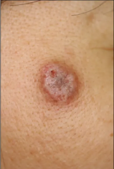
Fig. 1. A 1.5 cm in diameter, slightly erythematous to flesh colored, flat-topped hairy plaque on the right temple of the face.
A Case of Intradermal Melanocytic Nevus with Ossification (Nevus of Nanta)
Young Bok Lee, M.D., Kyung Ho Lee, M.D., Chul Jong Park, M.D.
Department of Dermatology, College of Medicine, The Catholic University of Korea, Seoul, Korea A 49-year-old woman presented with a 30-year history of asymptomatic plaque on her right temple. The histological examination revealed nests of nevus cells throughout the entire dermis.
Bony spicules were seen just beneath the nevus cell nests in the lower dermis. Cutaneous ossification is an unusual event. Herein, we present a case of intradermal melanocytic nevus with unusual ossification (nevus of Nanta). To the best of our knowledge, this is the first such case report in the Korean literature.
(Ann Dermatol (Seoul) 20(4) 197∼199, 2008) Key Words: Melanocytic nevus, Ossification
INTRODUCTION
Ossification within the skin may occur in a variety of conditions, including pilomatricoma, basal cell carcinoma, appendageal and fibrous prolifera- tion, inflammation and trauma1,2. The occurrence of ossification within a melanocytic nevus is an un- usual event3-5.
Herein, we present a case of intradermal melano- cytic nevus with unusual ossification (nevus of Nanta). To the best our knowledge, this is the first such case report in the Korean literature.
CASE REPORT
A 49-year-old woman presented with a 30-year history of asymptomatic plaque on her right temple.
Dermatological examination revealed a 1.5 cm in diameter, erythematous to flesh colored, flat topped plaque with hairs (Fig. 1). She had no history of
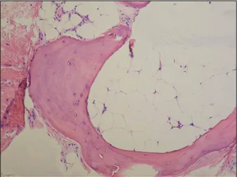
drug intake or medical illness. The histological examination showed a dense proliferation of benign nevus cells in the upper dermis. They were arranged in nests surrounding the hair follicles (Fig. 2). Bony spicules were seen in the lower dermis, underneath the nevus cell nests. Some of them were compact while others were surrounded by mature fatty tissue (Fig. 3).
Annals of Dermatology
198 YB Lee, et al. Vol. 20, No. 4, December 2008
Fig. 2. Nests of nevus cells were seen within the upper dermis, and bony spicules surrounding the mature fatty tissue were observed in the lower dermis, underneath the nevus cell nests (Hematoxylin-eosin, ×40).
Fig. 3. Bony spicules containing numerous osteocytes (Hematoxylin-eosin, ×100).
DISCUSSION
Bone formation within the skin may be a primary or secondary phenomenon. Primary cutaneous ossi- fication can occur in Albright's hereditary osteo- dystrophy and as primary osteoma cutis. Secondary metaplastic ossification occurs in association with a wide variety of conditions, including pilomatricoma, basal cell carcinoma, acne, melanocytic nevi, cel- lular blue nevus, scar, cutaneous mixed tumor, cylindroma, a cutaneous nodule of mixed connective tissue disease, dermatofibroma, ossifying plexiform tumor, pyogenic granuloma, cutis laxa-like pseudo- xanthoma elasticum, congenital plate-like osteoma cutis, organoid nevi, epidermal cyst and at the site of trauma or injection6. The melanocytic nevus with secondary ossification is referred to as 'nevus of Nanta'7. Melanocytic nevus with bone was first described by Heidingsfeld in 1908. Ever since Nanta reported on it in detail in 1911, it has been called osteo-nevus of Nanta7. The histopathology of nevus of Nanta is characterized as ossification within or
just beneath the nevus cell nests7.
To the best of our knowledge, the nevus of Nanta has not been reported on in the Korean medical literature, but there have been reports of large series in the international literature3,6,7. Both Conlin et al6 and Moulin et al3 reported that the nevus of Nanta was the most frequent cause of secondary ossifica- tion of the skin. The occurrence of nevus of Nanta was variable between 1.4%7 to 26%6 of all cutane- ous ossifications.
The origin of the ossification within the melano- cytic nevus is not precisely known. Many different theories have been proposed to explain this somewhat unusual finding. Burgdorf and Nasemann1 have suggested two mechanisms by which cutaneous ossification may occur: 1) transformation of primi- tive mesenchymal cells into osteoblasts that produce bone or 2) metaplastic transformation of other undetermined dermal cells that's stimulated by the appropriate cellular milieu. Keida et al8 suggested the involvement of transforming growth factor-β and connective tissue growth factor in the ossifi- cation of nevus of Nanta. Conlin et al6 reported that women were more commonly affected than men and they hypothesized there was an estrogenic effect on osteoblasts. Yet the exact reason why osteoma formation occurs remains to be determined.
In our case, the patient denied any preceding inflammation or trauma on the nevus, but it is difficult to exclude the possibility of previously un- recognized trauma or inflammation. We recom- mended getting an operation because there has
A Case of Intradermal Melanocytic Nevus with Ossification (Nevus of Nanta)
199been a reported case of malignant melanoma arising in a nevus of Nanta9, but the patient refused.
REFERENCES
1. Burgdorf W, Nasemann T. Cutaneous osteomas:
a clinical and histopathologic review. Arch Der- mato Res 1977;260:121-135.
2. Elder DE, Elenitsas R, Johnson BL Jr, Murphy GF.
Lever's Histopathology of the skin. 9th ed. Phila- delphia: Lippincott Williams & Wilkins, 2005:
1089-1093.
3. Moulin G, Souquet D, Balme B. Pigmented nevus and cutaneous ossifications. Apropos of 125 cases of osteonevi. Ann Dermatol Venereol 1991;118:
199-204.
4. Kanitakis J, Claudy A. Mummified ossified mela-
nocytic naevus. Eur J Dermatol 2000;10:466-467.
5. Orlow SJ, Watsky KL, Bolognia JL. Skin and bones. II. J Am Acad Dermatol 1991;25:447-462.
6. Conlin PA, Jimenez-Quintero LP, Rapini RP.
Osteomas of the skin revisited: a clinicopathologic review of 74 cases. Am J Dermatopathol 2002;
24:479-483.
7. Sasaki S, Mitsuhashi Y, Ito Y. Osteo-nevus of Nanta: a case report and review of the Japanese literature. J Dermatol 1999;26:183-188.
8. Keida T, Hayashi N, Kawakami M, Kawashima M.
Transforming growth factor beta and connective tissue growth factor are involved in the evolution of nevus of Nanta. J Dermatol 2005;32:442-445.
9. Culver W, Burgdorf WH. Malignant melanoma arising in a nevus of Nanta. J Cutan Pathol 1993;20:375-377.