서 론
견갑골 골절은 전체 골절의 0.5%–1%, 상지 골절 중 3%–5%를 차 지한다. 주위의 다양하고 두터운 근육층으로 인해 골절의 전위가 심하지 않은 경우가 많고 혈액 순환이 풍부해 골절의 유합이 빠
르며 불유합 혹은 부정유합이 발생해도 견관절 기능의 저하가 크 지 않다는 결과가 보고되어 오랜 기간 보존적 치료가 주축을 이 루었다.1,2) Judet3)을 시작으로 Magerl4)과 Hardegger 등5)이 견갑 골 체부 골절에서 수술적 치료를 보고한 이후로 수술 방법 및 결 과에 대한 연구가 늘어나고 있다. 그러나 국내에서는 수술적 치 료 결과의 증례보고6,7) 이외에는 논문이 많지 않고 보존적 치료가 주를 이루고 있다. 전위가 심한 견갑골 골절에서는 보존적 치료 후 부정유합 혹은 불유합의 가능성이 있으며 견갑-체간 관절의 이상 운동 및 견관절의 기능 저하로 이어질 수 있다.8) 이를 예방 하기 위해 전위가 심한 견갑골의 골절에서는 수술적 치료를 통한 해부학적 정복 및 골유합이 기능적 결과에 도움이 될 수 있다. 본
Copyright © 2020 by The Korean Orthopaedic Association
“This is an Open Access article distributed under the terms of the Creative Commons Attribution Non-Commercial License (http://creativecommons.org/licenses/by-nc/4.0/) which permits unrestricted non-commercial use, distribution, and reproduction in any medium, provided the original work is properly cited.”
The Journal of the Korean Orthopaedic Association Volume 55 Number 1 2020 Received September 5, 2018 Revised October 17, 2018
Accepted May 27, 2019
Correspondence to: Yoon-Min Lee, M.D., M.Sc.
Department of Orthopedic Surgery, Yeouido St. Mary's Hospital, College of Medicine, The Catholic University of Korea, 10 63-ro, Yeongdeungpo-gu, Seoul 07345, Korea
TEL: +82-2-3779-1192 FAX: +82-2-783-0252 E-mail: [email protected] ORCID: https://orcid.org/0000-0002-1404-4157
견갑골 체부 골절에서
외측 후방 금속판 고정술의 치료 결과
이윤민 • 여주동 • 송석환
가톨릭대학교 여의도성모병원 정형외과
Clinical Results of Lateral-Posterior Internal Fixation for the Treatment of Scapular Body Fractures
Yoon-Min Lee, M.D., M.Sc. , Joo-Dong Yeo, M.D., and Seok-Whan Song, M.D., Ph.D.
Department of Orthopedic Surgery, The Catholic University of Korea, Yeouido St. Mary's Hospital, Seoul, Korea
Purpose: Scapular body fractures have generally been treated with non-surgical methods. This study reports the clinical and radiological
outcomes after lateral-posterior internal fixation for treating displaced scapular body fractures.Materials and Methods:
From March 2007 to May 2017, out of 40 patients who underwent internal fixation for scapular fractures, 13 cases of lateral plate fixation of a scapular body fracture were reviewed retrospectively. Preoperative and postoperative displacement, angulation and glenopolar angle (GPA) were measured. The range of shoulder motion, visual analogue scale (VAS), and disabilities of the arm, shoulder, and hand (DASH), and Constant score were assessed at the last follow-up.Results: The mean follow-up period was 17.7 months (range, 6–45 months). The mean preoperative GPA was 23.3°±3.96° (range,
17.8°–28.1°) and the postoperative GPA was 31.1°±4.75° (range, 22.5°–40.1°). Injury to the suprascapular nerve, nonunion, fracture re- displacement, metallic failure, or infection did not occur. At the last follow-up, the mean range of motion was 150.5°±19.3° in forward flexion, 146.6°±2.34° in lateral abduction, 66.6°±19.1° in external rotation, and 61.6°±18.9° in internal rotation. The VAS, DASH, and Constant scores were 1.7±1.3, 6.2±2.4, and 86±7.9 points, respectively.Conclusion:
A scapular body fracture with severe displacement, angulation and marked decreased GPA can be stabilized by lateral- posterior plate fixation using the appropriate surgical technique with good functional and radiological results.Key words: scapula, body fracture, lateral-posterior, plate fixation, clinical results
47
Lateral-Posterior Plate Fixation for Scapular Body Fractures
연구에서는 전위된 견갑골 체부 골절에 대한 외측 금속판 고정술 시행 후 관절와-극간각(glenopolar angle, GPA)의 변화와 이와 관련된 기능적 치료 결과를 분석하고자 한다.
대상 및 방법
1. 연구 대상
본원에서 2007년 3월부터 2017년 5월까지 견갑골 골절로 수 술 받은 40명의 환자 중 견갑골 극(scapular spine), 오구돌기 (coracoid process) 및 견봉(acromion) 골절 환자를 제외하고, 체부 골절 혹은 관절와(glenoid) 골절을 포함하는 체부 골절 환 자에서 외측 금속판을 시행하고 수술 후 6개월 이상 추시한 13명 의 환자를 대상으로 후향적 연구를 진행하였다. 쇄골 골절과 견 갑골 관절와 경부 골절이 동반된 부유 견관절(floating shoulder) 은 제외하였다. 수상기전은 교통사고가 5명, 1.5 m 이상의 추락 사고가 7명, 단순 낙상이 1명이었다. 동반 손상으로는 단순 쇄골 골절이 6예, 상완골 골절이 1예, 늑골 골절이 2예, 요골두 골절이 1예, 경골 간부 골절척골이 1예였다. 견관절 단순 방사선 전후면 사진상 30° 미만의 GPA인 경우, 견갑골 측방 촬영에서 골편간의 접촉이 없이 전위되었거나 2 cm 이상의 전위가 있는 경우, 혹은 골편이 외측 경계(lateral border)에서 30°–40° 이상의 각 형성 중 하나 이상에 해당될 때 수술을 시행하였다(Table 1).9) 수술 전 견관절 전후면 사진에서 GPA를 측정하였고(Fig. 1A), 측방 촬영 (trans-scapular Y view)에서 골편의 전위 정도(Fig. 1B)와 각 변 형(Fig. 1C)을 확인하였다. 최종 추시 시 견관절의 운동범위(전방 굴곡, 외전, 신전, 내전, 외회전 및 내회전)와 시각통증점수(visual
analogue scale, VSA), disabilities of the arm, shoulder, and hand (DASH) 점수, Constant 점수를 확인하였고 최종 추시 시 GPA 및 전위의 정도를 평가하였다. 마지막 추시 시 방사선 사진 에서 견갑골 외측 피질골의 연속성이 회복되고 골절부위 간격 형 성이 사라진 경우 유합으로 판단하였다.
2. 수술 방법 및 재활
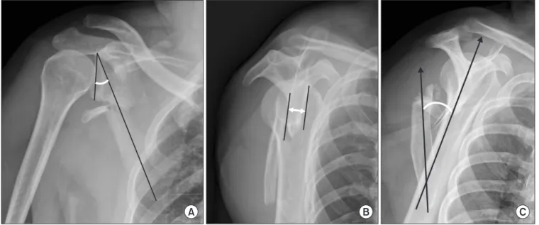
환자를 전신마취하에 측와위로 눕히고 견갑골 외측 경계에 금속 판 고정을 위하여 변형된 Judet 후방 접근법을 시행하였다(Fig.
2A). 견갑골의 내측 고정이 필요한 경우에는 저자들의 절개 보 다는 기본 Judet 후방 접근법을 사용하거나 추가적인 절개가 필 요하다. 극하근(infraspinatus muscle)과 소원근(teres minor muscle) 사이로 접근해 견갑골 외측부를 노출하였고(Fig. 2B) 극 하근을 과도하게 상방으로 당기지 않도록 주의해 극돌기 관절흔 (spinoglenoid notch)으로 분지되는 상견갑 신경 손상을 예방하 였다. 3.5 mm 재건 금속판 혹은 잠김 압박 금속판(reconstruc- tion plate or locking compression plate; Synthes, Oberdorf, Switzerland)을 사용해 내고정술을 시행하였다(Fig. 2C).
수술 후 3주까지 보조기(velpeau sling)를 착용하였고, 이후 3 주간 팔걸이(arm sling)를 착용하면서 수동적 견관절 재활운동을 권장하였다. 수술 9주부터 회전근개 및 견갑골 근력 강화 운동을 시작하였다.
통계 분석은 IBM SPSS statistics ver. 21.0 (IBM Corp., Ar- monk, NY, USA)을 이용해 실시하였다. 수술전, 후 및 정상측과 의 정규성 검증은 증례수가 적어 독립 표본 T 검증의 Wilcoxon signed-rank test를 사용하였다. p값이 0.05 이하인 경우 통계적
Table 1. Patients’ Demographic Data and Preoperative Radiological Parameters
Case No. Sex Age (yr) Side Injury mechanism Follow-up (mo) Associated injury
1 Male 50 Left Motorcycle TA 23 Clavicle fracture
2 Male 32 Left Industrial accident 15 None
3 Male 64 Left Industrial accident 16 Clavicle fracture
4 Male 57 Left Fall down 6 None
5 Male 59 Right Fall down 24 Rib fracture
6 Male 50 Left Drivers TA 12 Clavicle fracture
7 Female 53 Left Fall down 13 Vertebral fracture
8 Male 43 Left Fall down 24 None
9 Female 77 Left Slip down 35 Humerus fracture
10 Male 53 Right Pedestrian TA 11 Clavicle and radial head fracture
11 Male 53 Left Drivers TA 18 Clavicle and rib fracture
12 Female 50 Right Pedestrian TA 6 Clavicle fracture
13 Male 62 Left Fall down 12 Tibia fracture
TA, traffic accident.
으로 유의한 것으로 하였다. GPA의 정복 정도와 Constant 점수 간의 상관 관계를 확인하기 위해 Pearson correlation을 확인하 였다.
결 과
평균 수술 시간은 93.0±10.0분이었으며, 평균 실혈양은 115.2
±11.5 ml이었다. 평균 추시 기간은 평균 17.7±4.7개월(범위, 6–45개월)이었다. 1명의 환자는 내측 전위가 외측 고정 후에
도 남아 있어 추가 절개를 시행하고 내측에도 금속선 고정을 시 행하였고(Fig. 3), 나머지 12명의 환자에서는 외측 단일 금속판 및 금속선 고정만을 시행하였다(Fig. 4). 최종 추시 시 단순 방사 선 사진상 모두 골유합을 얻었다. 3명의 환자에서 평균 12개월 에 내고정물 제거술을 시행하였다. GPA는 수술 전 평균 23.3°
±3.96° (범위, 17.8°–28.1°)에서 수술 후 평균 31.1°±4.75° (범 위, 22.5°–40.1°)으로 정복되었고(p=0.001), 수술 후 GPA는 건 측(평균 35.6°±5.35°)과 비교하였을 때 통계적으로 차이가 없었 다(p=0.055). 골편 전위는 수술 전 평균 18.5 mm (범위, 8–27 Figure 2. Surgical techniques. (A) Illustration of incision line for modified Judet approach. (B) Inter-nervous approach between infraspinatus and teres minor. (C) Five-hole plate fixation at lateral posterior aspect of scapula.
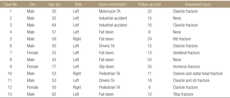
Figure 1. Measurements of the radiographical parameters. (A) The glenopolar angle (GPA) was measured on a true anteroposterior radiograph of the shoulder. A line is drawn from the superior pole of the glenoid fossa to the inferior pole. And other line is drawn from the superior pole of the glenoid fossa down to the most inferior pole of the scapular body. The angle formed by these two lines represents the GPA (normal range, 30°–40°). (B) Displacement was measured on a trans-scapular Y view. The distance between the anterior or posterior cortex of the proximal and distal fragments represents the displacement. (C) Angulation was measured on a trans-scapular Y view. One line was drawn though the proximal fragment parallel to the cortex and the other line was drawn though the distal fragment. The angle formed by these two intersecting lines represents the angulation of the fracture site.
49
Lateral-Posterior Plate Fixation for Scapular Body Fractures
mm)에서 수술 후 평균 5.1 mm (범위, 0–9 mm), 각 형성은 수 술 전 31.4° (범위, 5°–80°)에서 수술 후 6.3° (범위, 0°–15°)로 교 정되었다(Table 2). 관절운동 범위는 최종 추시 시 전방 거상 평 균 150.5°±19.3°, 외전 평균 146.6°±23.4°, 외회전 평균 66.6°
±19.1°, 내회전 평균 61.6°±18.9°로 측정되었으며 VAS는 1.7±
1.3점(범위, 0–4점), Constant 점수는 86±7.9(범위 78–95)점, DASH 점수는 6.2±2.4으로 측정되었다(Table 3). GPA와 Con- stant 점수의 Pearson correlation 점수는 0.750으로 나타났다.
외측 금속 고정 및 내측 금속선 고정을 시행한 환자에서만 내고 정물 제거술을 시행하였고, 나머지 12명의 환자는 제거 수술을 시행하지 않았다. 수술 후 상처 감염, 상견갑 신경 손상에 따른 회 전근개 기능 저하, 불유합, 골절 재전위, 내고정물 파손은 일어나 지 않았다.
고 찰
견갑골 체부 골절은 전위가 심하지 않거나 관절와 불합성이 없 는 경우 대부분 보존적 치료를 시도하며 양호한 치료 결과를 보 고한 연구들이 많았다. 견갑골 골절의 보존적인 치료로서 Goss10) 는 3–4주간의 sling이나 swathe로 관절을 고정한 후 조기에 관 절운동을 시작하면 6주 이후 골유합이 되고 6–12개월에 회복을 기대할 수 있다고 하였고 Wilber와 Evans11)는 30예의 견갑골 체 부 골절에서 보존적 치료 이후의 좋은 결과에 대해 보고하였다.
그러나 골절의 전위 및 각 형성이 심한 경우 불유합 혹은 부정유 합이 발생해 관절운동 범위 감소, 만성 통증 등 회복 후에도 기능 적인 측면에서의 저하가 발생할 가능성이 높고 수술의 적응증 및 술기에 대한 관심이 높아지면서 최근에는 체부 골절에 대한 수술 적 치료가 점차 증가하고 있다.
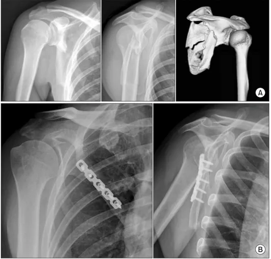
견관절은 운동은 관절와-상완 관절(glenohumeral joint), 견 봉-쇄골 관절(acromioclavicular joint), 흉쇄 관절(sternocla- Figure 3. A Fifty-year-old female, fracture of the right scapular body with comminution of lateral border. (A) Plain and computed tomography image showed a comminuted fracture with 26.2° of glenopolar angle (GPA) and 37 mm displacement. (B) After fixation of the lateral border with a dynamic compression plate and wiring for the medial border, GPA changed to 31.8° without displacement and angulation. (C) Radiographs after the removal operation.
Table 2. Changes in the Radiological Parameters
Case
No.
Glenopolar angle (°) Displacement (mm) Angulation (°)
Pre-op Contra-lateral Post-op p-value* Pre-op Post-op p-value* Pre-op Post-op p-value*
1 20.7 35.0 30.1 24 5 29.0 8.5
2 17.8 24.2 30.9 13 8 10.0 5.4
3 20.9 38.0 30.9 27 0 22.0 0
4 24.0 35.0 33.1 17 6 26.0 7.5
5 18.2 35.0 22.5 16 0 24.0 13.0
6 28.1 32.0 30.7 10 7 60.0 5.0
7 22.3 37.8 36.0 18 6 54.0 0
8 27.5 37.2 27.5 26 9 28.0 15.0
9 27.9 33.4 40.1 20 8 23.0 0
10 18.7 38.9 36.4 8 5 21.0 0
11 28.1 36.3 37.4 20 0 5.0 8.5
12 26.1 41.2 32.8 23 5 80.0 10.0
13 22.3 40.5 35.9 25 7 26.0 9.3
Mean (range)
23.3 (17.8–28.1)
35.6 (24.2–41.2)
31.1 (22.5–40.1)
0.001 19 (8–27)
5.1 (0–9)
0.013 31.4 (5–80)
6.3 (0–15)
0.008 Pre-op, preoperative; Post-op, postoperative. *p-value for Wilcoxon signed-rank test.
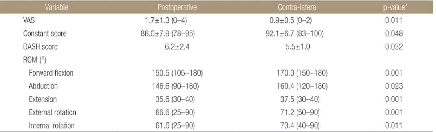
Figure 4. A 42-year-old male, fracture of the right scapula. (A) Preoperative images showed a comminuted body fracture with 22° in glenopolar angle (GPA) and 20 mm displacement. (B) GPA was reduced in the normal range (31.1°) after lateral dynamic compression plate fixation with 96 points of the Constant score and full range of shoulder motion.
51
Lateral-Posterior Plate Fixation for Scapular Body Fractures
vicular joint) 및 견갑-흉곽 관절(scapulothoracic joint)의 복 합적인 운동으로 이루어져 있어 불유합이나 부정유합으로 견 갑골의 골성 구조의 변화가 생기면, 견갑흉곽 관절의 이상운동 (dyskinesia), 탄발음 및 통증이 발생해 견관절의 기능 저하를 일 으킬 수 있다.8,12) Ada와 Miller13)도 전위된 견갑골 체부 골절에 서는 보존적 치료 후 만성적인 통증과 관절운동 저하가 발생하기 때문에 수술적인 치료가 필요하다고 주장하였다. 또한 견관절 불 안정성이 있는 환자에서도 견갑골 주위 강화 운동이 중요한 것처 럼 부정유합된 견갑골 보다는 해부학적으로 정복된 견갑골이 근 육 운동 회복에 영향을 미쳐 견관절 기능이 향상되는 결과를 보 일 수 있다.14,15)
관절 외 견갑골 골절에 대한 영상 검사에서 전후면 사진을 통 해 내외측 전위 및 GPA를 확인할 수 있으며, 견갑골 Y 사진에서 는 전위 및 각 형성을 평가할 수 있다.16,17) 이 중 GPA는 견갑골의 해부학적 정복 정도를 평가할 때 유용하며 견갑골 골절의 치료 결과에 영향을 미치는 요소로 알려져 있다.15,18,19) Morey 등20)은 체계적인 문헌 고찰(systematic review)에서 GPA의 정복 정도가 클수 록 높은 Constant 점수를 보였다고 보고하였고, 부유 견관절의 치료에 서 GPA와 임상 결과의 관계를 연구한 보고들에서도21,22) GPA의 정복 이 양호할수록 Constant 점수도 유의하게 높았다고 하였다. 본 연구 결과에서도 GPA는 수술 전 23.3°에서 수술 후 31.1°로 회복 되었다. Constant 점수와의 연관 계수도 0.750으로 GPA가 임상 결과에 영향을 미치는 방사선적 요소이었으며 이 각의 회복이 관 절와의 하부 경사(caudal tilt of glenoid)의 증가를 예방하여 견 관절의 기능 향상에 도움이 될 것이라고 생각한다.
견갑골 골절 시 보존적 치료가 수술적 치료보다 널리 받아들 여지는 이유는 보존적 치료에도 골유합이 잘 되며 기능도 양호 한 경우가 많을 뿐만 아니라 내고정 시 견갑골의 특이한 골성 구 조와 주위의 두꺼운 근육층 때문에 수술 시 접근 및 정복이 쉽지
않고 상견갑 신경(suprascapular nerve)과 액와신경(axillary nerve)의 손상 위험이 있기 때문이다. 그러나 견갑골의 외측 경 계는 피질골의 골질이 좋고 체부의 중앙이나 내측에 비해 두꺼워 약 14 mm 이상의 나사 삽입이 가능하다.14) 또한 잠김 금속판을 사용 한다면 골 두께나 골질로 인한 나사 삽입의 약점을 극복하여 안 정된 고정을 할 수 있다. 또한 상견갑 신경의 지배를 받는 극하건 과 액와 신경의 지배를 받는 소원근 사이로 접근하면 신경간 접 근법(internervous plane)으로 수술을 시행하여 신경 손상을 예 방할 수 있어 수술 후 회전근개의 근 위축을 예방할 수 있다.23,24) 본 연구는 후향적 연구이며 대상자의 수가 13예로 적고 이에 따라 추시 기간이 6개월인 증례도 포함되어 있다는 제한점이 있 다. 또한 견갑골 체부 골절의 보존적 치료 결과나 타 수술 방법과 의 비교 논문이 아니어서 해당 수술 술기의 우월성을 직접적으로 증명할 수 없다.
결 론
2 cm 이상 골절의 전위, 30°–40° 이상 각 형성 및 GPA가 22° 미 만으로 감소한 견갑골 체부 골절에서 외측 후방 금속판 고정은 적절한 수술 술기를 통해 골절의 정복 및 안정된 고정이 가능하 고, 방사선 사진상 만족스러운 골유합 및 방사선 지표의 호전과 함께 양호한 기능적 결과를 얻을 수 있다.
CONFLICTS OF INTEREST
The authors have nothing to disclose.
Table 3. Clinical Results
Variable Postoperative Contra-lateral p-value*
VAS 1.7±1.3 (0–4) 0.9±0.5 (0–2) 0.011
Constant score 86.0±7.9 (78–95) 92.1±6.7 (83–100) 0.048
DASH score 6.2±2.4 5.5±1.0 0.032
ROM (°)
Forward flexion 150.5 (105–180) 170.0 (150–180) 0.001
Abduction 146.6 (90–180) 160.4 (120–180) 0.023
Extension 35.6 (30–40) 37.5 (30–40) 0.001
External rotation 66.6 (25–90) 71.2 (50–90) 0.001
Internal rotation 61.6 (25–90) 73.4 (40–90) 0.011
Values are presented as mean±standard deviation (range) or mean (range). VAS, visual analogue scale; DASH, disabilities of the arm, shoulder, and hand; ROM, range of motion. *p-value for Wilcoxon signed-rank test.
ORCID
Yoon-Min Lee, https://orcid.org/0000-0002-1404-4157 Joo-Dong Yeo, https://orcid.org/0000-0002-6794-9267 Seok-Whan Song, https://orcid.org/0000-0002-8578-4697
REFERENCES
1. Nordqvist A, Petersson C. Fracture of the body, neck, or spine of the scapula. A long-term follow-up study. Clin Or- thop Relat Res. 1992;283:139-44.
2. You JW, Yoon HJ. Conservative treatment of scapular frac- tures. J Korean Soc Fract. 1994;7:364-70.
3. Judet R. [Surgical treatment of scapular fractures]. Acta Or- thop Belg. 1964;30:673-8. French.
4. Magerl F. [Osteosyntheses in the shoulder region. Pertuber- cular humeral fractures, scapular neck fractures]. Helv Chir Acta. 1974;41:225-32. German.
5. Hardegger FH, Simpson LA, Weber BG. The operative treat- ment of scapular fractures. J Bone Joint Surg Br. 1984;66:725- 31.
6. Shim DM, Kim JW, Kweon SH, Jeung UO, Lee JM. Surgical treatment of scapular fracture using by plate fixation: 4 cases report. J Korean Fract Soc. 2006;19:381-7.
7. Cole PA, Gauger EM, Schroder LK. Management of scapular fractures. J Am Acad Orthop Surg. 2012;20:130-41.
8. Chadwick EK, van Noort A, van der Helm FC. Biomechan- ical analysis of scapular neck malunion--a simulation study.
Clin Biomech (Bristol, Avon). 2004;19:906-12.
9. Armstrong CP, Van der Spuy J. The fractured scapula: im- portance and management based on a series of 62 patients.
Injury. 1984;15:324-9.
10. Goss TP. Fractures of the glenoid cavity. J Bone Joint Surg Am. 1992;74:299-305.
11. Wilber MC, Evans EB. Fractures of the scapula. An analysis of forty cases and a review of the literature. J Bone Joint Surg Am. 1977;59:358-62.
12. Kibler WB, McMullen J. Scapular dyskinesis and its relation to shoulder pain. J Am Acad Orthop Surg. 2003;11:142-51.
13. Ada JR, Miller ME. Scapular fractures. Analysis of 113 cases.
Clin Orthop Relat Res. 1991;269:174-80.
14. Bartoníček J, Frič V. Scapular body fractures: results of opera- tive treatment. Int Orthop. 2011;35:747-53.
15. Jones CB, Sietsema DL. Analysis of operative versus nonop- erative treatment of displaced scapular fractures. Clin Orthop Relat Res. 2011;469:3379-89.
16. Anavian J, Conflitti JM, Khanna G, Guthrie ST, Cole PA.
A reliable radiographic measurement technique for ex- tra-articular scapular fractures. Clin Orthop Relat Res.
2011;469:3371-8.
17. Wijdicks CA, Anavian J, Hill BW, Armitage BM, Vang S, Cole PA. The assessment of scapular radiographs: analysis of anteroposterior radiographs of the shoulder and the effect of rotational offset on the glenopolar angle. Bone Joint J.
2013;95-B:1114-20.
18. Bozkurt M, Can F, Kirdemir V, Erden Z, Demirkale I, Başbozkurt M. Conservative treatment of scapular neck frac- ture: the effect of stability and glenopolar angle on clinical outcome. Injury. 2005;36:1176-81.
19. Romero J, Schai P, Imhoff AB. Scapular neck fracture: the influence of permanent malalignment of the glenoid neck on clinical outcome. Arch Orthop Trauma Surg. 2001;121:313-6.
20. Morey VM, Chua KHZ, Ng ZD, Tan HMB, Kumar VP. Man- agement of the floating shoulder: does the glenopolar angle influence outcomes? A systematic review. Orthop Traumatol Surg Res. 2018;104:53-8.
21. Kim KC, Rhee KJ, Shin HD, Yang JY. Can the glenopolar an- gle be used to predict outcome and treatment of the floating shoulder? J Trauma. 2008;64:174-8.
22. Izadpanah K, Jaeger M, Maier D, Kubosch D, Hammer TO, Südkamp NP. The floating shoulder—clinical and radiologi- cal results after intramedullary stabilization of the clavicle in cases with minor displacement of the scapular neck fracture.
J Trauma Acute Care Surg. 2012;72:E8-13.
23. Noguchi T, Mautner JF, Duncan SFM. Dorsal plate fixation of scapular fracture. J Hand Surg Am. 2017;42:843.e1-843.e5.
24. Ao R, Yu B, Zhu Y, Jiang X, Shi J, Zhou J. Single lateral versus medial and lateral plates for treating displaced scapular body fractures: a retrospective comparative study. J Shoulder El- bow Surg. 2018;27:231-6.
견갑골 체부 골절에서
외측 후방 금속판 고정술의 치료 결과
이윤민 • 여주동 • 송석환
가톨릭대학교 여의도성모병원 정형외과
목적:
전위가 있는 견갑골 체부 골절에서 외측 후방 금속판을 이용한 내고정술 시행 후 방사선적 및 기능적 치료 결과를 보고하고 자 한다.대상 및 방법:
2007년 3월부터 2017년 5월까지 견갑골 골절로 수술을 받은 40명의 환자 중 견갑골 체부 골절에 대해 외측 후방 금 속판 고정을 사용한 13예의 환자를 후향적으로 연구하였다. 수술 전과 수술 후 골편 전위, 각 형성 및 관절와-극간각을 측정하였 고, 마지막 추시 시 관절운동 범위와 시각통증점수(visual analogue scale, VAS), disabilities of the arm, shoulder, and hand (DASH) 및 Constant 점수를 평가하였다.결과:
평균 추시 기간은 17.7개월(범위, 6–45개월)이었고, 견갑와-극간각은 수술 전 평균 23.3°±3.96° (범위, 17.8°–28.1°)에서 수술 후 평균 31.1°±4.75° (범위, 22.5°–40.1°)로 측정되었다. 수술 후 신경 및 혈관 손상, 불유합, 골절 전위, 내고정물 파손, 감 염은 일어나지 않았다. 마지막 추시 시 관절운동 범위는 전방 거상 평균 150.5°±19.3°, 외전 평균 146.6°±23.4°, 외회전 평균 66.6°±19.1°, 내회전 평균 61.6°±18.9°로 측정되었으며 VAS는 1.7±1.3점, DASH 점수는 6.2±2.4점, Constant 점수는 86±7.9 점으로 측정되었다.결론:
심한 골절의 전위, 각 형성 및 관절와-극간각의 감소가 뚜렷한 견갑골 체부 골절에서 외측 후방 금속판 고정은 적절한 수술 술기를 통해 골절의 정복 및 안정된 고정이 가능하고 방사선 사진상 만족스러운 골유합 및 방사선 지표의 호전과 양호한 기능적 결과를 얻을 수 있다.색인단어: 견갑골, 체부 골절, 외측 후방, 금속판 고정술, 임상 결과
접수일 2018년 9월 5일 수정일 2018년 10월 17일 게재확정일 2019년 5월 27일 책임저자 이윤민
07345, 서울시 영등포구 63로 10, 가톨릭대학교 의과대학 여의도성모병원 정형외과학교실
TEL 02-3779-1192, FAX 02-783-0252, E-mail [email protected], ORCID
https://orcid.org/0000-0002-1404-4157Original Article J Korean Orthop Assoc 2020; 55: 46-53 • https://doi.org/10.4055/jkoa.2020.55.1.46 www.jkoa.org pISSN : 1226-2102, eISSN : 2005-8918
53
Copyright © 2020 by The Korean Orthopaedic Association
“This is an Open Access article distributed under the terms of the Creative Commons Attribution Non-Commercial License (http://creativecommons.org/licenses/by-nc/4.0/) which permits unrestricted non-commercial use, distribution, and reproduction in any medium, provided the original work is properly cited.”