Evaluation of Post-
Neoadjuvant Chemotherapy Pathologic Complete
Response and Residual
Tumor Size of Breast Cancer:
Analysis on Accuracy of MRI and Affecting Factors
신보강화학요법 후 유방암의 병리학적 완전 관해 예측 및 잔류 암 평가: 유방자기공명영상의 정확도 및 영향인자 분석
Hyun Soo Ahn, MD
1, Yeong Yi An, MD
1* , Ye Won Jeon, MD
2, Young Jin Suh, MD
2, Hyun-Joo Choi, MD
31Department of Radiology, St. Vincent’s Hospital, College of Medicine, The Catholic University of Korea, Suwon, Korea
2Department of Surgery, Division of Breast & Thyroid Surgical Oncology, College of Medicine, The Catholic University of Korea, Seoul, Korea
3Department of Pathology, College of Medicine, The Catholic University of Korea, Seoul, Korea
Purpose To evaluate the accuracy of MRI in predicting the pathological complete response (pCR) and the residual tumor size of breast cancer after neoadjucant chemotherapy (NAC), and to determine the factors affecting the accuarcy.
Materials and Methods Eighty-eight breast cancer patients who underwent surgery after NAC at our center between 2010 and 2017 were included in this study. pCR was defined as the ab- sence of invasive cancer on pathological evaluation. The maximum diameter of the residual tu- mor on post-NAC MRI was compared with the tumor size of the surgical specimen measured pathologically. Statistical analysis was performed to elucidate the factors affecting pCR and the residual tumor size-discrepancy between the MRI and the pathological measurements.
Results The pCR rate was 10%. The diagnostic accuracy of MRI and the area under the curve for predicting pCR were 90.91% and 0.8017, respectively. The residual tumor sizes obtained using MRI and pathological measurements showed a strong correlation (r = 0.9, p < 0.001), especially in patients with a single mass lesion (p = 0.047). The size discrepancy between MRI and the path- ological measurements was significantly greater in patients with the luminal type (p = 0.023) and multifocal tumors/non-mass enhancement on pre-NAC MRI (p = 0.047).
Received June 11, 2020 Revised July 26, 2020 Accepted August 11, 2020
*Corresponding author Yeong Yi An, MD Department of Radiology, St. Vincent’s Hospital, College of Medicine,
The Catholic University of Korea, 93 Jungbu-daero, Paldal-gu, Suwon 16247, Korea.
Tel 82-31-249-8495 Fax 82-31-247-5713 E-mail [email protected] This is an Open Access article distributed under the terms of the Creative Commons Attribu- tion Non-Commercial License (https://creativecommons.org/
licenses/by-nc/4.0) which permits unrestricted non-commercial use, distribution, and reproduc- tion in any medium, provided the original work is properly cited.
ORCID iDs Hyun Soo Ahn https://
orcid.org/0000-0001-9167-709X Yeong Yi An
https://
orcid.org/0000-0003-0905-8498 Ye Won Jeon
https://
orcid.org/0000-0001-6578-5687 Young Jin Suh
https://
orcid.org/0000-0003-1033-3848 Hyun-Joo Choi
https://
orcid.org/0000-0003-2292-424X
Conclusion MRI is an accurate tool for evaluating pCR and residual tumor size in breast cancer pa- tients who receive NAC. Tumor subtype and initial MRI features affect the accuracy of MRI.
Index terms Breast Neoplasm; Neoadjuvant Therapy; Magnetic Resonance Imaging
서론
수술 전 신보강화학요법은 국소적 진행 유방암 환자뿐만 아니라 수술 가능한 병기의 유방암 환 자를 대상으로도 흔히 시행되고 있는 치료이다. 신보강화학요법은 수술 후 보조화학요법과 비등 한 무질병생존기간과 전체생존기간을 보일 뿐 아니라(1, 2), 병변의 크기를 감소시켜 유방 보존 수 술을 가능하게 하고, 미세전이를 통제하며, 생체 내에서의 반응 평가를 통해 질환의 예후 예측을 가능하게 하는 장점이 있다(3-5).
병리학적 완전 관해(pathological complete response)와 잔류 암(residual tumor)에 대한 정 확한 평가는 치료 및 수술 방침을 결정하고 예후를 예측하는 데 있어서 매우 중요하다. 그 이유는 신보강화학요법 후의 병리학적 완전 관해가 무질병생존기간과 전체생존기간에 대한 가장 확실한 대리 표지자이기 때문이다(4). 또한 신보강화학요법 종료 후에도 잔류 암이 남아있는 환자군에서 는 잔류 암의 완전 절제가 좋은 예후와 관련이 있기 때문에 수술적 절제가 필요한 부위를 정확히 파악하는 것이 필수적이라고 할 수 있다.
유방자기공명영상은 신보강화학요법 후의 병리학적 완전 관해와 잔류 암에 대한 평가에 있어서 가장 신뢰할 수 있는 검사 방법으로 알려져 있다(6-9). 하지만 유방자기공명영상도 완벽한 검사 방 법은 아니며, 영상과 병리 조직에서의 잔류 암의 크기 간에는 차이가 존재한다. 그리고 이로 인해 불필요하게 광범위하거나 또는 불충분한 수술이 이루어질 위험성이 있다.
따라서 이 연구는 1) 유방암의 임상병리학적 예후인자와 자기공명영상 소견 중 병리학적 완전 관해와 관련이 있는 인자들이 무엇인지 알아보고, 2) 병리학적 완전 관해를 예측하는 데 있어서 유 방자기공명영상의 정확도를 유방암의 아형에 따라 분석하고, 3) 자기공명영상과 병리 조직 간의 잔류 암 크기 차이와 이에 영향을 미치는 인자들이 무엇인지 알아보고자 한다.
대상과 방법
연구 대상 선정 및 자료조사
이 연구는 후향적 연구로 본원 임상시험심사위원회의 승인을 받았으며 동의서는 면제되었다
(IRB No. VC19RESI0092). 2010년부터 2017년까지 본원에서 유방암으로 진단받고 신보강화학요
법을 완료한 후 유방 보존 수술 또는 유방 전절제술을 시행 받은 88명의 환자를 대상으로 선정하
였다. 9명(10%)의 환자는 Adriamycin-Cyclophosphamide 화학요법, 24명(27%)은 Adriamycin-
Taxane 화학요법, 24명(27%)은 Fluorouracil-Adriamycin-Cyclophosphamide 화학요법, 그리
고 31명(35%)은 Docetaxel-Gemcitabine 화학요법을 받았다. Human epidermal growth factor
receptor-2 수용체(이하 HER2) 양성 환자들은 표적치료를 추가적으로 시행하였다. 선행화학요법 을 시작하기 전과 마지막 주기 화학요법을 시행한 후에 역동적 조영증강 자기공명영상을 시행하 였다. 항암화학요법을 완료한 후에 82명(93%)의 환자는 유방 보존 수술을 받았고, 6명(7%)의 환 자는 전절제술을 시행하였다. 수술은 모든 환자에서 시행되었으며 수술 전 마지막으로 자기공명 영상을 시행한 시점과 수술 시점 간의 차이는 평균 18일이었다(범위, 5~47).
영상검사의 획득
자기공명영상은 3.0-Tesla 자기공명영상 촬영기(Magnetom Verio; Siemens Medical Solu- tions, Erlangen, Germany)와 유방 전용 코일을 사용하여 엎드린 자세에서 양측 유방의 영상을 얻었다. 이용된 자기공명영상의 펄스 연쇄(pulse sequence)와 파라미터들은 다음과 같다. T2 강 조 축상면 영상(axial T2 weighted image), 확산 강조 영상(diffusion weighted image)을 얻었으 며, 역동적 조영증강 자기공명영상은 지방 억제 후 T1 강조 축상면 영상(T1-weighted flash 3D volumetric interpolated breath-hold sequence, repetition time/echo time: 4.4/1.7 ms, flip angle: 10, 1.2 mm slice thickness with no gap, acquisition time: 60 s)에서 조영제 0.1 mmoL/
kg gadopentetate dimeglumine (Dotarem; Guerbet, Roissy, France)을 주입 후 연속적으로 7, 67, 127, 187, 247, 367초 후에 6번의 영상을 얻었다. 후처리 과정을 통해 감산 영상(subtraction image)을 얻었고 다평면 재구성(multiplanar reconstruction) 하여 시상 입체 영상 및 최대 강조 투사 영상(maximal intensity projection)을 얻었다.
영상검사의 해석
환자의 임상정보, 자기공명영상 이외의 영상검사 결과, 병리 결과를 모르는 상태에서 10년 이상 의 경험이 있는 유방 영상 전문의와 한 명의 영상의학과 전공의가 후향적으로 자기공명영상 검사 를 판독하여 반응 양상을 분석하였으며, 차이가 있는 경우 토론 뒤에 합의 후 결론을 내었다. 반응 평가는 response evaluation criteria in solid tumors 1.1 (RECIST 1.1)에 의거하여 시행하였다.
신보강화학요법 후 시행한 유방자기공명영상에서 이전에 유방암이 있었던 부분에 남아있는 조영 증강이 없거나 미미하여 정상 주변조직과 구분되지 않는 경우를 임상적 완전 관해로 정의하였다.
조영증강을 보이는 병변이 있으면 잔류 암으로 간주하고 그 크기는 최대 강조 투사 영상에서 측정 한 최대 직경으로 정의하였다. 병변이 여러 개인 경우, 가장 큰 2개의 병변의 최대 직경의 합으로 반응 평가를 시행하였다.
병리학적 분석
20년의 경험이 있는 한 명의 유방 병리학 전문의가 신보강화학요법의 반응 평가를 위해 수술 후
얻은 병리 조직을 분석하였다. 병리 검사 결과를 이용하여 유방암의 조직학적 유형, 조직학적 분
화도, 액와림프절 전이, 림프관 침윤, 신경 주위 침윤, 혈관 침윤, Ki-67, 에스트로겐 수용체(estro-
gen receptor; 이하 ER), 프로게스테론 수용체(progesteron receptor; 이하 PR), HER2의 발현을
각각 기록하였다. 유방암은 면역조직화학 표지자를 이용하여 다음과 같이 4가지 주요 아형으로
분류하였다; 내강형 A (ER+, HER2-, Ki-67 < 14%), 내강형 B (ER+, HER2-, Ki-67 ≥ 14% or ER+, HER2+), HER2+ (ER-, PR-, HER2+), 삼중음성형(triple-negative breast cancer; 이하 TN) (ER-, PR-, HER2-). 병리학적 완전 관해는 병리 결과에서 침윤성 유방암이 발견되지 않는 것으로 정의하 였고, 유관상피내암의 유무는 고려하지 않았다. 병리학적 완전 관해 이외의 모든 소견은 병리적 불완전 관해로 정의하였다.
통계학적 분석
병리학적 완전 관해에 영향을 미치는 임상병리학적 예후인자와 자기공명영상 특징을 알아보기 위해 단변량 분석을 시행하였고, 연속적 변수는 independent two sample t-test, 비연속적 변수 는 Fisher’s exact test와 chi-square test를 이용하여 분석하였다. 단변량 분석에서 p 값이 0.05 미 만인 인자들을 통계적으로 유의한 것으로 보고, 해당 인자들을 대상으로 로지스틱 회귀 분석을 이 용한 다변량 분석을 시행하여 교차비(odds ratio)와 신뢰구간(confidence interval)을 계산하였다.
자기공명영상으로 병리학적 완전 관해를 예측하는 데 있어서의 진단적 정확도는 receiver op- erating characteristic curve를 이용하여 분석하였고, 유방암 전체 및 아형별로 정확도에 차이가 있는지에 대해 분석하였다. 곡선하부면적(area under the curve)이 0.7이 넘는 경우 우수한 판별 자로 생각하였다(10). 자기공명영상과 병리 조직에서의 잔류 암 크기의 연관성은 Pearson corre- lation을 이용하여 분석하였다. 자기공명영상과 병리 조직에서의 잔류 암 크기 차이에 영향을 미 치는 인자는 independent two sample t-test와 analysis of variance를 이용하여 분석하였다. 모 든 통계적 분석은 SAS version 9.4 (SAS Institute, Cary, NC, USA)를 이용하여 시행되었다.
결과
전체 88명의 환자들 중에서 9명(10%)이 병리학적 완전 관해에 도달하였고, 불완전 관해는 79명 (90%)이었다. 이외의 임상병리학적 소견들은 Table 1에 정리하였다. 병리학적 완전 관해와 병리 학적 불완전 관해 군 간에 통계적으로 유의미한 임상병리학적 차이는 없었다(Table 1). 유방암의 치료 전 자기공명영상 소견 중에서는 단초점 병변( p = 0.014), 단일 종괴(p = 0.0122), 테두리 조영 증강(rim enhancement)의 부재(p = 0.009), T2 강조영상 상 종양 내부 고신호강도의 부재(p = 0.0239)가 병리학적 완전 관해와 유의한 연관성이 있었다(Table 2). 하지만 위의 인자들을 대상으 로 다변량 로지스틱 회귀분석을 시행하였을 때 병리학적 완전 관해와 연관성을 보이는 독립적인 예측 인자는 없었다.
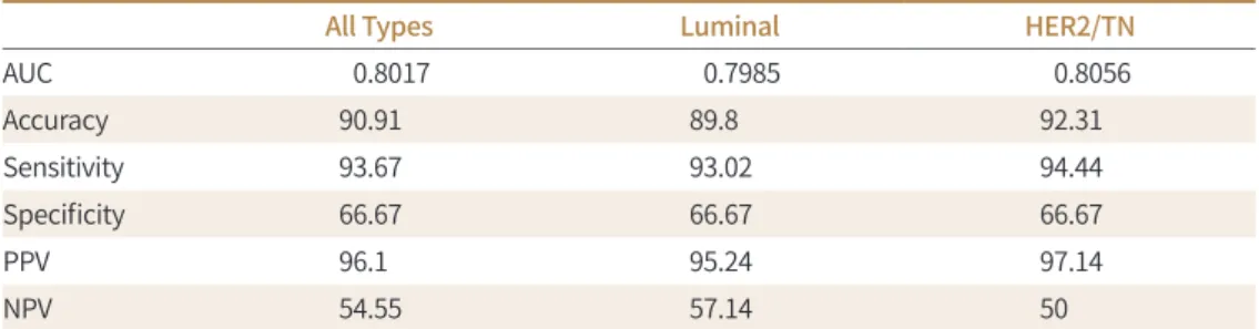
병리학적 완전 관해를 예측하는 데 있어서 유방자기공명영상의 진단 능력을 유방암의 아형에 따라 살펴본 결과, 전체 유방암 아형에서의 곡선하부면적 값은 0.8017, 내강형은 0.7985, HER2+/
TN 유형은 0.8056이었다. 모든 아형에서 곡선하부면적 값이 0.7이 넘어 자기공명영상은 병리학적
완전 관해 예측에 있어서 우수한 진단능력을 보였다. 그리고 민감도는 93.67로 높았지만 특이도는
66.67로 상대적으로 낮은 값을 보였으며, 정확도는 HER2+/TN 유형(92.31)에서 내강형보다(89.8)
조금 높게 나타났다(Table 3).
신보강화학요법 후 자기공명영상에서 측정한 잔류 암 크기와 실제 병리 조직에서의 잔류 암 크 기는 강한 연관성이 있었고( r = 0.9, p < 0.001) (Fig. 1), 유방암 아형과 치료 전 자기공명영상 소견 에 따라 다른 결과를 보였다. 잔류 암 크기의 차이는 HER2+/TN 유형보다 내강형에서 유의미하게
Table 1. Clinicopathologic Characteristics of the PatientsCharacteristic All Patients (n = 88)
Patients with pCR (n = 9)
Patients with Non-pCR*
(n = 79) p-Value
Age (years), mean ± SD 48.84 49.89 ± 8.43 48.72 ± 8.33 0.692
Pre NAC stage (AJCC 7th) 0.434
II 64 (72.73) 8 (88.89) 56 (70.89)
III 24 (27.27) 1 (11.11) 23 (29.11)
Chemotherapy regimen 0.370
AC 9 (10.23) 0 (0) 9 (11.39)
TA 24 (27.27) 1 (11.11) 23 (29.11)
FAC 24 (27.27) 2 (22.22) 22 (27.85)
DG 31 (35.23) 6 (66.67) 25 (31.65)
Pathology 0.345
IDC, NOS 71 (80.68) 9 (100) 62 (78.48)
ILC 4 (4.55) 0 (0) 4 (5.06)
Others 13 (14.77) 0 (0) 13 (16.46)
Axillary lymph node metastasis 0.485
No 45 (51.14) 6 (66.67) 39 (49.37)
Yes 43 (48.86) 3 (33.33) 40 (50.63)
Histologic grade† 0.676
Grade 1/2 64 (72.73) 6 (85.71) 58 (74.36)
Grade 3 21 (23.86) 1 (14.29) 20 (25.64)
Nuclear grade‡ 0.091
Grade 1/2 30 (34.09) 5 (71.43) 25 (32.05)
Grade 3 55 (62.50) 2 (28.57) 53 (67.95)
Tumor subtype 0.726
Luminal 49 (55.68) 6 (66.67) 43 (54.43)
TN/HER2 39 (44.32) 3 (33.33) 36 (45.57)
Ki-67 (%) 0.502
Low (≤ 40) 49 (55.68) 4 (44.44) 45 (56.96)
High (> 40) 39 (44.32) 5 (55.56) 34 (43.04)
Resected margin 0.678
Negative 68 (77.27) 8 (88.89) 60 (75.95)
Positive 20 (22.73) 1 (11.11) 19 (24.05)
Data are number (%) of patients.
*Patients with non-pCR were defined as patients with a response other than pCR.
†Histologic grade data were available for a total of 85 patients.
‡Nuclear grade data were available for a total of 85 patients.
A = adriamycin, AJCC = American Joint Committee on Cancer, C = cyclophosphamide, D = docetaxel, F = flu- orouracil, G = gemcitabine, HER2 = human epidermal growth factor receptor 2, IDC = invasive ductal cancer, ILC = invasive lobular cancer, NAC = neoadjuvant chemotherapy, NOS = not otherwise specified, pCR = patho- logic complete response, SD = standard deviation, T = taxane, TN = triple negative
Table 2. Pre-Treatment MRI Characteristics of the Patients Characteristic Patients with pCR
(n = 9)
Patients with Non-pCR*
(n = 79) p-Value
Amount of fibroglandular tissue† 0.710
A or B 3 (33.33) 22 (27.85)
C or D 6 (66.67) 57 (72.15)
Background parenchymal enhancement 0.344
Minimal, mild 9 (100) 65 (82.28)
Moderate, marked 0 (0) 14 (17.72)
Multifocality 0.014
Unifocal 8 (88.89) 35 (44.3)
Multiple 1 (11.11) 44 (55.7)
Lesion type 0.012
Single mass 8 (88.89) 34 (43.04)
Multifocal/NME 1 (11.11) 45 (56.96)
Shape 0.271
Oval/round 1 (11.11) 25 (31.65)
Irregular 8 (88.89) 54 (68.35)
Margin > 0.999
Circumscribed 2 (22.22) 21 (26.58)
Irregular/spiculated 7 (77.78) 58 (73.42)
Enhancement 0.009
Rim (-) 9 (100) 41 (51.9)
Rim (+) 0 (0) 38 (48.1)
Kinetics‡ 0.125
Type I, II 3 (33.33) 10 (12.66)
Type III 6 (66.67) 69 (87.34)
Intratumoral high SI 0.024
Negative 9 (100) 48 (60.76)
Positive 0 (0) 31 (39.24)
Peritumoral edema 0.484
Negative 4 (44.44) 26 (32.91)
Positive 5 (55.56) 53 (67.09)
Pre-NAC ADC value 905 ± 165.5 925 ± 184.8 0.770
Response pattern 0.344
Concentric shrinkage 9 (100) 65 (82.28)
Splitted shrinkage 0 (0) 14 (17.72)
Data are number (%) of patients.
*Patients with non-pCR were defined as patients with a response other than pCR.
†For amount of FGT. A = almost entirely fat, B = scattered fibroglandular tissue, C = heterogeneous fibro- glandular tissue, D = extreme fibroglandular tissue.
‡For kinetics. Type I = persistent enhancement, Type II = plateau enhancement, Type III = washout enhance- ment.
ADC = apparent diffusion coefficient, NAC = neoadjuvant chemotherapy, NME = non-mass enhancement, pCR = pathologic complete response, SI = signal intensity
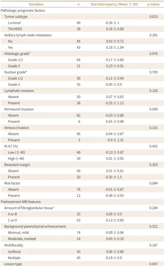
더 컸고( p = 0.023) (Table 4, Figs. 2, 3), 영상에서 다발성/비종괴성 조영증강으로 보이는 군에서 단일 종괴로 보이는 군보다 차이가 유의미하게 크게 나타났다( p = 0.047) (Figs. 2, 3). 다른 임상병 리적 인자와 자기공명영상 소견들은 통계적으로 유의미한 차이를 보이지 않았다.
고찰
본 연구에서 비록 다변량 분석에서 통계적 의의는 없었지만 단변량 분석에서 유방암의 치료 전 자기공명영상 소견 중 단초점 병변, 단일 종괴, 종양 중심부의 괴사를 의미하는 테두리 조영증강 의 부재 및 T2 강조영상에서 종괴 내 고신호의 부재가 완전 관해와 관련이 있었다. 치료 전 완전
Table 3. Diagnostic Performance of MRI for Predicting pCR According to Tumor SubtypesAll Types Luminal HER2/TN
AUC 0.8017 0.7985 0.8056
Accuracy 90.91 89.8 92.31
Sensitivity 93.67 93.02 94.44
Specificity 66.67 66.67 66.67
PPV 96.1 95.24 97.14
NPV 54.55 57.14 50
AUC = area under the curve, HER2 = human epidermal growth factor receptor 2, NPV = negative predictive value, pCR = pathologic complete response, PPV = positive predictive value, TN = triple negative
16 14 12 10 8 6 4 2 0
Surgical pathology size (cm)
0 1 2 3 4 5 6 7 8 R2 = 0.3454
MRI size (cm) MRI pathology size correlation
Fig. 1. Correlation between post-neoadjuvant chemotherapy MRI and pathological measurements of resid- ual tumor size.
Table 4. Factors Affecting the Difference between the Size of the Residual Tumor of the Surgical Specimen and that Determined by MRI
Variables n Size Discrepancy (Mean ± SD) p-Value
Pathologic prognostic factors
Tumor subtype 0.023
Luminal 49 -0.26 ± 1
TN/HER2 39 0.16 ± 0.68
Axillary lymph node metastasis 0.281
No 45 0.03 ± 0.72
Yes 43 0.18 ± 1.04
Histologic grade* 0.076
Grade 1/2 64 -0.17 ± 0.89
Grade 3 21 0.23 ± 0.91
Nuclear grade* 0.795
Grade 1/2 30 -0.11 ± 0.94
Grade 3 55 -0.05 ± 0.9
Lymphatic invasion 0.120
Absent 50 0.07 ± 0.65
Present 38 -0.25 ± 1.12
Perineural invasion 0.099
Absent 82 -0.03 ± 0.88
Present 6 -0.65 ± 0.98
Venous invasion 0.101
Absent 85 -0.04 ± 0.87
Present 3 -0.9 ± 1.31
Ki-67 (%) 0.452
Low (≤ 40) 49 -0.13 ± 0.87
High (> 40) 39 0.01 ± 0.92
Resected margin 0.303
Absent 68 0.01 ± 0.61
Present 20 -0.35 ± 1.5
Risk factor 0.084
Absent 76 -0.01 ± 0.87
Present 12 -0.48 ± 0.93
Pretreatment MRI features
Amount of fibroglandular tissue† 0.284
A or B 25 0.09 ± 0.9
C or D 63 -0.13 ± 0.89
Background parenchymal enhancement 0.321
Minimal, mild 74 -0.09 ± 0.96
Moderate, marked 14 0.05 ± 0.33
Multifocality 0.187
Unifocal 43 0.06 ± 0.88
Multiple 45 -0.19 ± 0.9
Lesion type 0.047
관해 여부를 예측할 수 있는 원발암의 특징에 대한 이전 연구들에서도 경계가 좋은 종괴, T2 강조 영상 상 종양 내부 고신호강도의 부재가 병리학적 완전 관해와 연관이 있다고 보고된 바 있어 본 연구의 결과와 유사하였다(11, 12). 우리 연구에서는 완전 관해 군과 불완전 관해 군 간에 임상병 리학적인자에서 유의미한 차이가 없었지만 일부 이전 연구들에서는 내강형보다 HER2+/TN 유형 에서 병리학적 완전 관해 비율이 높다는 보고가 있었다(13-15). 단초점 병변 및 단일 종괴 형태는 삼중음성암에서 흔히 보이는 소견으로(2), 단초점 및 단일 종괴 병변이 완전 관해와 연관성을 보인 본 연구의 결과를 설명할 수 있을 것으로 생각한다. 한편, 일부 이전 연구들은 신보강화학요법 후 자기공명영상에서 종양의 반응 양상과 병리학적 완전 관해와의 관계에 주목하였고 동심성 위축 이 병리학적 완전 관해와 관련이 있다고 하였지만(16, 17), 본 연구에서는 종양의 반응 양상과 완
Table 4. Factors Affecting the Difference between the Size of the Residual Tumor of the Surgical Specimen and that Determined by MRI (Continued)Variables n Size Discrepancy, (mean ± SD) p-Value
Single mass 42 0.12 ± 0.69
Multifocal/NME 46 -0.25 ± 1.02
Mass shape 0.128
Oval/Round 26 0.12 ± 0.63
Irregular 62 -0.15 ± 0.97
Margin 0.165
Circumscribed 23 0.15 ± 0.66
Irregular/spiculated 65 -0.15 ± 0.95
Enhancement 0.340
Rim (-) 50 -0.15 ± 0.97
Rim (+) 38 0.03 ± 0.77
Kinetics‡ 0.635
Type I, II 13 -0.24 ± 1.43
Type III 75 -0.04 ± 0.77
Intratumoral high SI 0.252
Absent 57 -0.14 ± 1
Present 31 0.06 ± 0.65
Peritumoral edema 0.637
Absent 30 -0.13 ± 0.94
Present 58 -0.04 ± 0.87
MR clinical response
Response pattern 0.652
Concentric 74 -0.04 ± 0.76
Splitted 14 -0.22 ± 1.42
*Histologic grade and nuclear grade data were available for a total of 85 patients.
†For amount of FGT. A = almost entirely fat, B = scattered fibroglandular tissue, C = heterogeneous fibro- glandular tissue, D = extreme fibroglandular tissue.
‡For kinetics. Type I = persistent enhancement, Type II = plateau enhancement, Type III = washout enhance- ment.
HER2 = human epidermal growth factor receptor 2, SD = standard deviation, SI = signal intensity, TN = triple negative
전 관해 간에는 유의미한 연관성을 찾을 수 없었다.
신보강화학치료의 발전으로 일부 유방암 아형에서는 상당히 높은 확률로 병리학적 완전 관해가 나타나고 있고, 이러한 상황은 병리학적 완전 관해가 된 환자들에서도 유방의 수술적 절제가 필요 한지, 아니면 방사선 치료만으로 충분한지 대한 논의의 필요성을 대두시켰다(18). 이러한 배경에 서, 신보강화학요법을 받은 환자들에서 자기공명영상의 병리학적 완전 관해 예측 능력에 관련된 주제는 지난 수년간 많은 관심을 받았다. 우리 연구에서 완전 관해를 예측하는 데 있어서 자기공 명영상의 진단적 정확도는 90.91%, 곡선하부면적은 0.8017이었으며 유방암 아형 간의 진단적 정 확도는 HER2+/TN 유형에서 내강형보다 조금 더 높은 값을 보였다(각각 92.31%, 89.8%). 그리고 모든 유방암 아형에서 양성예측도(95.24~97.14 범위)에 비해 음성예측도(50~57.1 범위)가 낮은 값 을 보였으며, 내강형에서 HER2+/TN 유형에 비해 다소 높은 음성예측도 값을 보였다(각각 57.1, 50). 이전 연구들에서 자기공명영상의 병리학적 완전 관해 예측 정확도는 73.1~88.6%(10, 19-23)
Fig. 2. 40-year-old female with IDC, TN type, single mass, Discrepancy (MRI size - pathology size) = 0 A, B. Pre-NAC T1-weighted axial dynamic image and sagittal contrast-enhanced MIP image show a single ir- regular heterogeneously enhancing mass in the left breast.C, D. Post-NAC T1-weighted axial dynamic image and sagittal contrast-enhanced MIP image show no en- hancing lesion at the previous lesion site.
The response was classified as a complete clinical response, and surgery revealed that there was no re- maining tumor.
In this case, the size discrepancy between MRI and pathology was 0.
IDC = invasive ductal cancer, MIP = maximum intensity projection, NAC = neoadjuvant chemotherapy, TN = triple-negative
A
C
B
D
의 범위였으며 본 연구의 결과에 비해 다소 낮았으나 큰 차이는 보이지 않았고, 곡선하부면적은 이전 연구들에서 0.77~0.84 (10, 23)의 범위였고 본 연구의 결과도 이 범주 내에 있었다. 또한 본 연 구에서는 HER2+/TN 유형에서 병리학적 완전 관해 예측 정확도가 다소 높았으나 통계적으로 유 의미한 차이는 나지 않았다. 이전 연구들에 비추어 볼 때도 유방암 아형에 따른 정확도의 차이에 대해서는 일관적인 결과가 도출되지 않은 상태이며, 계속적인 연구가 필요할 것으로 보인다. 그리 고 일부 연구들은(10, 20) 양성예측도에 비해 음성예측도가 낮은 값을 보인다고 하였으며 특히, 내 강형에서 HER2+/TN 유형에 비해 낮은 음성예측도 값을 보인다고 하였다. 본 연구에서도 양성예 측도가 음성예측도 보다 상당히 높은 값을 보여 이 점은 이전 연구 결과들에 부합하였으나, 음성 예측도의 유방암 아형별 비교에 있어서는 이전 연구들과 달리 내강형의 결과값이 더 높았다. 하지 만 본 연구의 유방암 아형별 음성예측도 차이는 크지 않아서 아형별로 유의미한 차이가 있다고 보 는 데는 한계가 있을 것으로 생각된다. 양성예측도는 잔류 암 크기의 예측과 관련이 있고 음성예
Fig. 3. 42-year-old female with IDC, luminal type, multiple/NME. Discrepancy (MRI size - pathology size) = 3.8 - 6.2 = -2.4.A, B. Pre-NAC T1-weighted axial dynamic image and sagittal contrast-enhanced MIP image show a regional NME in the outer portion of the right breast.
C, D. Post-NAC T1-weighted axial dynamic and sagittal contrast-enhanced MIP images show that the lesion decreased in size to 3.8 cm and manifested as multifocal split lesions.
Surgery revealed a 6.2-cm residual invasive tumor, and the size discrepancy between MRI and pathology was -2.4 cm.
IDC = invasive ductal cancer, MIP = maximum intensity projection, NAC = neoadjuvant chemotherapy A
C
B
D
측도는 병리학적 완전 관해 예측과 관련이 있는 통계량이므로, 자기공명영상으로 병리학적 완전 관해를 예측하는 것은 잔류 암 평가에 비해 상대적으로 신뢰도가 떨어진다고 할 수 있다.
한편 신보강항암요법 후 완전 관해 예측뿐만 아니라, 잔류 암이 남은 경우 그 크기를 정확히 평 가하는 것도 중요한데 그 이유는 수술을 계획하고 예후를 예측하는 데 도움이 되기 때문이다. 본 연 구에서 자기공명영상과 수술 병리 조직의 잔류 암 크기는 매우 강한 연관성을 보였다( r = 0.9, p <
0.001). 이전 연구들에서는 둘 간의 연관성은 0.465~0.92의 범위를 보였고 한 연구(23)를 제외하면 모두 0.6을 넘는 강한 연관성이 있다고 보고하였다(12, 19, 22-24). 본 연구의 결과 값은 기존 연구 의 범위 내에 있고, 따라서 자기공명영상은 잔류 암 크기 예측에 있어서 상당히 정확한 검사 방법 이라 할 수 있다.
또한 이 연구에서는 유방암 아형과 치료 전 자기공명영상 소견이 자기공명영상과 수술 병리 조 직의 잔류 암 크기 차이에 영향을 미친다는 결과가 나왔다. 즉, HER2+/TN 유형보다 내강형에서 자기공명영상과 수술 병리 조직의 잔류 암 크기 차이가 유의미하게 더 컸고(p = 0.023) (Table 4), 치료 전 자기공명영상에서 다발성/비종괴성 조영증강으로 보이는 군에서 단일 종괴로 보이는 군 보다 유의미하게 차이가 크게 나타났다(p = 0.047). 하지만 신보강화학요법 후 반응 양상에 따라서 는 잔류 암 크기 차이가 유의미하게 다르지 않았다. 내강형 그리고 치료 전 자기공명영상에서 다 발성/비종괴성 조영증강으로 보이는 경우에 잔류 암 크기 차이가 더 크게 나타난다는 점은 기존 연구들의 결과와 일치하였다(22-29). 하지만 신보강화학요법 후의 반응 양상에 대해서는 이전 연 구들이 동심성 위축보다 가지돌기성 위축을 보이는 환자에서 잔류 암의 크기를 과대 또는 과소평 가하는 경우가 많다고 보고했던 것에 반하여(24, 30) 이 연구에서는 두 군 간에 통계적으로 유의미 한 차이가 없었다.
본 연구 결과에 따르면 자기공명영상은 병리 조직의 잔류 암을 면밀히 반영하는 검사 방법이기
는 하지만 위양성과 위음성 사례들도 있었다. 대표적인 위음성 사례로는 가지돌기성 위축을 보이
는 환자에서 잔류 암의 크기가 과소평가되는 것이었고(Fig. 3), 이것은 이전 연구들에서도 보고되
었던 위음성 사례의 형태이다. 기존 연구들에 따르면 잔류 암 평가에 있어서 자기공명영상의 대표
적 위음성 요인은 작은 잔류 암을 발견하지 못하고 병리학적 완전 관해로 판단하거나 잔류 암의
형태가 가지돌기성 위축과 같은 작은 다발성 병변으로 나타나게 되어 일부 병변을 발견하지 못하
고 크기를 과소평가하게 되는 경우이다(19, 22). 한편, 신보강화학요법 후 병리학적 완전 관해를
평가할 때 가장 논란이 되는 점은 잔류 유관상피내암이 있어도 병리학적 완전 관해로 보아야 하는
가에 대한 것이다. 병리학적 TNM 병기 분류법에서는 침윤성 유방암만을 잔류 암으로 정의하고
있으므로 본 연구에서는 이를 따라 침윤성 유방암만을 잔류 암으로 정의하였고, 이것이 위양성 사
례의 원인이 되었다. 이전 연구들에서도 대표적 위양성 요인으로 잔류 유관상피내암이 있거나 병
변이 비국소성(segmental, regional, diffuse) 인 경우를 들고 있다(22, 28, 31). 유관상피내암은 침
윤성 유방암과 영상에서 구분이 되지 않으며 다초점성/다발성/비종괴성 조영증강 형태로 보여 정
확한 크기를 측정하기 어려워 잔류 암의 크기가 과대평가될 수 있다(22, 23).
제한점
본 연구의 제한점은 다음과 같다. 첫째, 단일기관에서 시행된 후향적 연구로서 대상 환자의 수 가 적어 일반화하는 데 어려움이 있다. 둘째, 대상 환자군의 신보강화학요법의 종류 및 기간에 다 양성이 존재하였고, 이것이 병변의 반응 양상에 영향을 미쳤을 수 있다. 특히 우리 연구에서는 Docetaxel-Gemcitabine 요법으로 치료한 환자군이 상당수 포함되어 있고(35.23%), Docetaxel- Gemcitabine 화학요법은 Anthracycline 및 Taxane을 기반으로 하는 화학요법에 비해 병리적 완전 관해 비율이 다소 떨어질 수 있다(32). 또한 Taxane 화학요법은 잔류 암의 조영증강을 감소 시켜 치료에 대한 반응을 과대평가하게 하고 위음성 사례의 원인이 될 수 있다고 알려져 있다(33).
셋째, 유관상피내암은 병리학적 완전 관해의 정의에 포함되지 않았고 이것이 자기공명영상의 정 확도에 영향을 주었을 수 있다.
결론적으로, 자기공명영상은 신보강화학요법 후에 항암제에 대한 완전 관해 및 잔류 암 크기의 예측에 있어서 비교적 정확도가 높은 신뢰할 만한 검사 방법이며 치료 전 자기공명영상의 원발암 소견이 정확도에 영향을 미친다. 특히 내강형과 다초점 종괴 및 비종괴성 조영증강의 경우 실제 잔류 암과 자기공명영상에서의 크기에 차이가 있을 수 있어 해석에 주의해야 한다.
Author Contributions
Conceptualization, A.Y.Y.; data curation, A.H.S., A.Y.Y.; formal analysis, A.H.S., A.Y.Y.; funding acqui- sition, A.Y.Y.; investigation, A.H.S., A.Y.Y.; methodology, all authors; project administration, A.H.S., A.Y.Y.; resources, all authors; software, A.H.S., A.Y.Y.; supervision, A.Y.Y.; validation, A.H.S., A.Y.Y.; vi- sualization, A.H.S., A.Y.Y.; writing—original draft, A.H.S., A.Y.Y.; and writing—review & editing, all au- thors.
Conflicts of Interest
The authors have no potential conflicts of interest to disclose.
Funding None
REFERENCES
1. Mauri D, Pavlidis N, Ioannidis JP. Neoadjuvant versus adjuvant systemic treatment in breast cancer: a meta- analysis. J Natl Cancer Inst 2005;97:188-194
2. Trop I, LeBlanc SM, David J, Lalonde L, Tran-Thanh D, Labelle M, et al. Molecular classification of infiltrating breast cancer: toward personalized therapy. Radiographics 2014;34:1178-1195
3. Kaufmann M, Von Minckwitz G, Bear HD, Buzdar A, McGale P, Bonnefoi H, et al. Recommendations from an international expert panel on the use of neoadjuvant (primary) systemic treatment of operable breast can- cer: new perspectives 2006. Ann Oncol 2007;18:1927-1934
4. Mathew J, Asgeirsson KS, Cheung KL, Chan S, Dahda A, Robertson JF. Neoadjuvant chemotherapy for local- ly advanced breast cancer: a review of the literature and future directions. Eur J Surg Oncol 2009;35:113-122 5. Rauch GM, Adrada BE, Kuerer HM, Van la Parra RF, Leung JW, Yang WT. Multimodality imaging for evaluating
response to neoadjuvant chemotherapy in breast cancer. AJR Am J Roentgenol 2017;208:290-299
6. Chagpar AB, Middleton LP, Sahin AA, Dempsey P, Buzdar AU, Mirza AN, et al. Accuracy of physical examina- tion, ultrasonography, and mammography in predicting residual pathologic tumor size in patients treated with neoadjuvant chemotherapy. Ann Surg 2006;243:257-264
7. Fangberget A, Nilsen LB, Hole KH, Holmen MM, Engebraaten O, Naume B, et al. Neoadjuvant chemotherapy in breast cancer-response evaluation and prediction of response to treatment using dynamic contrast-en-
hanced and diffusion-weighted MR imaging. Eur Radiol 2011;21:1188-1199
8. Marinovich ML, Houssami N, Macaskill P, Sardanelli F, Irwig L, Mamounas EP, et al. Meta-analysis of magnet- ic resonance imaging in detecting residual breast cancer after neoadjuvant therapy. J Natl Cancer Inst 2013;
105:321-333
9. Montemurro F, Martincich L, De Rosa G, Cirillo S, Marra V, Biglia N, et al. Dynamic contrast-enhanced MRI and sonography in patients receiving primary chemotherapy for breast cancer. Eur Radiol 2005;15:1224- 1233
10. McGuire KP, Toro-Burguete J, Dang H, Young J, Soran A, Zuley M, et al. MRI staging after neoadjuvant che- motherapy for breast cancer: does tumor biology affect accuracy? Ann Surg Oncol 2011;18:3149-3154 11. Bae MS, Shin SU, Ryu HS, Han W, Im SA, Park IA, et al. Pretreatment MR imaging features of triple-negative
breast cancer: association with response to neoadjuvant chemotherapy and recurrence-free survival. Radi- ology 2016;281:392-400
12. Esserman L, Kaplan E, Partridge S, Tripathy D, Rugo H, Park J, et al. MRI phenotype is associated with re- sponse to doxorubicin and cyclophosphamide neoadjuvant chemotherapy in stage III breast cancer. Ann Surg Oncol 2001;8:549-559
13. Colleoni M, Viale G, Zahrieh D, Pruneri G, Gentilini O, Veronesi P, et al. Chemotherapy is more effective in pa- tients with breast cancer not expressing steroid hormone receptors: a study of preoperative treatment. Clin Cancer Res 2004;10:6622-6628
14. Houssami N, Macaskill P, Von Minckwitz G, Marinovich ML, Mamounas E. Meta-analysis of the association of breast cancer subtype and pathologic complete response to neoadjuvant chemotherapy. Eur J Cancer 2012;48:3342-3354
15. Kawashima H, Inokuchi M, Furukawa H, Ikeda H, Kitamura S. Magnetic resonance imaging features of breast cancer according to intrinsic subtypes: correlations with neoadjuvant chemotherapy effects. Springerplus 2014;3:240
16. Ballesio L, Gigli S, Di Pastena F, Giraldi G, Manganaro L, Anastasi E, et al. Magnetic resonance imaging tumor regression shrinkage patterns after neoadjuvant chemotherapy in patients with locally advanced breast cancer: correlation with tumor biological subtypes and pathological response after therapy. Tumour Biol 2017;39:1010428317694540
17. Eom HJ, Cha JH, Choi WJ, Chae EY, Shin HJ, Kim HH. Predictive clinicopathologic and dynamic contrast-en- hanced MRI findings for tumor response to neoadjuvant chemotherapy in triple-negative breast cancer. AJR Am J Roentgenol 2017;208:W225-W230
18. De Los Santos J, Bernreuter W, Keene K, Krontiras H, Carpenter J, Bland K, et al. Accuracy of breast magnetic resonance imaging in predicting pathologic response in patients treated with neoadjuvant chemotherapy.
Clin Breast Cancer 2011;11:312-319
19. Chen JH, Feig B, Agrawal G, Yu H, Carpenter PM, Mehta RS, et al. MRI evaluation of pathologically complete response and residual tumors in breast cancer after neoadjuvant chemotherapy. Cancer 2008;112:17-26 20. De Los Santos JF, Cantor A, Amos KD, Forero A, Golshan M, Horton JK, et al. Magnetic resonance imaging as
a predictor of pathologic response in patients treated with neoadjuvant systemic treatment for operable breast cancer. Translational Breast Cancer Research Consortium trial 017. Cancer 2013;119:1776-1783 21. Hayashi Y, Takei H, Nozu S, Tochigi Y, Ichikawa A, Kobayashi N, et al. Analysis of complete response by MRI
following neoadjuvant chemotherapy predicts pathological tumor responses differently for molecular sub- types of breast cancer. Oncol Lett 2013;5:83-89
22. Ko ES, Han BK, Kim RB, Ko EY, Shin JH, Hahn SY, et al. Analysis of factors that influence the accuracy of mag- netic resonance imaging for predicting response after neoadjuvant chemotherapy in locally advanced breast cancer. Ann Surg Oncol 2013;20:2562-2568
23. Loo CE, Straver ME, Rodenhuis S, Muller SH, Wesseling J, Vrancken Peeters MJ, et al. Magnetic resonance imaging response monitoring of breast cancer during neoadjuvant chemotherapy: relevance of breast can- cer subtype. J Clin Oncol 2011;29:660-666
24. Kim HJ, Im YH, Han BK, Choi N, Lee J, Kim JH, et al. Accuracy of MRI for estimating residual tumor size after neoadjuvant chemotherapy in locally advanced breast cancer: relation to response patterns on MRI. Acta Oncol 2007;46:996-1003
25. Bouzón A, Acea B, Soler R, Iglesias Á, Santiago P, Mosquera J, et al. Diagnostic accuracy of MRI to evaluate tumour response and residual tumour size after neoadjuvant chemotherapy in breast cancer patients. Ra-
diol Oncol 2016;50:73-79
26. Akashi-Tanaka S, Fukutomi T, Watanabe T, Katsumata N, Nanasawa T, Matsuo K, et al. Accuracy of contrast- enhanced computed tomography in the prediction of residual breast cancer after neoadjuvant chemother- apy. Int J Cancer 2001;96:66-73
27. Belli P, Costantini M, Malaspina C, Magistrelli A, LaTorre G, Bonomo L. MRI accuracy in residual disease eval- uation in breast cancer patients treated with neoadjuvant chemotherapy. Clin Radiol 2006;61:946-953 28. Rosen EL, Blackwell KL, Baker JA, Soo MS, Bentley RC, Yu D, et al. Accuracy of MRI in the detection of residu-
al breast cancer after neoadjuvant chemotherapy. AJR Am J Roentgenol 2003;181:1275-1282
29. Yeh E, Slanetz P, Kopans DB, Rafferty E, Georgian-Smith D, Moy L, et al. Prospective comparison of mam- mography, sonography, and MRI in patients undergoing neoadjuvant chemotherapy for palpable breast cancer. AJR Am J Roentgenol 2005;184:868-877
30. Bahri S, Chen JH, Mehta RS, Carpenter PM, Nie K, Kwon SY, et al. Residual breast cancer diagnosed by MRI in patients receiving neoadjuvant chemotherapy with and without bevacizumab. Ann Surg Oncol 2009;16:
1619-1628
31. Partridge SC, Gibbs JE, Lu Y, Esserman LJ, Sudilovsky D, Hylton NM. Accuracy of MR imaging for revealing re- sidual breast cancer in patients who have undergone neoadjuvant chemotherapy. AJR Am J Roentgenol 2002;179:1193-1199
32. Jeon YW, Kim TH, Youn HJ, Han S, Jung Y, Gwak G, et al. Multicenter phase II trial of neoadjuvant chemo- therapy with docetaxel and gemcitabine in locally advanced breast cancer. J Breast Cancer 2017;20:340-346 33. Schrading S, Kuhl CK. Breast cancer: influence of taxanes on response assessment with dynamic contrast-
enhanced MR imaging. Radiology 2015;277:687-696
신보강화학요법 후 유방암의 병리학적 완전 관해 예측 및 잔류 암 평가: 유방자기공명영상의 정확도 및 영향인자 분석
안현수
1· 안영이
1* · 전예원
2· 서영진
2· 최현주
3목적 신보강화학요법을 시행한 유방암 환자에서 병리학적 관해와 잔류 암의 크기를 평가하 는 데 있어 유방자기공명영상의 정확도를 분석하고 이에 영향을 미치는 인자들이 무엇인지 알아본다.
대상과 방법 2010년부터 2017년까지 본원에서 신보강화학요법 후 수술을 시행한 88명의 유 방암 환자를 대상으로 하였다. 병리학적 관해는 수술 병리 결과에서 침윤성 유방암이 발견되 지 않는 것으로 정의하였고 자기공명영상과 병리 조직의 잔류 암 크기 차이는 최대 직경으로 비교하였다. 병리학적 관해 및 자기공명영상과 병리 조직에서의 잔류 암 크기 차이에 영향을 미치는 인자를 알아보기 위해 통계분석을 시행하였다.
결과 전체 환자의 10%가 병리학적 관해에 도달하였다. 자기공명영상으로 관해를 예측할 때 의 정확도와 곡선하부면적은 각각 90.91%, 0.8017이었다. 신보강화학요법 시행 후 유방자기 공명영상과 병리 조직에서 측정한 잔류 암의 크기는 매우 강한 연관성을 보였고(r = 0.9, p <
0.001), 특히 영상에서 단일 종괴로 보였던 병변에서( p = 0.047) 그러하였다. 자기공명영상과 병리 조직 간의 잔류 암 크기는 내강형(p = 0.023), 그리고 자기공명영상에서 다초점 종괴 및 비종괴성 조영증강을 보인( p = 0.047) 환자군에서 유의미하게 큰 차이를 보였다.
결론 자기공명영상은 유방암의 병리학적 완전 관해와 잔류 암 크기의 평가에 있어서 정확도 가 높은 검사이다. 유방암 아형과 병변의 영상의학적 소견이 자기공명영상의 정확도에 영향 을 미친다.
1가톨릭대학교 의과대학 성빈센트병원 영상의학과,
2가톨릭대학교 의과대학 외과 유방갑상선종양분과,
3가톨릭대학교 의과대학 병리과