Systemic Manifestations of Immunoglobulin G4-Related Disease: A Pictorial Essay
다양한 인체 장기에서 보일 수 있는 면역글로불린 G4 관련 질환: 임상화보
Kyungri Park, MD
1, Yo Won Choi, MD
1, Bo-Kyeong Kang, MD
1, Ji Young Lee, MD
1, Jeong Seon Park, MD
1,
Su-Jin Shin, MD
2,3, Hye Ryoung Koo, MD
1*
Departments of 1Radiology and 2Pathology, Hanyang University College of Medicine, Hanyang University Hospital, Seoul, Korea
3Department of Pathology, Yonsei University College of Medicine, Gangnam Severance Hospital, Seoul, Korea
Immunoglobulin G4 (IgG4)-related disease is a systemic fibro-inflammatory disease character- ized by pathologic findings in various organs. Imaging is critical for the diagnosis and treatment assessment of patients with IgG4-related disease. In this pictorial essay, we review the key fea- tures of multiple imaging modalities, typical pathologic findings, and differential diagnosis of IgG4-related disease. This systematic pictorial review can further our understanding of the broad-spectrum manifestations of this disease.
Index terms Immunoglobulin G4-Related Disease; Ultrasonography;
Computed Tomography, X-Ray; Magnetic Resonance Imaging
INTRODUCTION
Immunoglobulin G4 (IgG4)-related disease is a chronic fibro-inflammatory condition that has unique histopathological features (Fig. 1). IgG4-related disease seems to be ca- pable of affecting every organ other than well-established concepts of autoimmune pancreatitis (AIP). Common imaging findings of IgG4-related disease include swelling, nodules, and/or wall thickening, which may mimic existing conditions in each organ, especially malignant conditions. In order to understand the whole picture of IgG4-relat- ed disease, radiologists need to clarify the precise spectrum of each organ involvement.
Received July 28, 2020 Revised September 1, 2020 Accepted September 30, 2020
*Corresponding author Hye Ryoung Koo, MD Department of Radiology, Hanyang University College of Medicine, Hanyang University Hospital, 222-1 Wangsimni-ro, Seongdong-gu, Seoul 04763, Korea.
Tel 82-2-2290-9156 Fax 82-2-2293-2111
E-mail [email protected] This is an Open Access article distributed under the terms of the Creative Commons Attribu- tion Non-Commercial License (https://creativecommons.org/
licenses/by-nc/4.0) which permits unrestricted non-commercial use, distribution, and reproduc- tion in any medium, provided the original work is properly cited.
ORCID iDs Kyungri Park https://
orcid.org/0000-0003-4884-3421 Yo Won Choi
https://
orcid.org/0000-0003-0508-9430 Bo-Kyeong Kang
https://
orcid.org/0000-0001-6834-3825 Ji Young Lee
https://
orcid.org/0000-0003-1181-8070 Jeong Seon Park
https://
orcid.org/0000-0002-5550-6645 Su-Jin Shin
https://
orcid.org/0000-0001-9114-8438 Hye Ryoung Koo
https://
orcid.org/0000-0002-7562-2256
IgG4-RELATED AIP
AIP is the most commonly known manifestation of IgG4-related disease. AIP can be cate- gorized into two main radiologic patterns; diffuse and focal (1).
Diffuse disease is well-known as uniformly enlarged sausage-shaped pancreas. The peri- pancreatic hypoattenuating capsule on CT, the so-called “peripancreatic halo”, is thought to be a much more specific finding, and is the result of phlegmon, fibrotic tissue and fluid. The areas where AIP is involved demonstrate hypointense T1 and hyperintense T2 signals; on the other hand, peripancreatic capsules show both hypointense signal intensity in both T1 and T2-weighted sequences (Fig. 2) (2).
The focal type, also called mass-forming AIP, is less common than the diffuse type and is hard to differentiate from pancreatic carcinoma. Focal disease usually involves the pancreat- ic head. The multifocal type can also be present. The fact that AIP does not show upstream pancreatic duct dilatation substantially differentiates it from pancreatic adenocarcinoma.
The affected parenchyma exhibits hypointense T1 and hyperintense T2 signals, similar to the
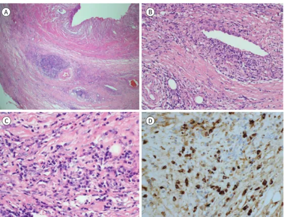
Fig. 1. Characteristic histological features of IgG4-related disease in retroperitoneal fibrosis (Fig. 1 and Fig. 5 are representative of the same patient).
A. IgG4-related disease is typically characterized by deposition of dense and wire-like strands of fibrotic col- lagen (H&E stain, × 100).
B. The vein is partially obliterated by lymphocyte and plasma cell infiltration (H&E stain, × 200).
C. The involved organ is extensively infiltrated by inflammatory infiltrates including lymphocytes and plas- ma cells (H&E stain, × 400).
D. Several plasma cells are positive for IgG4 (immunohistochemical stain, × 400).
H&E = hematoxylin & eosin, IgG4 = immunoglobulin G4
A B
D C
Fig. 2. Diffuse autoimmune pancreatitis.
A 67-year-old male with abdominal discomfort and dyspepsia. A diffusely enlarged pancreas is surrounded by a low-attenuation halo on the axial contrast-enhanced portal-phase CT image (arrows) and a hypointense capsule-like rim can be seen on the T2-weighted MR image (arrowheads).
diffuse type. AIP is reported to display high diffusion-weighted image (DWI) signal in a more longitudinal shape and have much lower apparent diffusion coefficient (ADC) value than pancreatic cancer (Fig. 3) (3).
IgG4-RELATED SCLEROSING CHOLANGITIS
Biliary involvement occurs simultaneously in almost 90% of AIP cases. The main differen- tial diagnosis that should be kept in mind is primary sclerosing cholangitis. Biliary sclerosis in IgG4-related disease is typically observed as a long expansion and smooth contour with upstream duct dilatation, resulting in obstructive jaundice. Sclerosis may occur anywhere, but the most common site is the intrapancreatic common bile duct (Fig. 4). On the other hand, primary sclerosing cholangitis presents a so-called “beaded appearance” with multifo- cal, short and band-like strictures (4).
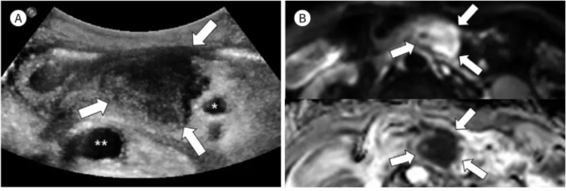
Fig. 3. Focal autoimmune pancreatitis.
A 77-year-old male with jaundice. His serum immunoglobulin G4 concentration was markedly increased (1565 mg/dL).
A. There is an abnormal focal hypoechoic mass (arrows) at the pancreatic head on the ultrasound image (*Superior mesenteric artery, **Aorta).
B. A mass on the pancreatic head exhibits striking hyper signal intensity on the diffusion-weighted image (ar- rows, b value = 800 s/mm2, upper panel) with a markedly low apparent diffusion coefficient value (arrows, lower panel ).
A B
IgG4-RELATED RETROPERITONEAL FIBROSIS
Retroperitoneal fibrosis a rare fibrotic reaction in the retroperitoneum that typically occurs with ureteric obstruction. Approximately 70% of cases of retroperitoneal fibrosis are related to infiltration of IgG4-positive cells (previously known as idiopathic retroperitoneal fibrosis), while the rest of them can be secondary to several other causes including malignancy. Malig- nant retroperitoneal fibrosis is result from severe desmoplastic response to metastasis from prostate, breast and colon cancer or retroperitoneal primary tumor such as lymphoma or sarcoma (5). It is crucial to distinguish whether it is result from IgG4-related disease or malig- nant cause because the prognosis is completely different. In terms of extent, IgG4-related retroperitoneal fibrosis is usually distributed below the renal hilum level and spares the pos- terior aspect of the aorta (Fig. 5) (6). Thus, IgG4-related retroperitoneal fibrosis does not push the aorta forward, but the malignant tumor forms a bulky mass, displacing the aorta anteri- orly (7). MRI can be helpful for differential diagnosis. ADC value of chronic form is signifi- cantly higher than that of active or malignant retroperitoneal fibrosis (8). A retroperitoneal mass that shows low signal intensity on T2-weighted image is suggestive of benign lesion in the late inactive stage (9). However, diagnostic confirmation should be made with biopsy be- cause it is not always correct to generalize benign or malignant by imaging features (8-10).
IgG4-RELATED RENAL DISEASE
Takahashi et al. (11) reported several patterns of renal involvement: round or wedge- Fig. 4. Immunoglobulin G4-related sclerosing cholangitis.
A 58-year-old male with abdominal discomfort and dyspepsia.
A. MR cholangiopancreatography shows proportional smooth biliary tree dilatation with abrupt intrapan- creatic narrowing of the CBD (arrows).
B. Narrowed CBD is surrounded by hypointense rim on T2-weighted image (arrowheads).
CBD = common bile duct A
B
A
B
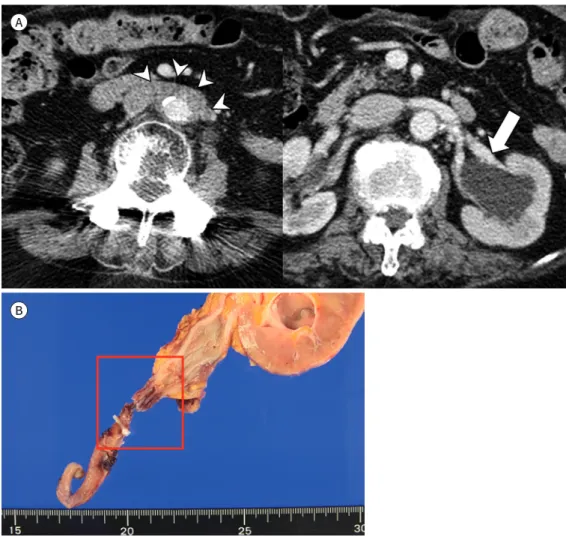
Fig. 5. IgG4-related retroperitoneal fibrosis.
A 77-year-old female who experienced excessive urinary frequency for 12 years.
A. Axial contrast-enhanced portal-phase CT shows mildly enhanced soft tissue mass (arrowheads) sur- rounding the aorta, inferior vena cava, and left hydronephrosis (arrow).
B. The gross pathologic specimen demonstrates a stricture in the mid portion of the ureter, narrowed by fi- brosis and a thickened wall (red box). Histologic specimen obtained by left nephroureterectomy confirmed a ureteral stricture with IgG4 cells > 140/high-power field, consistent with IgG4-related disease.
IgG4 = immunoglobulin G4
shaped lesions located in bilateral and peripheral cortical locations, soft tissue rind or nod- ules in both renal sinuses and enhanced wall thickening of the renal pelvis. Lesions of the re- nal sinus and renal pelvis are much more rare than renal cortical lesions. On CT, renal cortical lesions are typically isoattenuated relative to the normal renal cortex on noncontrast scans but hypointense on corticomedullary phase imaging before progressive enhancement on delayed scans (12). On MRI, the cortical lesions exhibit T2 hypointensity and T1 isointen- sity on conventional sequences and present the same dynamic enhancement patterns as CT (13). In addition, these lesions demonstrate abnormal restricted diffusion on DWI sequences.
The DWI sequence is known to be useful for the early detection of diseases because it is sig- nificantly more sensitive for lesion detection than T2-weighted images (Figs. 6, 7) (13). The main differential diagnosis includes metastatic disease, lymphoma and pyelonephritis.
IgG4-RELATED LUNG DISEASE
Inoue et al. (14) demonstrated that the spectrum of IgG4-related lung involvement can be categorized into 4 categories according to the radiologic abnormality: solid nodular type, multiple round-shaped ground-glass opacities, alveolar interstitial type and bronchovascular type (Figs. 8, 9). Sixty-nine percent of patients had more than three of the above radiologic findings. In addition to lung parenchymal lesions, IgG4-related disease might involve intra- thoracic lymph nodes, pleura, mediastinum in the chest (Fig. 10) (15).
SALIVARY AND LACRIMAL GLAND INVOLVEMENT
Salivary and lacrimal gland involvement are also common in IgG4-related disease. It may Fig. 7. Immunoglobulin G4-related disease with kidney involvement (soft tissue rind in both renal sinuses).
A 67-year-old male (the same patient as in Fig. 2) presented with synchronous pancreatic and renal mani- festations. Ill-defined soft-tissue rinds encase the bilateral renal pelvis without hydronephrosis. The soft tis- sue rinds are enhanced homogeneously on the contrast-enhanced CT (white arrows). The soft tissue rinds are hypointense on T2-weighted MR images (black arrows) and hyperintense on diffusion-weighted imag- ing (arrowheads, b value = 800 s/mm2).
Fig. 6. Immunoglobulin G4-related disease with kidney involvement (peripheral cortical lesions).
A 52-year-old asymptomatic female. Axial contrast-enhanced delayed phase CT image shows multiple well-defined nodules with delayed en- hancement in both kidneys (white arrows). The nodules in both kidneys are hypointense in T2-weighted MR images (black arrows) and obviously hyperintense on diffusion-weighted imaging (arrowheads, b = 800 s/mm2).
Fig. 8. Asymptomatic 48-year-old female with IgG4-related pulmonary disease.
A well-defined, ovoid-shaped solitary pulmonary nodule in the left lower lobe was incidentally detected on the chest CT (white arrow). The lesion exhibits mild hypermetabolic activity on 18F-fluorodeoxyglucose PET/
CT, which raised the suspicion of a malignancy (black arrow). Wedge resection of the left lower lobe was per- formed, and histology confirmed IgG4- related disease.
IgG4 = immunoglobulin G4
A
C
B
D Fig. 9. Immunoglobulin G4-related pulmonary disease.
A 55-year-old male with chest discomfort, dyspnea, and febrile sensation.
A. There are complex radiologic features in the right upper lobe, including bronchovascular thickening (black arrowhead), ground-glass opacity (white arrow), and interlobular septal thickening (white arrow- head) on the CT.
B. On the axial scan obtained at the left atrial level, an ill-defined solid nodule (white arrow) and an air bron- chogram (arrowhead) are present in the right lower lobe superior segment. Right lower interlobar lymph node enlargement is also noted (black arrow).
C. The axial scan obtained at the ventricular level shows a 2.5 cm nodule with a spiculated margin at the basal portion of the right lower lobe (arrow).
D. A follow-up chest CT 5 months after treatment with corticosteroids demonstrates near-complete interval resolution of the mass in the right lower lobe (arrow).
Fig. 10. IgG4-related sclerosing mediastinitis.
A 64-year-old female with sudden-onset dyspnea.
A. Contrast-enhanced CT shows a partially lobulated, homogeneously enhancing mass in the anterior me- diastinum (arrow). The pericardium is also markedly thickened and enhanced on the contrast-enhanced CT (arrowheads).
B. Photograph of the surgical specimen demonstrates a partly lobulated, yellowish anterior mediastinal mass. The peeled pericardium exhibits a yellowish and buckling appearance. Histologic analysis confirmed IgG4-related disease.
IgG4 = immunoglobulin G4
A B
A B
Fig. 11. Immunoglobulin G4-related disease manifestations in the submandibular and lacrimal glands.
A 76-year-old male with sudden onset xerostomia.
A. Contrast-enhanced CT images show bilateral, symmetrical enlargement with homogeneous enhancement of the submandibular (white ar- rows) and lacrimal glands (black arrows).
B. Both submandibular glands are enlarged and have bilateral hypoechoic nodular areas with considerable vascularization as seen on the ul- trasound images.
Fig. 12. A 79-year-old male with unilateral immunoglobulin G4-related disease manifestation in the salivary gland.
The same patient as in Fig. 3 presented with a non-tender palpable neck mass. The enlarged left submandibular gland exhibits diffuse hetero- geneous enhancement and mild intraglandular duct dilatation on the contrast-enhanced CT (arrow). The left submandibular gland was com- pletely replaced by an abnormal hypoechoic mass on the ultrasound. Net-like hyperechoic lines and prominent internal vascularity are pres- ent in the mass.
be unilateral or bilateral. Unilateral submandibular enlargement (Kuttner tumor) presents as a hard swelling that is difficult to differentiate from a neoplasm (Fig. 11) (16). Swelling of bi- lateral salivary and submandibular glands is another pattern of the disease (Mikulicz dis- ease). On US, hypoechoic nodular areas with increased vascularity are detected in the bilat- eral salivary glands (Fig. 12) (17). Overall CT and MR image characteristics are similar to other organ involvement. The main differential diagnosis are lymphoma, sarcoidosis, Sjo- gren disease, and Wegener granulomatosis.
ORBITAL INVOLVEMENT
In the orbital region, IgG4-related disease primarily involves the lacrimal gland. Extraocu- lar muscle involvement is also considered to be on the extension of the IgG4-related disease spectrum. Unilateral lacrimal gland lesion sometimes occurs, and the differentiation be- tween infectious dacryoadenitis and tumor may be difficult with imaging alone. In patients with extraocular muscle involvement, the lateral rectus is the most enlarged and tendinous portions are usually spared (Fig. 13). Graves’ disease and idiopathic inflammatory orbital pseudotumor should be included in the differential diagnosis (18).
IgG4-RELATED SCLEROSING MASTITIS
Clinical manifestations of IgG4-related sclerosing mastitis appear as solitary or multiple palpable and nonpainful lumps. According to the few cases reported, focal asymmetries, not accompanied by calcifications, are typical on mammography. On ultrasound, hypoechoic and ill-defined masses appear to be the common findings (Fig. 14). As these findings are so nonspecific, surgery or biopsy would be required at the next step (19).
IgG4-RELATED LYMPHADENOPATHY
This condition is usually found incidentally while performing the imaging work-up in pa-
Fig. 13. Immunoglobulin G4-related disease manifestations in the lacrimal gland and extraocular muscle.
A 40-year-old female with a painful eyelid mass. Gadolinium-enhanced T1-weighted MRI images show a unilateral left lacrimal gland enlargement with homogeneous enhancement (white arrow) and thickened mid-portion of the left lateral rectus muscle (black arrow).
A
C
B
D
Fig. 14. A 33-year-old female with IgG4-related disease manifestations in the synchronous lacrimal gland, breast, and lymph nodes. Her main symptom was a palpable left orbital mass without pain that was detect- ed 7 months prior.
A. Enlarged bilateral lacrimal glands exhibit low signal intensity on both T1 (white arrows) and T2 weighted axial images (black arrows), indicative of fibrosis.
B. There are multiple enlarged cervical lymph nodes with mild enhancement on the contrast-enhanced CT (arrows).
C. A small enhanced mass in the lower outer quadrant of the right breast is detected incidentally on the contrast-enhanced abdominal CT (arrow).
D. Ultrasound image shows a circumscribed hypoechoic mass measuring approximately 2 cm in the lower outer quadrant of the right breast (arrowheads), matched with the lesion on the abdominal CT. Histologic analysis indicated an IgG4+/IgG+ cell ratio > 40% per high-power field.
IgG4 = immunoglobulin G4
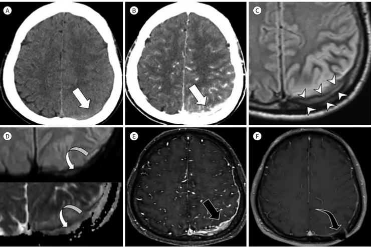
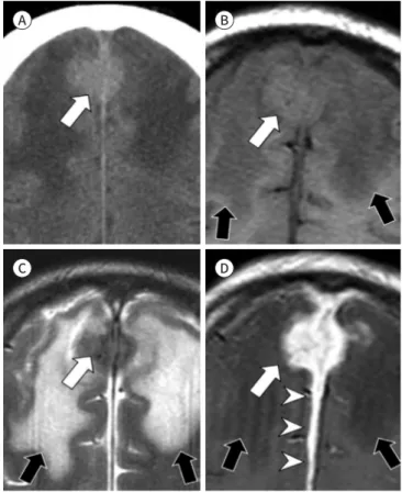
Fig. 15. Immunoglobulin G4-related hypertrophic pachymeningitis (linear dural thickening).
A 52-year-old male with sudden-onset motor aphasia.
A, B. Noncontrast (A) and contrast-enhanced CT (B) show a hyperdense lesion with intense contrast enhancement at the left parietal convexi- ty (arrows).
C. The lesion exhibits intermediate signal intensity with peripheral rims of low signal intensity on the MR fluid-attenuated inversion recovery images (arrowheads).
D. The diffusion‐weighted imaging (b value = 1000 s/mm2) and ADC map show abnormal restricted diffusion with low ADC values for the left parietal cortex (curved arrows).
E. Gadolinium-enhanced T1-weighted MRI shows localized dural thickening and enhancement on CT (arrow).
F. MR image obtained 1 year after steroid treatment shows marked decreased enhancement and thickness of the dura of the left parietal por- tion (curved arrow).
ADC = apparent diffusion coefficient
tients with IgG4- related disease in other organs. Lymphadenopathy in IgG4-related disease tends to be mild, and the lymph nodes are generally measured to be less than 2 cm in size (Fig. 14) (20).
IgG4-RELATED HYPERTROPHIC PACHYMENINGITIS
The clinical presentations of IgG4-related hypertrophic pachymeningitis include neurolog- ical symptoms originating from mechanical compression of neurovascular structures, result- ing in functional deficits. On imaging studies, the disease manifests as localized or diffuse linear dural thickening or mass-like lesions (Figs. 15, 16) (21).
A
D
B
E
C
F
CONCLUSION
Imaging is critical for the diagnosis and treatment assessment of patients with IgG4-related disease. The pancreas is most well-known organ involvement, but extrapancreatic manifes- tations have been increasingly reported. Radiologists need to understand the imaging fea- tures of various organ involvement since imaging plays an integral role in providing a timely diagnosis and avoiding unnecessary invasive diagnostic approaches.
Author Contributions
Conceptualization, K.H.R.; data curation, all authors; investigation, all authors; methodology, K.H.R.; project administration, K.H.R.; resources, all authors; supervision, K.H.R.; validation, K.H.R.;
writing—original draft, K.H.R, P.K.
Conflicts of Interest
Yo Won Choi has been an Section Editor of Journal of the Korean Society of Radiology since 2015;
however, he was not involved in the peer reviewer selection, evaluation, or decision process of this ar- ticle. Otherwise, no other potential conflicts of interest relevant to this article were reported.
Funding None
REFERENCES
1. Sahani DV, Kalva SP, Farrell J, Maher MM, Saini S, Mueller PR, et al. Autoimmune pancreatitis: imaging fea- tures. Radiology 2004;233:345-352
Fig. 16. Immunoglobulin G4-related hypertrophic pachymeningitis (mimicking meningioma). A 59-year-old male presenting with seizure.
A. Noncontrast CT shows a high-density lobulating mass at the anterior falx, mimicking meningioma (white arrow).
B, C. The mass (white arrow) shows intermediate signal intensity on T1-weighted image (B) and low signal intensity on T2-weighted image (C) with severe perilesional edema (black arrows).
D. The lesion displays long and thick flax enhancement similar to the dural tail (arrowheads) on gadolinium-enhanced T1-weighted image.
The mass also shows homogenously intense enhancement (white ar- row) with severe perilesional edema (black arrows).
A
C
B
D
2. Irie H, Honda H, Baba S, Kuroiwa T, Yoshimitsu K, Tajima T, et al. Autoimmune pancreatitis: CT and MR characteristics. AJR Am J Roentgenol 1998;170:1323-1327
3. Muhi A, Ichikawa T, Motosugi U, Sou H, Sano K, Tsukamoto T, et al. Mass-forming autoimmune pancreatitis and pancreatic carcinoma: differential diagnosis on the basis of computed tomography and magnetic reso- nance cholangiopancreatography, and diffusion-weighted imaging findings. J Magn Reson Imaging 2012;
35:827-836
4. Madhusudhan KS, Das P, Gunjan D, Srivastava DN, Garg PK. IgG4-related sclerosing cholangitis: a clinical and imaging review. AJR Am J Roentgenol 2019;213:1221-1231
5. Vaglio A, Salvarani C, Buzio C. Retroperitoneal fibrosis. Lancet 2006;367:241-251
6. Brun B, Laursen K, Sørensen IN, Lorentzen JE, Kristensen JK. CT in retroperitoneal fibrosis. AJR Am J Roentgenol 1981;137:535-538
7. Cronin CG, Lohan DG, Blake MA, Roche C, McCarthy P, Murphy JM. Retroperitoneal fibrosis: a review of clinical features and imaging findings. AJR Am J Roentgenol 2008;191:423-431
8. Bakir B, Yilmaz F, Turkay R, Ozel S, Bilgiç B, Velioglu A, et al. Role of diffusion-weighted MR imaging in the differentiation of benign retroperitoneal fibrosis from malignant neoplasm: preliminary study. Radiology 2014;272:438-445
9. Arrivé L, Hricak H, Tavares NJ, Miller TR. Malignant versus nonmalignant retroperitoneal fibrosis: differenti- ation with MR imaging. Radiology 1989;172:139-143
10. Rubenstein WA, Gray G, Auh YH, Honig CL, Thorbjarnarson B, Williams JJ, et al. CT of fibrous tissues and tumors with sonographic correlation. AJR Am J Roentgenol 1986;147:1067-1074
11. Takahashi N, Kawashima A, Fletcher JG, Chari ST. Renal involvement in patients with autoimmune pancre- atitis: CT and MR imaging findings. Radiology 2007;242:791-801
12. Triantopoulou C, Malachias G, Maniatis P, Anastopoulos J, Siafas I, Papailiou J. Renal lesions associated with autoimmune pancreatitis: CT findings. Acta Radiol 2010;51:702-707
13. Kim B, Kim JH, Byun JH, Kim HJ, Lee SS, Kim SY, et al. IgG4-related kidney disease: MRI findings with em- phasis on the usefulness of diffusion-weighted imaging. Eur J Radiol 2014;83:1057-1062
14. Inoue D, Zen Y, Abo H, Gabata T, Demachi H, Kobayashi T, et al. Immunoglobulin G4-related lung disease:
CT findings with pathologic correlations. Radiology 2009;251:260-270
15. Ryu JH, Sekiguchi H, Yi ES. Pulmonary manifestations of immunoglobulin G4-related sclerosing disease.
Eur Respir J 2012;39:180-186
16. Geyer JT, Ferry JA, Harris NL, Stone JH, Zukerberg LR, Lauwers GY, et al. Chronic sclerosing sialadenitis (Küttner tumor) is an IgG4-associated disease. Am J Surg Pathol 2010;34:202-210
17. Yamamoto M, Takahashi H, Ohara M, Suzuki C, Naishiro Y, Yamamoto H, et al. A new conceptualization for Mikulicz’s disease as an IgG4-related plasmacytic disease. Mod Rheumatol 2006;16:335-340
18. Tiegs-Heiden CA, Eckel LJ, Hunt CH, Diehn FE, Schwartz KM, Kallmes DF, et al. Immunoglobulin G4-related disease of the orbit: imaging features in 27 patients. AJNR Am J Neuroradiol 2014;35:1393-1397
19. Zalaquett E, Razmilic D, Oddo D. Immunoglobulin G4-related sclerosing mastitis: AIRP best cases in radio- logic-pathologic correlation. Radiographics 2016;36:959-962
20. Cheuk W, Yuen HK, Chu SY, Chiu EK, Lam LK, Chan JK. Lymphadenopathy of IgG4-related sclerosing dis- ease. Am J Surg Pathol 2008;32:671-681
21. Lu LX, Della-Torre E, Stone JH, Clark SW. IgG4-related hypertrophic pachymeningitis: clinical features, diag- nostic criteria, and treatment. JAMA Neurol 2014;71:785-793
다양한 인체 장기에서 보일 수 있는 면역글로불린 G4 관련 질환: 임상화보
박경리
1· 최요원
1· 강보경
1· 이지영
1· 박정선
1· 신수진
2,3· 구혜령
1*
면역글로불린 G4 (immunoglobulin G4; 이하 IgG4) 관련 질환은 특징적인 병리 소견을 동 반한 섬유화를 일으키는 전신 염증성 질환이다. 영상검사는 IgG4 관련 질환의 진단 및 치료 반응 평가에 필수적이다. 본 임상화보는 IgG4 관련 질환의 다양한 임상 증례를 통해 영상 소 견 및 감별진단을 제시함으로써 IgG4 관련 질환에 대한 이해의 폭을 넓히고자 한다.
1한양대학교 의과대학 한양대학교병원 1영상의학과, 2병리과,
3연세대학교 의과대학 강남세브란스병원 병리과