Correlations between the CagA Antigen and Serum Levels of Anti-Helicobacter pylori IgG and IgA in Children
We tested correlations between anti-Helicobacter pylori IgG and IgA levels and the urease test, anti-CagA protein antibody, degree of gastritis, and age. In total, 509 children (0-15 years) were enrolled. Subjects were stratified as 0-4 years (n = 132), 5-9 years (n = 274), and 10-15 years (n = 103) and subjected to the urease test, histopathology, ELISA, and Western blot using whole-cell lysates of H. pylori strain 51. The positivity rate in the urease test (P = 0.003), the degree of chronic gastritis (P = 0.021), and H. pylori infiltration (P < 0.001) increased with age. The median titer for anti-H. pylori IgG was 732.5 IU/mL at 0-4 years, 689.0 IU/mL at 5-9 years, and 966.0 IU/mL at 10-15 years (P < 0.001); the median titer for anti-H. pylori IgA was 61.0 IU/mL at 0-4 years, 63.5 IU/mL at 5-9 years, and 75.0 IU/mL at 10-15 years (P < 0.001). The CagA-positivity rate was 26.5% at 0-4 years, 36.5% at 5-9 years, and 46.6% at 10-15 years for IgG (P = 0.036), and 11.3% at 0-4 years, 18.6% at 5-9 years, and 23.3% at 10-15 years for IgA (P < 0.001). Anti- H. pylori IgG and IgA titers increased with the urease test grade, chronic gastritis degree, active gastritis, and H. pylori infiltration. Presence of CagA-positivity is well correlated with a high urease test grade and high anti-H. pylori IgG/IgA levels.
Keywords: Antibodies IgG/IgA; Helicobacter pylori; CagA Protein; Urease; Children Ji-Hyun Seo,1 Chun Woo Lim,1
Ji Sook Park,1 Jung Sook Yeom,1 Jae-Young Lim,1 Jin-Su Jun,1 Hyang-Ok Woo,1 Hee-Shang Youn,1 Seung-Chul Baik,2 Woo-Kon Lee,2 Myung-Je Cho,2 and Kwang-Ho Rhee2
1Department of Pediatrics, Gyeongsang National University School of Medicine, Gyeongsang Institute of Health Science, Jinju, Korea; 2Department of Microbiology, Gyeongsang National University School of Medicine, Gyeongsang Institute of Health Science, Jinju, Korea
Received: 1 September 2015 Accepted: 10 December 2015 Address for Correspondence:
Hee-Shang Youn, MD
Department of Pediatrics, Gyeongsang National University School of Medicine, 15 Jinju-daero 816-beon-gil, Jinju 52727, Korea
E-mail: [email protected]
Funding: This study was supported by a grant from the National R&D Program for Cancer Control of the Ministry of Health &
Welfare of the Republic of Korea (0820050).
http://dx.doi.org/10.3346/jkms.2016.31.3.417 • J Korean Med Sci 2016; 31: 417-422
INTRODUCTION
Helicobacter pylori is an important etiological factor for acute and chronic gastritis, gastric and duodenal ulcers, and gastric adenocarcinoma (1). The severity of H. pylori infection depends on the strain virulence, host susceptibility, and environmental factors (2). The measurement of specific antibodies in serum has been used as a noninvasive method for detecting H. pylori infection (3) and over 90% of H. pylori-infected patients have detectable serum IgG antibodies (4). Serological tests are com- mercially available, which are easy to perform and inexpensive, but studies on children indicate a high sensitivity range of 50%- 90% and the specificity ranges from 83% to 100% (5-8).
The number of immunoreactive bands significantly increas- es with age and reactions to the VacA and CagA antigens are more frequently found in older children (9). In our previous study, we found that more than 80% of the seropositive enzyme- linked immunosorbent assay (ELISA) results were CagA positive, whereas the other 20% of the seropositive results using ELISA could be attributed to its reaction with another H. pylori anti- gen (10). Therefore, the use of whole-cell lysates of H. pylori strain 51 in ELISA may increase the yield when detecting anti-
H. pylori antibodies in the Korean population.
Several studies have investigated the relationship between antibody titers and the pathogenesis of H. pylori, but the results are inconclusive (3,11). Quantitative evaluations of anti-H. py- lori antibodies or against H. pylori recombinant purified pro- teins have been performed in some human diseases where H.
pylori infections may play a role in their pathogenesis (9,12-14).
However, the clinical significance of high antibody levels to H.
pylori according to quantitative ELISA has not been established and high anti-H. pylori antibody levels have not been demon- strated to be predictive of the severity of gastroduodenal diseas- es or the density of H. pylori colonization.
Thus, to help identify factors that correlate with antibody lev- els in children, we evaluated the correlations between the levels of anti-H. pylori IgG and IgA antibodies and the urease test grade, presence of CagA antigen, degree of gastritis, and age.
MATERIALS AND METHODS Study population
As a member of the National Biobank of Korea, Gyeongsang National University Hospital (GNUH) collects serum samples Gastroenterology & Hepatology
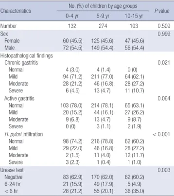
Table 1. Baseline and clinical characteristics
Characteristics No. (%) of children by age groups
P value
0-4 yr 5-9 yr 10-15 yr
Number 132 274 103 0.509
Sex Female
Male 60 (45.5)
72 (54.5) 125 (45.6)
149 (54.4) 47 (45.6) 56 (54.4)
0.999
Histopathological findings Chronic gastritis Normal Mild Moderate Severe
4 (3.0) 94 (71.2) 28 (21.2) 6 (4.5)
4 (1.4) 211 (77.0)
46 (16.8) 13 (4.7)
0 (0) 64 (62.1) 28 (27.2) 11 (10.7)
0.021
Active gastritis Normal Mild Moderate Severe
103 (78.0) 20 (15.2) 9 (6.8) 0 (0)
214 (78.1) 44 (16.1) 13 (4.7)
3 (1.1)
65 (63.1) 27 (26.2) 9 (8.7) 2 (1.9)
0.064
H. pylori infiltration Normal Mild Moderate Severe
98 (74.2) 29 (22.0) 2 (1.5) 3 (2.3)
216 (78.8) 46 (16.8) 11 (4.0)
1 (0.4)
62 (60.2) 28 (27.2) 12 (11.7) 1 (1.0)
< 0.001
Urease test Negative 6-24 hr < 6 hr
83 (62.9) 21 (15.9) 28 (21.2)
170 (62.0) 49 (17.9) 55 (20.1)
62 (60.2) 5 (4.9) 36 (35.0)
0.003
Fig. 1. The immunoblot assay results were classified into four patterns on the basis of immunoreactive bands. Only pattern I, with reactivity against 120-kDa antigens and other H. pylori antigens, was considered to be a specific marker of H. pylori in- fection in this study. Panel A shows the Ponceau S‐stained nitrocellulose membrane onto which the marker proteins and separated H. pylori antigen were transferred.
Pattern I II III IV
150 kDa 10075
50 37 25 2015 10
from random patients and stores them at -80°C. Among the samples collected over 21 years, we examined those from 509 children who underwent upper gastroduodenoscopy at GNUH during 1991-2010. Thus, we enrolled 509 children and we re- viewed the results of urease test and the histopathological find- ings, and tested the reserved serum. The sera were stratified into three age groups: 0-4 years (n = 132), 5-9 years (n = 274), and 10-15 years (n = 103) (Table 1).
Urease tests and histopathological findings
Three gastric endoscopic biopsies taken from the gastric prepy- loric antrum with an Olympus GIF-XP endoscope with pediat- ric forceps were first subjected to urease tests, which were per- formed in the endoscopy room. Based on the rapidity of the color change, the subjects were designated as grades 0 (nega- tive, no color change), 1 (color change between 6 and 24 hours), or 2 (color change within 6 hours).
Three biopsy specimens each from the gastric antrum and gastric body were stained with hematoxylin-eosin for the histo- logical analyses. The histology results were interpreted accord- ing to the Updated Sydney System. All of the histopathological slides that we reviewed had been prepared and donated by the GNUH.
ELISA and western blot analysis
Anti-H. pylori IgG and IgA titers were measured by ELISA (10) using the coated with the prepared whole cell proteins of H. py- lori strain 51 (10 μg/mL and 50 μL per well diluted with coating
buffer). Diluted sera (IgG 1:400, and IgA 1:100) were added to antigen-coated wells (50 μL per well).
Anti-CagA IgG and IgA antibodies were evaluated by West- ern blot using whole-cell lysates of H. pylori strain 51 (15). The western blot patterns were assigned to four categories on the basis of CagA (pattern I), urease without CagA (pattern II), oth- er proteins except CagA and urease (pattern III), and no band (pattern IV) (Fig. 1).
Statistical analysis
The statistical analyses were performed using IBM SPSS Statis- tics 21 (IBM, Chicago, IL, USA). We tested whether the antibody titer had significant correlations with the urease test results, an- tigen patterns, or age. We used bivariate correlation (Spearman’s rho), paired samples t-tests, and nonparametric tests to analyze the differences in the antibody titers between CagA-positive and-negative sera, among the three urease test grades, and in the three age groups. The numbers and sex ratios in the three age groups were different, so GLM regression analysis was used for correction. Post-hoc analysis using Scheffé’s method was applied when significant differences were detected among three groups. Statistically significant differences were accepted at P < 0.05.
Ethics statement
The study protocol was reviewed and approved by the institu- tional review board of GNUH (GNUHIRB-2015-08-020). Inform- ed consent was exempted by the board.
RESULTS Study population
Table 1 shows the number and sex distribution of the subjects according to age. There were no significant differences in the sex distributions among the age groups.
Results of the urease test and histopathological findings The positivity rates for the urease test were 37.1% at 0-4 years, 38.0% at 5-9 years, and 39.9% at 10-15 years (P = 0.003). The de- grees of chronic gastritis (P = 0.021), active gastritis (P = 0.064), and H. pylori infiltration (P < 0.001) increased with age (Table 1).
Anti-H. pylori IgG and IgA titers
The median titers for anti-H. pylori IgG were 732.5 IU/mL at 0-4 years, 689.0 IU/mL at 5-9 years, and 966.0 IU/mL at 10-15 years (P < 0.001). The median titers for anti-H. pylori IgA were 61.0 IU/mL at 0-4 years, 63.5 IU/mL at 5-9 years, and 75.0 IU/mL at 10-15 years (P < 0.001). The anti-H. pylori IgG titers were higher at 10-15 years than those at 1-5 years and 6-10 years (P = 0.006), but there was no significant difference in the anti-H. pylori IgA titers among the three age groups (P = 0.454).
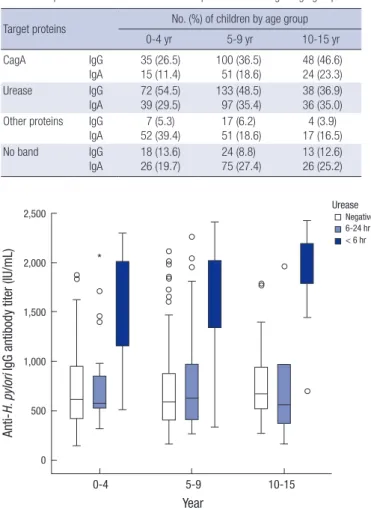
Western blot patterns according to age groups
The proportions of IgG positivity according to the four western blot patterns (I-IV) were 36.0%, 47.7%, 5.5%, and 10.8%, respec- tively (P = 0.008). The proportions of IgA positivity according to the four patterns were 17.7%, 33.8%, 23.6%, and 25.0%, respec- tively (P = 0.221). The CagA-positivity rates were 26.5% at 0-4 years, 36.5% at 5-9 years, and 46.6% at 10-15 years for IgG (P = 0.036, Fig. 2), and 11.4% at 0-4 years, 18.6% at 5-9 years, and 23.3%
at 10-15 years for IgA (P < 0.001) (Table 2). The western blot neg- ative (no band) rates for IgG were 13.6%, 8.8%, and 12.6% at 0-4 years, 5-9 years, and 10-15 years, respectively. Post-hoc analysis
using Scheffé’s method detected no differences in the propor- tions of the four Western blot patterns among the three age groups for IgG (P = 0.094) and for IgA (P = 0.161).
Correlations between the urease test grade, degree of histopathological findings, western blot patterns, and antibody titers for IgG and IgA
The titers of anti-H. pylori IgG antibodies increased with the ure- ase test grade (r = 0.527, P < 0.001), chronic gastritis (r = 0.613, P < 0.001), active gastritis (r = 0.545, P < 0.001), and the degree of H. pylori infiltration (r = 0.593, P < 0.001). The anti-H. pylori IgA titers also increased with the urease test grade (r = 0.450, P < 0.001), degree of chronic gastritis (r = 0.523, P < 0.001), ac- tive gastritis (r = 0.453, P < 0.001), and H. pylori infiltration (r = 0.480, P < 0.001). In the urease test, the anti-H. pylori IgG (Fig. 3) and IgA (Fig. 4) antibody titers were higher with grade I than the other grades (P < 0.001), regardless of age. According to the four western blot patterns, the anti-H. pylori IgG (Fig. 5) and IgA (Fig. 6) titers were higher with the CagA-positive pattern (P <
0.001), regardless of age.
Table 2. Proportions with the four western blot patterns according to age groups Target proteins No. (%) of children by age group
0-4 yr 5-9 yr 10-15 yr
CagA IgG
IgA
35 (26.5) 15 (11.4)
100 (36.5) 51 (18.6)
48 (46.6) 24 (23.3)
Urease IgG
IgA 72 (54.5)
39 (29.5) 133 (48.5)
97 (35.4) 38 (36.9) 36 (35.0) Other proteins IgG
IgA
7 (5.3) 52 (39.4)
17 (6.2) 51 (18.6)
4 (3.9) 17 (16.5)
No band IgG
IgA
18 (13.6) 26 (19.7)
24 (8.8) 75 (27.4)
13 (12.6) 26 (25.2)
Year
0-4 5-9 10-15
100 90 80 70 60 50 40 30 20 10 0 (%)
No band Other proteins Urease CagA
Fig. 2. Proportions with the four western blot patterns according to age groups. The CagA-positivity rate was 26.5% at 0-4 years, 36.5% at 5-9 years, and 46.6% at 10- 15 years for IgG (P = 0.036). Post-hoc analysis using Scheffé’s method detected no differences in the proportions with the four western blot pattern among the three age groups for IgG (P = 0.094).
Fig. 3. Anti-H. pylori IgG antibody titers according to age groups and the urease test grade. The anti-H. pylori IgG antibody titers were higher with grade I (positive within 6 hours) than the other grades (P < 0.001) in all age groups.
Anti-H. pylori IgG antibody titer (IU/mL)
Year
0-4 5-9 10-15
2,500
2,000
1,500
1,000
500
0
*
Urease Negative 6-24 hr
< 6 hr
DISCUSSION
In the present study, we found that the presence of the CagA antigen was the major factor related to high levels of anti-H. py- lori IgG and IgA antibodies, regardless of age. CagA is known to be an important virulence factor in H. pylori (14) and antibod- ies against CagA have been observed in gastritis, gastroduode- nal ulcer, and gastric cancer patients (13,16,17). In the early 2000s,
80%-100% of H. pylori strains possessed the cagA gene in East Asia (18,19) and 94% were cagA-positive in H. pylori DNA ex- tracts from 33 Korean children (20). In Japan, the CagA was the most reactive antigen recognized by all the H. pylori positive sera even from children under the age of 3 year (21). Therefore, the regional CagA antigens for serodiagnosis of H. pylori would be important, which could affect the rate of seropositivity (21).
Thus, a positive test result for anti-CagA antibody was regarded as an H. pylori infection in Korean studies (15,22). In the pres- ent study, the highest positivity rate for CagA was 46.6% among the 10-15 years group, although this CagA-positivity rate is low- er than the seroprevalence (59.6%) of H. pylori infections in a recent study of the general population in Korea (23), as well as the seroprevalence rate (68.0% CagA-positive) in children aged 6-15 years using the same immunoblot analysis during 1998- 1999 (17). Recent seroprevalence studies of H. pylori infection suggest that the decrease in the seroprevalence of H. pylori may be related to the improved socioeconomic status of Koreans (23,24).
Young children may have a different immune response to H.
pylori, with preferences for specific antigens, as well as lower ti- ters than adults (8). A lower sensitivity has been reported based on serological H. pylori tests in children compared with adults (25). Using commercial ELISA kits, false-negative results were found more often in children aged younger than 5 years (8,26).
In our previous study, we showed that the Genedia IgG ELISA kit, which uses H. pylori antigen obtained from a Korean H. py- lori strain, achieved a higher seropositivity rate than other ELI- SA kits (e.g., GAP IgG, HM-CAP, and Pyloriset EIA-G obtained from USA and Finland) (17). There are differences in the anti- genicity of multiple H. pylori strains and even among different Fig. 4. Anti-H. pylori IgA antibody titers according to age groups and the urease test
grade. The anti-H. pylori IgA antibody titers were higher with grade I (positive within 6 hours) than the other grades (P < 0.001) in all age groups.
Anti-H. pylori IgA antibody titer (IU/mL)
Year
0-4 5-9 10-15
500
400
300
200
100
0
*
*
**
***
*
*
*
Urease Negative 6-24 hr
< 6 hr
Fig. 5. Anti-H. pylori IgG antibody titers according to the western blot patterns and age. The anti-H. pylori IgG antibody titers were higher with the CagA-positive pattern (P < 0.001) regardless of age.
Anti-H. pylori IgG antibody titer (IU/mL)
Year
0-4 5-9 10-15
2,500
2,000
1,500
1,000
500
0
*
CagA (+) Urease (+), CagA (-) CagA and Urease (-) No band
Fig. 6. Anti-H. pylori IgA antibody titers according to the western blot patterns and age. The anti-H. pylori IgA antibody titers were higher with the CagA-positive pattern (P < 0.001) regardless of age.
Anti-H. pylori IgA antibody titer (IU/mL)
Year
0-4 5-9 10-15
500
400
300
200
100
0
*
Caa A (+) Urease (+), CagA (-) CagA and Urease (-) No band
**
antigens in the same strain (9). In the present study, the median levels of anti-H. pylori IgG and IgA antibody also increased with age but the antibody titers were higher in the CagA-positive cas- es than those in the CagA-negative cases, even in children aged under 5 years.
Age is strongly related to H. pylori infections (27). In the pres- ent study, the degree of chronic gastritis, active gastritis, and H.
pylori infiltration and the positive urease test rate increased sig- nificantly with age. The CagA-positivity rate increased with age but not significantly. Previously, the grades of active and chron- ic inflammation, atrophy, lymphoid follicles, and H. pylori den- sity were correlated with the IgG antibody levels in the antral mucosa (3,11,28). The serum antibody response to H. pylori also depends on the severity of H. pylori-associated diseases (29). In the present study, the anti-H. pylori IgG and IgA anti- body levels were correlated with the degree of chronic gastritis and H. pylori infiltration. The degrees of the histopathological findings and the anti-H. pylori IgG and IgA antibody titers were also correlated with the ages of the children.
A positive urease test within 6 hours was another factor that affect ed the anti-H. pylori IgG and IgA antibody levels regard- less of age. Buffered urease tests require at least 1,000 organisms to generate a positive reaction (30) and a higher degree of H.
pylori infiltration is correlated with a faster positive reaction in the urease test (31,32). Therefore, a rapid reaction in the urease test may be related to a high density of bacteria. In the present study, there was no significant correlation between the presence of anti-urease antibody, the titer of anti-H. pylori antibodies, and the urease test grade. The change in H. pylori strains from CagA-positive to CagA-negative in Korea should be considered when evaluating the seroepidemiology of H. pylori infections.
The current study had some limitations as follows. We con- ducted the current study with a retrospective design. We simply analyzed the results of urease tests and histopathological find- ings in this study, and we did not evaluate the clinical histories of the children.
In summary, we found that the anti-H. pylori IgG and IgA an- tibody titers were higher in the CagA-positive sera of children regardless of age, while higher IgG and IgA titers were observed with a higher degree of active gastritis and H. pylori infiltration, higher urease test grade, and greater age. In conclusion, the pres- ence of CagA antigen is the main factor that affects the levels of anti-H. pylori IgG and IgA antibodies regardless of age. The ELI- SA test is a valuable diagnostic tool for diagnosing CagA-posi- tive H. pylori infections in children. However, further studies are needed of CagA-negative strain infections and the levels of anti-H. pylori IgG and IgA antibodies.
ACKNOWLEDGMENT
The serum samples used in this study were provided by the
Gyeongsang National University Hospital, which is a member of the National Biobank of Korea, which is supported by the Ministry of Health, Welfare and Family Affairs. All samples de- rived from the National Biobank of Korea were obtained with informed consent under institutional review board approved protocols.
DISCLOSURE
The authors have no potential conflicts of interest to disclose.
AUTHOR CONTRIBUTION
Research conception & design: Seo JH, Youn HS, Cho MJ, Rhee KH. Performing the experiments: Lim CW, Jun JS, Baik SC, Lee WK. Data acquisition: Seo JH, Park JS, Yeom JS, Lim JY, Woo HO, Youn HS. Data analysis and interpretation: Seo JH, Cho MJ. Sta- tistical analysis: Seo JH, Youn HS. Drafting of the manuscript:
Seo JH, Lim CW, Jun JS, Youn HS. Critical revision of the manu- script: Seo JH, Woo HO, Youn HS. Receiving grant: Youn HS.
Approval of final manuscript: all authors.
ORCID
Ji-Hyun Seo http://orcid.org/0000-0002-0691-3957 Chun Woo Lim http://orcid.org/0000-0003-4146-4349 Ji Sook Park http://orcid.org/0000-0002-4704-2246 Jung Sook Yeom http://orcid.org/0000-0003-0688-7493 Jae-Young Lim http://orcid.org/0000-0001-5205-202X Jin-Su Jun http://orcid.org/0000-0002-6382-6286 Hyang-Ok Woo http://orcid.org/0000-0001-8849-9341 Hee-Shang Youn http://orcid.org/0000-0002-5498-838X Seung-Chul Baik http://orcid.org/0000-0001-6033-4078 Woo-Kon Lee http://orcid.org/0000-0003-3913-2265 Myung-Je Cho http://orcid.org/ 0000-0002-4958-9827 Kwang-Ho Rhee http://orcid.org/0000-0002-4422-4992 REFERENCES
1. McColl KE. Clinical practice. Helicobacter pylori infection. N Engl J Med 2010; 362: 1597-604.
2. Malaty HM. Epidemiology of Helicobacter pylori infection. Best Pract Res Clin Gastroenterol 2007; 21: 205-14.
3. Chen TS, Li FY, Chang FY, Lee SD. Immunoglobulin G antibody against Helicobacter pylori: clinical implications of levels found in serum. Clin Diagn Lab Immunol 2002; 9: 1044-8.
4. Schumann C, Triantafilou K, Rasche FM, Möricke A, Vogt K, Triantafilou M, Hahn P, Schneider EM, Lepper PM. Serum antibody positivity for dis- tinct Helicobacter pylori antigens in benign and malignant gastroduode- nal disease. Int J Med Microbiol 2006; 296: 223-8.
5. de Oliveira AM, Rocha GA, Queiroz DM, Mendes EN, de Carvalho AS, Fer- rari TC, Nogueira AM. Evaluation of enzyme-linked immunosorbent as-
say for the diagnosis of Helicobacter pylori infection in children from dif- ferent age groups with and without duodenal ulcer. J Pediatr Gastroen- terol Nutr 1999; 28: 157-61.
6. Raymond J, Kalach N, Bergeret M, Barbet JP, Benhamou PH, Gendrel D, Dupont C. Evaluation of a serological test for diagnosis of Helicobacter pylori infection in children. Eur J Clin Microbiol Infect Dis 1996; 15: 415-7.
7. Raymond J, Sauvestre C, Kalach N, Bergeret M, Dupont C. Immunoblot- ting and serology for diagnosis of Helicobacter pylori infection in children.
Pediatr Infect Dis J 2000; 19: 118-21.
8. Kindermann A, Konstantopoulos N, Lehn N, Demmelmair H, Koletzko S.
Evaluation of two commercial enzyme immunoassays, testing immuno- globulin G (IgG) and IgA responses, for diagnosis of Helicobacter pylori infection in children. J Clin Microbiol 2001; 39: 3591-6.
9. Rocha GA, Oliveira AM, Queiroz DM, Carvalho AS, Nogueira AM. Immu- noblot analysis of humoral immune response to Helicobacter pylori in children with and without duodenal ulcer. J Clin Microbiol 2000; 38: 1777- 81.
10. Seo JH, Jun JS, Youn HS, Yeom JS, Park JS, Park CH, Woo HO, Lee WK, Cho MJ, Rhee KH. Development of an ELISA for quantitative detection of im- munoglobulin G (IgG) and IgA antibodies to Helicobacter pylori for use in Korean pediatric patients with H. pylori-associated diseases. Gut Liver 2013; 7: 437-42.
11. Sheu BS, Shiesh SC, Yang HB, Su IJ, Chen CY, Lin XZ. Implications of He- licobacter pylori serological titer for the histological severity of antral gas- tritis. Endoscopy 1997; 29: 27-30.
12. Yokota S, Amano K, Fujii N, Yokochi T. Comparison of serum antibody ti- ters to Helicobacter pylori lipopolysaccharides, CagA, VacA and partially purified cellular extracts in a Japanese population. FEMS Microbiol Lett 2000; 185: 193-8.
13. Gao L, Michel A, Weck MN, Arndt V, Pawlita M, Brenner H. Helicobacter pylori infection and gastric cancer risk: evaluation of 15 H. pylori proteins determined by novel multiplex serology. Cancer Res 2009; 69: 6164-70.
14. Satomi S, Yamakawa A, Matsunaga S, Masaki R, Inagaki T, Okuda T, Suto H, Ito Y, Yamazaki Y, Kuriyama M, et al. Relationship between the diversi- ty of the cagA gene of Helicobacter pylori and gastric cancer in Okinawa, Japan. J Gastroenterol 2006; 41: 668-73.
15. Jeong HL, Jung YS, Jun JS, Yeom JS, Park JS, Seo JH, Lim JY, Park CH, Woo HO, Youn HS, et al. Comparison of four commercial ELISA kits and in- house immunoblotting for diagnosis of Helicobacter pylori infection. Pe- diatr Gastroenterol Hepatol Nutr 2012; 15: 85-90.
16. Holtmann G, Talley NJ, Mitchell H, Hazell S. Antibody response to specif- ic H. pylori antigens in functional dyspepsia, duodenal ulcer disease, and health. Am J Gastroenterol 1998; 93: 1222-7.
17. Blaser MJ, Perez-Perez GI, Kleanthous H, Cover TL, Peek RM, Chyou PH, Stemmermann GN, Nomura A. Infection with Helicobacter pylori strains possessing cagA is associated with an increased risk of developing ade- nocarcinoma of the stomach. Cancer Res 1995; 55: 2111-5.
18. Yamazaki S, Yamakawa A, Okuda T, Ohtani M, Suto H, Ito Y, Yamazaki Y,
Keida Y, Higashi H, Hatakeyama M, et al. Distinct diversity of vacA, cagA, and cagE genes of Helicobacter pylori associated with peptic ulcer in Ja- pan. J Clin Microbiol 2005; 43: 3906-16.
19. Wong BC, Yin Y, Berg DE, Xia HH, Zhang JZ, Wang WH, Wong WM, Huang XR, Tang VS, Lam SK. Distribution of distinct vacA, cagA and iceA alleles in Helicobacter pylori in Hong Kong. Helicobacter 2001; 6: 317-24.
20. Ko JS, Kim KM, Oh YL, Seo JK. cagA, vacA, and iceA genotypes of Helico- bacter pylori in Korean children. Pediatr Int 2008; 50: 628-31.
21. Akada J, Okuda M, Hiramoto N, Kitagawa T, Zhang X, Kamei S, Ito A, Na- kamura M, Uchida T, Hiwatani T, et al. Proteomic characterization of He- licobacter pylori CagA antigen recognized by child serum antibodies and its epitope mapping by peptide array. PLoS One 2014; 9: e104611.
22. Kim EA, Kim YO, Lim JY, Jung YS, Park CH, Woo HO, Youn HS, Ko GH, Baik SC, Lee WK, et al. Antibody response of infants to Helicobacter py- lori infection. Korean J Gastroenterol 2000; 35: 704-15.
23. Yim JY, Kim N, Choi SH, Kim YS, Cho KR, Kim SS, Seo GS, Kim HU, Baik GH, Sin CS, et al. Seroprevalence of Helicobacter pylori in South Korea.
Helicobacter 2007; 12: 333-40.
24. Kim HY, Kim N, Kim SM, Seo JH, Park EH, Lee DH. Seroprevalence of He- licobacter pylori infection in Korean health personnel. Gut Liver 2013; 7:
648-54.
25. Crabtree JE, Mahony MJ, Taylor JD, Heatley RV, Littlewood JM, Tompkins DS. Immune responses to Helicobacter pylori in children with recurrent abdominal pain. J Clin Pathol 1991; 44: 768-71.
26. Corvaglia L, Bontems P, Devaster JM, Heimann P, Glupczynski Y, Keppens E, Cadranel S. Accuracy of serology and 13C-urea breath test for detection of Helicobacter pylori in children. Pediatr Infect Dis J 1999; 18: 976-9.
27. Pounder RE, Ng D. The prevalence of Helicobacter pylori infection in dif- ferent countries. Aliment Pharmacol Ther 1995; 9 Suppl 2: 33-9.
28. Hsu PI, Lai KH, Tseng HH, Liu YC, Yen MY, Lin CK, Lo GH, Huang RL, Huang JS, Cheng JS, et al. Correlation of serum immunoglobulin G Heli- cobacter pylori antibody titers with histologic and endoscopic findings in patients with dyspepsia. J Clin Gastroenterol 1997; 25: 587-91.
29. Chomvarin C, Ottiwet O, Hahnvajanawong C, Intapan PM, Wongwajana S. Seroreactivity to specific antigens of Helicobacter pylori infection is as- sociated with an increased risk of the dyspeptic gastrointestinal diseases.
Int J Infect Dis 2009; 13: 647-54.
30. Graham DY. Helicobacter pylori and the endoscopist: whether to diag- nose. Gastrointest Endosc 1991; 37: 577-9.
31. Seo JH, Youn HS, Park JJ, Yeom JS, Park JS, Jun JS, Lim JY, Park CH, Woo HO, Ko GH, et al. Influencing factors to results of the urease test: age, sam- pling site, histopahtologic findings, and density of Helicobacter pylori.
Pediatr Gastroenterol Hepatol Nutr 2013; 16: 34-40.
32. Seo JH, Park JS, Yeom JS, Lim JY, Park CH, Woo HO, Baik SC, Lee WK, Cho MJ, Rhee KH, et al. Correlation between positive rate and number of bi- opsy samples on urease test in childhood Helicobacter pylori infection. J Korean Med Sci 2014; 29: 106-9.