J Korean Soc Radiol 2017;76(6):395-402 https://doi.org/10.3348/jksr.2017.76.6.395
INTRODUCTION
Aberrant systemic arterial supply to the normal lung (ASANL) is a rare disease. Although most patients with ASANL are asymp- tomatic, the presenting symptoms are cardiac murmur in child- hood, and recurrent hemoptysis, dyspnea on exertion, and con- gestive heart failure in adults (1). Diagnosis is relatively easy with chest computed tomography (CT) (1, 2). However, treat- ment options vary, including surgical resection of the involved lung and surgical ligation of the aberrant artery, and endovas- cular treatment has been recently introduced (3). We reported a case of a 24-year-old man with ASANL who was treated by the endovascular method. In addition, we reviewed the relevant lit- erature.
CASE REPORT
A 24-year-old man presented with dyspnea on exertion and intermittent blood-tinged sputum. Dyspnea had started in his childhood, and was aggravated with intermittent blood-tinged sputum for the past 2–3 years. He was a social smoker (1 cigarette a day), and had no specific history.
Chest CT showed large tortuous vessels arising from the de- scending thoracic aorta in the basal segments of the left lower lobe (Fig. 1A). The largest diameter of the vessel at the origin was 1.4 cm. There were no findings to suggest hypertrophied sys- temic artery associated with chronic inflammation. In contrast to scimitar syndrome, the lungs were not hypogenetic, and the left lower lobar pulmonary vein was dilated and normally drained to the left atrium (Fig. 1B). Courses of the bronchial tree were normal, and thickening of the interstitial markings with ground- glass opacities were observed. These findings were compatible
Transarterial Embolization Treatment for Aberrant Systemic Arterial Supply to the Normal Lung: A Case Report and Literature Review
정상 폐에 공급하는 비정상 체동맥에 대한 경동맥 색전술 치료:증례 보고 및 문헌 고찰
Bo Ra Kim, MD, Jeong-Hyun Jo, MD, Byeong-Ho Park, MD*
Department of Radiology, Dong-A University Hospital, Dong-A University College of Medicine, Busan, Korea
A 24-year-old man presented with dyspnea on exertion and intermittent blood-tinged sputum. He was diagnosed with aberrant systemic arterial supply to the normal lung (ASANL) based on the results of imaging studies. The patient was successfully treated with transarterial embolization using coils and a vascular plug and his symptoms dis- appeared during the follow-up. Herein, we reported the imaging findings of ASANL, differential diagnoses, and its treatment options. In addition, we reviewed the relevant literature.
Index terms
Vascular Malformations Embolization, Therapeutic Lung
Received August 22, 2016 Revised December 2, 2016 Accepted December 19, 2016
*Corresponding author: Byeong-Ho Park, MD Department of Radiology, Dong-A University Hospital, Dong-A University College of Medicine,
26 Daesingongwon-ro, Seo-gu, Busan 49201, Korea.
Tel. 82-51-240-5371 Fax. 82-51-253-4931 E-mail: [email protected]
This is an Open Access article distributed under the terms of the Creative Commons Attribution Non-Commercial License (http://creativecommons.org/licenses/by-nc/4.0) which permits unrestricted non-commercial use, distri- bution, and reproduction in any medium, provided the original work is properly cited.
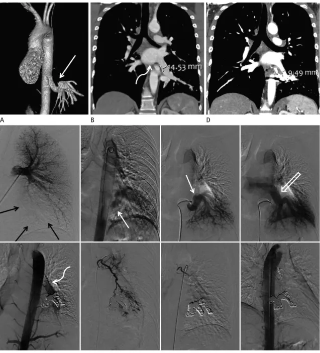
Fig. 1. Aberrant systemic arterial supply to the normal lung treated with transarterial embolization in a 24-year-old man.
A, B. Initial chest CT. Volume rendering image (A) shows a 14-mm-sized, large tortuous vessel (arrow) arising from the descending thoracic aorta and supplying the basal segments of the left lower lobe. On lung window setting image, thickening of the interstitial markings with ground glass opacities, consistent with pulmonary congestion, was noted in the left lower lobe; courses of the bronchial tree in both lungs were normal (not shown). On coronal reformatted image (B), the left lower lobar pulmonary vein is dilated, as compared to the right lower lobar pulmonary vein (curved arrow), and normally drains to the left atrium.
C. Images of conventional angiography (upper panel) and transarterial embolization (lower panel). Left pulmonary angiography shows hypoplasia or absence of the left lower pulmonary artery and decreased perfusion in the left lower lung zone (black arrows). Initial thoracic aortography and selective angiography of the aberrant systemic artery reveal an aberrant systemic artery (white arrows) arising from the descending thoracic aor- ta and supplying the left lower lung zone. On delayed phase, the enlarged left lower pulmonary vein (open arrow) that drains to the left atrium, is observed. On post-embolization aortography, the aberrant systemic artery is no longer visualized, and hypertrophied left bronchial artery (curved arrow) with arteriovenous shunt in the left lower lung zone, which was not revealed on the initial thoracic aortography, is observed. The left bronchial artery was selected, and embolization was conducted using microcoils and gelfoam particles. On final post-embolization aortography, the aberrant systemic artery and the hypertrophied bronchial artery are obliterated.
D. Follow-up chest CT image at 4 months after treatment. On the coronal reformatted image after treatment, the left lower lobar pulmonary vein is decreased in diameter.
A B D
C
with ASANL.
For evaluation of cardiac and pulmonary complications, echo- cardiography, lung perfusion scan, and pulmonary function test were conducted. Echocardiography indicated mild left ventric- ular enlargement with normal ejection fraction. Lung perfusion scan showed severe hypoperfusion in the left lower lobe. Mild ob- structive lung disease was suspected on pulmonary function test.
Treatment options include surgical treatment such as wedge resection of the involved lung, and surgical ligation of the aber- rant systemic artery (ASA), and endovascular treatment. Endo- vascular treatment was chosen considering his young age and potential for early recovery, and informed consent was obtained.
The right femoral artery and the left femoral vein were punc- tured under local anesthesia. Left pulmonary angiography sh- owed a hypoplastic left lower pulmonary artery with decreased perfusion in the left lower lung zone (LLLZ). On thoracic aortog- raphy, an ASA supplying the LLLZ was observed. A 5 Fr Yashiro
catheter (Radifocus®; Terumo Corp., Tokyo, Japan) was advanced to the origin of the ASA, and selective aberrant arteriography showed large arterial bran-ches supplying the LLLZ. We also observed an enlarged left lower pulmonary vein, which drained to the left atrium. However, there was no direct shunt vessel.
Transarterial embolization (TAE) was performed from the distal branches of the ASA, using twelve 6-mm, and eleven 8-mm platinum coils (Nester®; Cook, Bloomington, IN, USA) and one 10-mm fibered IDC coil (Interlock®; Boston Scientific, Natick, MA, USA). After coil embolization, the proximal portion of the ASA was embolized using a 14-mm Amplatzer Vascular Plug® type II (AGA Medical Corporation, Golden Valley, MN, USA), to cover the largest portion of the ASA at its origin. Post-embo- lization thoracic aortography indicated absence of blood flow from the ASA to the left lower pulmonary vein. However, new development of hypertrophied left bronchial artery with an ar- teriovenous shunt was noted in the LLLZ. This artery was tar-
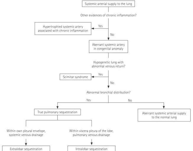
Fig. 2. A simplified algorithm for differential diagnosis of systemic arterial supply to the lung. The presence or absence of ancillary findings of chronic inflammation, abnormal venous return, and courses of the bronchial tree should be checked for differential diagnosis.
Scimitar syndrome
True pulmonary sequestration
Extralobar sequestration Intralobar sequestration Hypertrophied systemic artery
associated with chronic inflammation
Aberrant systemic artery in congenital anomaly
Aberrant systemic arterial supply to the normal lung Abnormal bronchial distribution?
Yes
Yes
Yes
No
No
No Systemic arterial supply to the lung
Other evidences of chronic inflammation?
Hypogenetic lung with abnormal venous return?
Within own pleural envelope,
systemic venous drainage Within viscera pleura of the lobe, pulmonary venous drainage
geted with a 1.98 Fr microcatheter and embolized using two 3-mm microcoils (Nester) and 1000–1400-µm gelfoam parti- cles (Cali-Gel®; Alicon Pharm SCI&TEC Co., Ltd., Hangzhou, China). After embolization, the hypertrophied left bronchial artery was not observed (Fig. 1C).
The patient complained of chest pain, fever, and chills for 6 days, which improved with supportive care. One week after TAE, his symptoms disappeared and he was discharged.
The patient was asymptomatic during the 1-year follow-up.
Follow-up chest CT at 4-months post-TAE showed decreased dilation of the distal branches of the ASA, and decreased size of the left lower lobar pulmonary vein (Fig. 1D).
DISCUSSION
ASANL is a rare congenital anomaly (1). Diagnosis of ASANL is relatively easy by imaging studies, especially CT (1, 2). Vascu- lar structures from the descending thoracic aorta in the left low- er lobe with normal bronchial distribution are common findings of ASANL (2). On conventional angiography, a dilated ASA sup- plies the LLLZ and drains into the left atrium through the inferi- or pulmonary vein in patients with ASANL (3, 4). The pulmo- nary arterial supply in the affected segments may be normal or absent (5).
In systemic arterial supply to the lungs, especially in adults, hypertrophied normal systemic arteries associated with chronic inflammation and aberrant systemic arteries in congenital anom- alies are considered as differential diagnoses (6), for e.g., pseu- dosequestration shows normal bronchial tree and pulmonary artery with other evidences of chronic inflammation. Congenital anomalies with ASA include scimitar syndrome, bronchopul- monary sequestration, and ASANL. Anomalous venous return with hypogenetic lung are suggestive of scimitar syndrome;
whereas, bronchopulmonary sequestration and ASANL are dif- ferentiated based on the occurrence of abnormal or normal br- onchial tree, respectively. Fig. 2 shows a simplified algorithm for the differential diagnosis.
Treatment is recommended in symptomatic and asymptomat- ic patients with ASANL because of the risk of congestive heart failure, recurrent severe infections, and hemorrhagic complica- tions (1, 2). Conventionally, surgical treatment has been the stan- dard therapeutic option (2). In pulmonary sequestration, resec-
tion of a sequestered lung is favored because of the risk of recu- rrent infection; whereas, surgical ligation of the ASA alone may be sufficient in cases of ASANL. Because TAE is a well-estab- lished treatment in patients with hemoptysis, and surgical liga- tion of the ASA is indicated in ASANL, embolization can be adjusted in ASANL (3, 5). Since Brühlmann et al. (3) first de- scribed a 51-year-old man with ASANL who was successfully treated by coil embolization, 19 cases treated with TAE have been reported in the English literature (Table 1). Brühlmann et al. (3) report a low risk of post-embolization pulmonary infarc- tion because of dual arterial supply to the affected lung paren- chyma. Abe et al. (5) reported cases of the complete type of ASANL that lack normal pulmonary arterial supply to the in- volved area, of which, only one case showed asymptomatic tran- sient pulmonary infarction. Saida et al. (7) suggest that the em- bolized area has low risk of infarction because tissue permeability or microcirculation through the persistent bronchial arteries preserves the perfusion of tissues. Nevertheless, proximal occlu- sion with coils or vascular plugs is advocated. Distal emboliza- tion using particles, such as gelatin sponge particles or polyvinyl alcohol, should be avoided to minimize the risk of ischemic injury (5). In our case, the ASA was embolized with large sized coils and proximal occlusion was performed using a vascular plug.
A rare case of ASANL with two feeders including a normal sys- temic artery and ASA supplying the affected lung zone was re- ported (4). Unlike previously reported ASANL cases, we detected a newly developed hypertrophied bronchial artery soon after the ASA-embolization. Similar phenomena have been reported in the literature on embolization of pulmonary arteriovenous malforma- tions (PAVMs) (8, 9). In our case, the hypertrophied bronchial ar- tery was possibly a preexisting lesion exaggerated by relative isch- emia, or a new lesion caused by procedure-related local ischemia.
It is likely that the bronchial artery to the involved area underwent compensatory hypertrophy to preserve tissue perfusion. As re- ported in PAVMs, a hypertrophied bronchial artery can cause re- current hemoptysis, and therefore, requires treatment. In our pa- tient, the proximal portion of the suddenly developed bronchial artery was embolized with coils and large-diameter gelfoam parti- cles to preserve tissue perfusion through collaterals and minimize the risk of ischemic injury.
Post-embolization syndrome is the most common complica- tion of TAE of ASANL, and can be controlled conservatively.
Table 1. Reported Cases of Aberrant Systemic Arterial Supply to the Normal Lung which were Treated with TAE in the English Literature AuthorsYear of PublicationSex/AgeClinical ManifestationsOrigins of Systemic Vessels Pulmonary Artery in Involved Lung Site
Site of Drainage of LungManagementFollow-Up PeriodComplication after TAE Jariwala et al. (4)2014F/7 moFailure to thriveIMA, descending thoracic aortaNormalEntire left lung2-staged TAE with coils and vascular plugs6 monthsNone Kim et al. (1)2014M/22Recurrent hemoptysisDescending thoracic aortaNot mentionedRight lower lobeTAE with vascular plugs28 monthsPost-embolization syndrome Sugihara et al.2013F/67Incidental finding on CTDescending thoracic aortaAbsentLeft lower lobeTAE with coils under balloon occlusion12 monthsNone Anil et al. (10)2012M/23Recurrent hemoptysisDescending thoracic aortaAbsentLeft lower lobeTAE with coils and cyanoacrylate glue6 monthsChest discomfort for 2 days Canyigit et al.2011M/53Acute chest painDescending thoracic aortaAbsentLeft lower lobeTAE with coils and vascular plugs10 monthsPost-embolization syndrome Abe et al. (5)2011F/24Recurrent hemoptysisDescending thoracic aortaAbsentLeft lower lobeTAE with coils1 yearChest pain for 3 days Lim et al.2009M/26Recurrent hemoptysisDescending thoracic aortaNot mentionedLeft lower lobeTAE with coils6 monthsPost-embolization syndrome Muñoz et al. (2)2008M/29HemoptysisAbdominal aortaNormalRight lower lobeTAE with coils12 monthsNone Kosutic et al.2007M/3 moRespiratory failureDescending thoracic aorta (2 separated origins)
HypoplasticRight upper lobeOne: TAE with coils The other: spontaneously closed after cardiac catheterization
6 monthsSevere respiratory failure, significant hypercapnia, and multiresistant Pseudomonas infection, which were treated successfully Saida et al. (7)2006F/41Recurrent hemoptysisDescending thoracic aortaAbsentLeft lower lobeTAE with coils6 yearsNone Huang et al.2005F/3 moRespiratory distress, cardiac murmurDescending thoracic aortaAbsentLeft lower lobeTAE with coils6 monthsNone F/13Recurrent hemoptysisDescending thoracic aortaAbsentLeft lower lobeTAE with coils18 monthsResidual arterial supply at 6 months, successfully treated by re-embolization
Table 1. Reported Cases of Aberrant Systemic Arterial Supply to the Normal Lung which were Treated with TAE in the English Literature (Continued) AuthorsYear of PublicationSex/AgeClinical ManifestationsOrigins of Systemic Vessels Pulmonary Artery in Involved Lung Site
Site of Drainage of LungManagementFollow-Up PeriodComplication after TAE Abdulhamid et al.2004M/13Chest pain, hemoptysisSeveral arterial collaterals from various levels of left IMA, and the midthoracic right intercostal artery
Not mentionedRight lung and pericardiumTAE with coils18 monthsNone F/15Recurrent hemoptysisRight innominate a, transverse aortic arch, proximal descending aorta, right IMA
Not mentionedRight middle and lower lobesTAE with coils18 monthsCough and wheezing for 2 days M/7Recurrent hemoptysisAortaNot mentionedRight lungTAE with coils2 yearsNone F/10Hemoptysis, chokingAortaNot mentionedNot mentionedTAE with coils9 monthsRecurred bleeding due to collateral vessel at 5 months, successfully treated by re-embolization Chabbert et al.2002M/17Acute chest painAbdominal aortaNormalRight lower lobeTAE with coils12 monthsPost-embolization syndrome Izillo et al.2000M/34Recurrent hemoptysisCeliac trunkAbsentRight lower lobeTAE with coils2 yearsTransient and asymptomatic pulmonary infarction Brühlmann et al. (3)1998M/51Massive hemoptysisDescending thoracic aortaNormal; small caliberLeft lower lobeTAE with coils10 monthsNone IMA = internal mammary artery, TAE = transarterial embolization
Other reported complications included recurrent bleeding due to developed collaterals or residual arterial supply, and some re- spiratory symptoms, which were successfully treated. Along with pulmonary infarction, secondary infection and fistula are theoretically possible, but have not been reported to date (10).
Long-term results after TAE for ASANL remain unclear. Pre- vious reports indicate that patients show improvement during the follow-up periods ranging from 6 months to 6 years. Addi- tional long-term studies are clearly required.
Our patient was successfully treated with TAE. Brillet et al.
(9) reported development of a systemic collateral supply after embolotherapy of PAVM. They showed that enlargement of a systemic artery was observed more frequently in patients with clinical and/or radiological features suggesting post-treatment lung infarction, than in those without these features. Although our patient has been asymptomatic for 1 year, close and long- term follow-up is required.
REFERENCES
1. Kim JH, Kim SS, Ha KS, Bae J, Park Y. Anomalous arterial supply to normal basal segment of the right lower lobe:
endovascular treatment with the amplatzer vascular plug.
Tuberc Respir Dis (Seoul) 2014;76:295-298
2. Muñoz JJ, García JA, Bentabol M, Padín MI, Serrano F. En- dovascular treatment of hemoptysis by abnormal systemic pulmonary artery supply. Cardiovasc Intervent Radiol 2008;
31:427-430
3. Brühlmann W, Weishaupt D, Goebel N, Imhof E. Therapeu- tic embolization of a systemic arterialization of lung with-
out sequestration. Eur Radiol 1998;8:355-358
4. Jariwala P, Ramesh G, Sarat Chandra K. Congenital anom- alous/aberrant systemic artery to pulmonary venous fistu- la: closure with vascular plugs & coil embolization. Indian Heart J 2014;66:95-103
5. Abe T, Mori K, Shiigai M, Okura N, Okamoto Y, Saida T, et al.
Systemic arterial supply to the normal basal segments of the left lower lobe of the lung--treatment by coil emboli- zation--and a literature review. Cardiovasc Intervent Radiol 2011;34 Suppl 2:S117-S121
6. Do KH, Goo JM, Im JG, Kim KW, Chung JW, Park JH. Sys- temic arterial supply to the lungs in adults: spiral CT find- ings. Radiographics 2001;21:387-402
7. Saida T, Ninomiya H, Hojo F, Nakayama M, Yamauchi T, Saida Y. Systemic arterial supply to the normal basal seg- ments of the left lower lobe treated by coil embolization, with long-term follow-up. Radiat Med 2006;24:365-368 8. Pollak JS, Saluja S, Thabet A, Henderson KJ, Denbow N,
White RI Jr. Clinical and anatomic outcomes after embolo- therapy of pulmonary arteriovenous malformations. J Vasc Interv Radiol 2006;17:35-44; quiz 45
9. Brillet PY, Dumont P, Bouaziz N, Duhamel A, Laurent F, Remy J, et al. Pulmonary arteriovenous malformation treat- ed with embolotherapy: systemic collateral supply at multi- detector CT angiography after 2-20-year follow-up. Radiol- ogy 2007;242:267-276
10. Anil G, Taneja M, Tan AG. Endovascular treatment of isolated systemic arterial supply to normal lung with coil and glue embolisation. Br J Radiol 2012;85:e83-e86
정상 폐에 공급하는 비정상 체동맥에 대한 경동맥 색전술 치료:
증례 보고 및 문헌 고찰
김보라 · 조정현 · 박병호*
운동성 호흡곤란과 간헐적인 혈성 객혈을 주소로 내원한 24세 남자 환자로 영상 검사를 통해 정상 폐에 공급하는 비정상 적인 체동맥으로 진단되었다. 코일과 혈관 플러그를 이용한 경동맥 색전술을 시행하여 성공적으로 치료하였다. 본 증례에 서 정상 폐에 공급하는 비정상적인 체동맥의 영상 소견과 감별 진단 및 여러 치료 방법에 대해 이야기하고, 이전 문헌들을 검토해 보고자 한다.
동아대학교 의과대학 동아대학교병원 영상의학과