Introduction
In cases of multiple tooth loss, treatment option can be removable partial denture, complete denture, overdenture, or implant sup- ported prosthesis. Among them, implant supported fixed pros- thesis slows down resorption of alveolar ridges and rehabilitates approx- imately full function of the natural teeth,1,2hence achieving relatively higher satisfaction on patient's behalf, compared to the conventional removable dentures.3By providing more stable occlusal conditions, implant supported fixed partial denture can be a good alternative, when the patient shows lack of neuromuscular control for remov- able denture.4
The aim of this case report was to show the functionally and esthet- ically satisfying full mouth rehabilitation with implant supported all- ceramic and porcelain-fused-to-gold prostheses.
Case report
A 56-year-old female patient was referred to the Department of Prosthodontics at Seoul National University Dental Hospital. She complained discomfort derived from the fractured maxillary pros-
theses, and the lower removable partial denture she had been using. She wanted the prosthetic treatment using implant sup- ported fixed partial denture. When presented, #16, 17, 25-27, 31- 36, 41-47 (FDI system) were missing and mandibular removable par- tial denture showed excessive occlusal wear so that the vertical dimen- sion was decreased (Fig. 1).
In clinical and radiological examination after removing all spoiled prostheses, preservation of all the remaining teeth, #13, 15, 23, 24, 37, was planned. In the upper part, after caries treatment, tem- porary bridges were fabricated from #15 to #24. In the lower, by tem- porary complete denture, vertical dimension was re-established. During adjusting the prostheses, the patient was satisfied by its function and esthetics.
A CT scan was ordered to evaluate the presence of sufficient bone volume. A radiographic implant CT stent was fabricated by dupli- cating the temporary denture. Implant position was determined as
#12, 16, 22, 25, 26 area in maxilla, and #33, 34, 36, 43, 44, 46 area in mandible. In the upper esthetic zone, from #12 to #22 area, bone grafting was added to treatment plan to compensate bone resorption.
After one month of adjusting period with temporary prostheses, the implant surgery was performed under IV sedation. The whole
Functional and esthetical full mouth rehabilitation with implant supported prostheses:
A case report
Jae-Woong Yeon, Young-Jun Lim, Ho-Beom Kwon, Myung-Joo Kim*
Department of Prosthodontics, School of Dentistry, Seoul National University, Seoul, Republic of Korea
This report describes the prosthetic treatment of a patient with multiple missing teeth. Installation of five fixtures on maxilla with sinus lift and six fixtures on mandible with ramal bone graft were performed. With implant supported all-ceramic with zirconia core using CAD/CAM technology and porcelain-fused-to-gold prosthesis, treatment with positive outcome which satisfies both functional and esthetical aspect was obtained. (J Korean Acad Prosthodont 2015;53:81-5)
Key words: Dental implants; CAD-CAM; Full mouth rehabilitation
c cc
2015 The Korean Academy of Prosthodontics
This is an Open Access article distributed under the terms of the Creative Commons Attribution Non-Commercial License (http://creativecommons.org/licens- es/by-nc/3.0) which permits unrestricted non-commercial use, distribution, and reproduction in any medium, provided the original work is properly cited.
*Corresponding Author: Myung-Joo Kim
Department of Prosthodontics and Dental Research Institute, School of Dentistry, Seoul National University, 101 Daehak-ro, Jongno-gu, Seoul 110-744, Republic of Korea +82 2 2072 0815: e-mail, [email protected]
Article history: Received December 10, 2014 / Last Revision December 22, 2014 / Accepted January 5, 2015
procedure was done during one-day hospitalization in the Department of Oral and Maxillofacial Surgery at Seoul National University Dental Hospital. The radiographic stent was converted to a surgical stent to guide the placement of the implant fixtures. The graft bone for esthetic zone was harvested from mandibular right ramus as a block bone, and sinus elevation was done on maxillary left poste- rior area. Four Straumann� implants (Institut Straumann AG,
Waldenburg, Switzerland) were placed in the maxilla and 6 Straumann �implants were installed in the mandible. In maxillary right lateral incisor area, implant was inserted 5 months later. The implant diameters and length were summarized in Table 1.
Osseointegration was clinically and radiographically confirmed 6 months later from the surgery. The irreversible hydrocolloid impression material (Aroma Fine Plus; GC Corp., Tokyo, Japan) was used for preliminary impression. Mandibular recording base was fab- ricated with acrylic resin (Quicky; Nissin Dental Products Inc., Kyoto, Japan) and wax rim was mounted on it. Definitive impression was taken using the pick-up impression copings (Straumann, Waldenburg, Switzerland) on #16i, 25i, 26i, 33i, 34i, 36i, 43i, 44i, 46i with vinyl polysiloxane impression material (Examixfine, GC Corp., Tokyo, Japan). Centric relation record was obtained after determining the vertical dimension by recording base and wax rim.
In this process, silicone bite registration material (Exabite II; GC Corp., Tokyo, Japan) was used. Casts were mounted on semi-adjustable artic- ulator arbitrarily.
Abutment selection was performed on the master casts using Straumann prosthetic planning kit. Cement-retained metal ceram- ic bridge was planned on #33i-43i area, considering the location of screw hole. For #16i, 25i, 26i, 34i-36i, 44-46i area, screw-retained metal ceramic bridges were planned. To fulfill the esthetic and func- tional requirement, porcelain-fused-to-gold was chosen for this case putting gold occlusal surface on #16i, 25i, 26i, 36i, 46i area, con- sidering the usual Korean dietary habit.
Gold copings were tried-in to ensure passive fitness. Coping for
#44i-46i was sectioned and reconnection and soldering procedure was accomplished using duralay resin (Reliance Dental Mfg., Worth, IL, USA). Periapical radiographs confirmed complete adaptation. To obtain accurate centric relation record, bite-registration was done with the coping installed, using duralay resin. After building up veneering porcelain, definitive prostheses were deliv-
연제웅∙임영준∙권호범∙김명주 고정성 임플란트 보철물을 이용한 완전구강회복 증례
Table 1. Implant placement data
Implant location Type of implant Implant diameter (mm) Implant length (mm)
Maxilla right lateral incisor Straumann�Bone level NC 3.3 10
right first molar Straumann�Tapered Effect RN 4.1 8
left lateral incisor Straumann�Bone level NC 3.3 8
left second premolar Straumann�Tapered Effect RN 4.1 10
left first molar Straumann�Tapered Effect RN 4.1 8
Mandible right canine Straumann�Bone level NC 3.3 10
right first premolar Straumann�Bone level NC 3.3 8
right first molar Straumann�Standard Plus RN 4.1 8
left canine Straumann�Bone level NC 3.3 10
left first premolar Straumann�Bone level NC 3.3 8
left first molar Straumann�Standard Plus RN 4.1 8
NC: Narrow CrossFit�, RN: Regular Neck.
Fig. 1. (A) Pretreatment intraoral view, (B) Pretreatment panoramic radiograph.
A
B
ered. The cement-retained metal ceramic bridge in mandibular anterior was cemented with noneugenol temporary resin cement (Premier Implant Cement; Premier Dental Products Co., Plymouth Meeting, PA, USA).
Three months later after the fixture in maxillary left lateral incisor was implanted, definitive impression taking with vinyl polysiloxane impression material (Examixfine, GC Corp., Tokyo, Japan) was done by placing pick-up copings (Institut Straumann AG, Waldenburg, Switzerland) on #12i, 22i, and using double cord technique on #13, 15, 23, 24. The soft tissue of maxillary anterior area was managed with ovate pontic through the adjusting period, and acrylic resin (Quicky; Nissin Dental Products Inc., Kyoto, Japan) jig was made for duplicating the anterior guidance of tem- porary prostheses.
All ceramic fixed partial denture with zirconia coping was cho- sen for #15-24 area to satisfy the patient’s esthetic concern. Three- unit porcelain-fused-to-zirconia bridge for #13-15, 4-unit porcelain-
fused-to-zirconia bridge for #12i-22i, and porcelain-fused-to-zirconia single crowns for #23, 24 were planned.
Zirconia core (ZirPremium, Acucera Inc., Korea) was fabricat- ed using CAD/CAM technology, and tried-in to check for fitness.
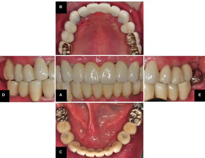
After building up veneering porcelain, the bridge for #13-15, the crowns for #23, 24 were cemented with resin cement (Rely-X UniCem, 3M ESPE, Seefeld, Germany), and the bridge for #12i-22i was cement- ed with noneugenol temporary resin cement (Premier Implant Cement; Premier Dental Products Co., Plymouth Meeting, PA, USA) (Fig. 2). The prostheses were designed using mutually protected occlu- sion concept. The anterior teeth protected the posterior teeth from excursive force and wear, and posterior teeth supported the bite force.
Oral hygiene instruction and regular check-up were administered.
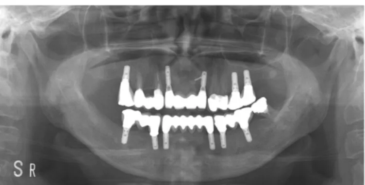
During 18 months of follow-up period, clinical and radiological conditions of the osseointegrated implants and prostheses were sta- ble without any signs of inflammation or unexpected bone loss around the implants (Fig. 3).
Fig. 2. Postoperative intraoral view. (A) Frontal view, (B, C) Occlusal view, (D, E) Buccal view.
B
A
D E
C
Discussion
In this case, as all has been acknowledged as a treatment of choice for function and esthetics, implant supported fixed partial denture achieved good satisfaction on patient's behalf, resulting in increased psychological confidence and social activity. With fixed reconstruc- tions, it is particularly important that there is sufficient interocclusal space to ensure rigidity.4
For the framework of coping material, high noble alloys, base met- als, and zirconia or alumina ceramics can be used. Metal sub- structures have the advantage of the ability to section and reconnect but their reduced light transmission hinders getting natural appear- ance. In this case, gold framework made adjusting its fitness by sec- tioning and reconnecting. Zirconium oxide, also known as zirconia, has gained increasing popularity in contemporary dentistry due to its biocompatibility, high flexural strength, toughness, and esthet- ic properties.5,6As CAD/CAM technology gets ubiquitous, zir- conia is currently being used for the fabrication of all ceramic copings and frameworks for both fixed prosthodontics and implant dentistry.5,6
Several studies have tried to evaluate the feasibility of using dif- ferent restorative materials in fabrication of implant supported prostheses.7-9 In this case, molar area was restored with gold
occlusal surface to fulfill the usual dietary habit of Koreans, and the rest with porcelain occlusal surface for esthetics. CAD/CAM tech- nology was applied to the esthetic zone and premolar area.
In conclusion, implant supported prostheses satisfied the patient's esthetic, functional desire in the case of multiple tooth loss. For the anterior and premolar area, CAD/CAM technology achieved pre- dictable, successful outcome with the help of proper combina- tion of conventional restorative materials.
References
1. Adell R, Lekholm U, Rockler B, Bra�nemark PI. A 15-year study of osseointegrated implants in the treatment of the edentulous jaw. Int J Oral Surg 1981;10:387-416.
2. van Steenberghe D, Lekholm U, Bolender C, Folmer T, Henry P, Herrmann I, Higuchi K, Laney W, Linden U, Astrand P.
Applicability of osseointegrated oral implants in the rehabilita- tion of partial edentulism: a prospective multicenter study on 558 fixtures. Int J Oral Maxillofac Implants 1990;5:272-81.
3. Pjetursson BE, Karoussis I, Bu¨rgin W, Bra¨gger U, Lang NP. Patients' satisfaction following implant therapy. A 10-year prospective cohort study. Clin Oral Implants Res 2005;16:185-93.
4. Budtz-Jo¨rgensen E. Restoration of the partially edentulous mouth-a comparison of overdentures, removable partial dentures, fixed partial dentures and implant treatment. J Dent 1996;24:
237-44.
5. Raigrodski AJ. Contemporary materials and technologies for all- ceramic fixed partial dentures: a review of the literature. J Prosthet Dent 2004;92:557-62.
6. Conrad HJ, Seong WJ, Pesun IJ. Current ceramic materials and systems with clinical recommendations: a systematic review.
J Prosthet Dent 2007;98:389-404.
7. Pjetursson BE, Tan K, Lang NP, Bra¨gger U, Egger M, Zwahlen M. A systematic review of the survival and complication rates of fixed partial dentures (FPDs) after an observation period of at least 5 years. Clin Oral Implants Res 2004;15:625-42.
8. Bozini T, Petridis H, Garefis K, Garefis P. A meta-analysis of prosthodontic complication rates of implant-supported fixed dental prostheses in edentulous patients after an observation period of at least 5 years. Int J Oral Maxillofac Implants 2011;
26:304-18.
9. Triwatana P, Nagaviroj N, Tulapornchai C. Clinical perfor- mance and failures of zirconia-based fixed partial dentures: a review literature. J Adv Prosthodont 2012;4:76-83.
연제웅∙임영준∙권호범∙김명주 고정성 임플란트 보철물을 이용한 완전구강회복 증례
Fig. 3. Panoramic radiograph 18 months after placement of definitive prosthesis.
고정성 임플란트 보철물을 이용한 완전구강회복 증례
연제웅∙임영준∙권호범∙김명주*
서울대학교 치과대학 치과보철학교실
본 증례의 환자는 상악 전치부 보철물의 파절 및 기존에 사용하던 하악 의치의 불편감 때문에 임플란트를 이용한 고정성 보철을 원한다는 주소로 내원하였다. 상악동 거상술, 하악지 골이식과 함께 상악에 5개, 하악에 6개의 임플란트를 식립하였다. 전치부와 양쪽 구치부의 세 부분으로 나누어 임플란트 지지형 고정성 보철물을 장착하였다. 이상과 같은 과정을 통해 적절한 심미적, 기능적 결과를 얻었기에 이를 보고하고자 한다. (대한치과 보철학회지 2015;53:81-5)
주요단어: 임플란트; CAD-CAM; 완전구강회복술
*교신저자: 김명주
110-744 서울 종로구 대학로 101 서울대학교 치과대학 치과보철학교실 02-2072-0815: e-mail, [email protected]
원고접수일: 2014년 12월 10일 / 원고최종수정일: 2014년 12월 22일 / 원고채택일:
2015년 1월 5일
2015 대한치과보철학회
이 글은 크리에이티브 커먼즈 코리아 저작자표시-비영리 3.0 대한민국 라이선스에 따라 이용하실 수 있습니다.
c cc