서론
Crohn병은 장벽 전 층을 침범하는 만성 육아종성 염증으로 구강에서 항문까지 소화관의 어떤 부위에서도 발생할 수 있으 나, 환자의 80% 정도가 소장에 병변을 보이며, 그 중에서도 말 단회장이 가장 호발 부위이다. 침범부위는 다발성이며, 침범부 위 사이에 정상부위가 끼여 있는 형태로 분포한다(1).
Crohn병을 처음 진단할 때에는 환자의 임상증상, 혈액학적 검사소견, 병리조직학적 소견, 영상의학적 소견을 모두 종합하 여 판단하게 되며 특히, 영상소견의 특징이나 분포가 진단과 치 료에 매우 중요한 역할을 하게 된다. 2011년에 발표된 Ameri-
can College of Radiology (ACR)에 따르면 CT 소장조영술은 Crohn병을 진단받은 환자에서 급성 악화나 합병증이 의심되는 상황에서 평가하는데 현재까지 알려진 영상의학검사 중 가장 좋은 검사로 알려져 있다.
Crohn병 치료의 성공여부는 질병의 활성도나 침범 정도에 달 려 있으며, 따라서 Crohn병이 의심되는 환자의 영상소견을 분 석할 때에는 Crohn병의 진단뿐 아니라 질병의 활성도, 침범 정 도, 중증도를 평가하는 것이 매우 중요하다. 즉, Crohn병으로 인한 소장 내경의 감소가 있을 경우에, 급성기 염증소견으로 인 한 분절성 소장 내경 감소인 경우는 약물치료에도 잘 반응하지 만, 섬유성 협착에 의한 소장 내경이 감소한 경우 수술적 치료
J Korean Soc Radiol 2013;68(2):107-115
Received August 20, 2012; Accepted November 1, 2012 Corresponding author: Seong Jin Park, MD Department of Diagnostic Radiology, Kyung Hee University Hospital, 23 Kyungheedae-ro, Dongdaemun-gu, Seoul 130-872, Korea.
Tel. 82-2-958-8610 Fax. 82-2-968-0787 E-mail: [email protected]
Copyrights © 2013 The Korean Society of Radiology
Purpose: To retrospectively compare the accuracy of axial and coronal planes of CT
enterography for detection of pathologic findings of Crohn disease.Materials and Methods: 168 patients who were suspected of having Crohn dis-
ease underwent CT enterography. 66 patients who were diagnosed Crohn disease were retrospectively evaluated (endoscopic biopsy of terminal ileum: 12 patients, segmental resection of small bowel: 6 patients, diagnosed based on a combination of clinical, histopathological and imaging findings: 48 patients). 2 radiologists re- viewed axial planes of CT enterography and one month later reviewed coronal planes. CT enterography findings of active phase, chronic phase and complications of Crohn disease were evaluated and then compared with axial and coronal planes by using chi-square test.Results: Mucosal hyperenhancement, wall thickening, and mesenteric fat stranding
were more detected on axial planes, which were CT findings of active Crohn disease.Pseudosacculation, fibrotic strictures, fistulas, abscesses were more detected on coronal planes, which were CT findings of chronic Crohn disease or complications.
In particular, pseudosacculation and fibrotic strictures were significantly more de- tected on coronal planes.
Conclusion: When evaluating CT enterography in Crohn disease, coronal planes pro-
vide more useful diagnostic information of pseudosacculation and fibrotic strictures.Index terms
Crohn Disease CTCT Enterography
Comparative Study between Axial and Coronal Planes of CT
Enterography in Evaluation of Disease Activity and Complications of Crohn Disease
1Crohn병의 진단과 질병 활성도, 합병증 평가에 있어서 CT 소장조영술의 횡단영상과 관상영상의 유용성 비교
1Sung Eun Ahn, MD
1, Seong Jin Park, MD
1, Soung Kyung Moon, MD
1, Joo Won Lim, MD
1, Dong Ho Lee, MD
1, Young Tae Ko, MD
1, Hyo Jong Kim, MD
2Departments of 1Diagnostic Radiology, 2Gastroenterology, Kyung Hee University Hospital, Seoul, Korea
대상과 방법
환자
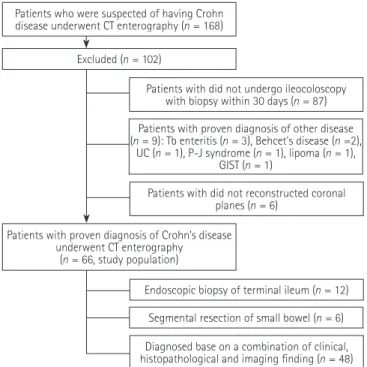
2010년 11월부터 2011년 12월까지 본원에서 소장의 Crohn 병이 의심되어 CT 소장조영술을 시행한 168명의 환자를 대상 으로 하였다(Fig. 1).
168명의 환자 중 연구에 포함된 환자의 포함기준은 다음과 같았다. 1) CT 소장조영술을 시행한 30일 이내에 대장내시경과 생검을 시행한 환자, 2) 수술 혹은 내시경 생검을 통한 병리조 직소견으로 Crohn병으로 확진된 환자나 환자의 임상증상, 내시 경 생검을 통한 병리조직학적 소견, 영상의학적 소견을 모두 종 합하여 판단하여 Crohn병으로 진단된 환자, 3) CT 소장조영술 의 관상영상이 재구성된 환자를 연구의 포함기준으로 하였다.
168명의 환자 중 102명의 환자가 다음과 같은 이유로 조사 에서 제외되었다. CT 소장조영술을 시행한 30일 이내에 대장 내시경과 생검을 시행하지 않은 환자 87명과, Crohn병 이외의 질병으로 진단받은 환자 9명(결핵성 장염 3명, 베체트병 2명, 궤양성 대장염 1명, 포이츠-예거증후군 1명, 지방종 1명, 위장 관 간질종양 1명), 그리고 관상영상이 재구성되지 않는 6명이 조사에서 제외되었다.
총 66명의 환자가 최종 연구에 포함되었으며, 49명의 남자 와 17명의 여자로 구성되었고 평균연령은 28.5세(12~62세)였 다. 이들은 모두 CT 소장조영술을 시행한 30일 이내에 대장내 시경과 생검을 시행하였으며, Crohn병으로 진단된 환자였다.
12명의 환자가 말단회장에서 내시경 생검을 시행하여 Crohn병 으로 확진되었고 6명의 환자는 소장이나 말단회장의 수술을 통해 Crohn병으로 확진되었다(회장 분절 절제술 3명; 말단회 장, 막창자 절제술 2명; 회장, 맹장, 대장 절제술 1명). 48명의 환자는 말단회장에서 내시경 생검을 시행하여 만성 육아종성 염증소견이 있었으며 환자의 임상증상과 영상의학적 소견을 종 합하여 판단하여 Crohn병으로 진단되었다.
CT 소장조영술
환자들은 CT 소장조영술을 시행하기 전 40분 동안 poly- ethyleneglycol (Colyte-FⓇ, Taejoon, Seoul, Korea) 2 L를 10 분간의 간격을 두고 5차례에 나누어, 검사 시행하기 전 40분, 30분, 20분, 10분, 0분에 각각 400 mL씩 마시게 하여 소장내 강에 polyethyleneglycol을 채워 소장의 내강과 소장벽이 대비 되도록 전처치하였다. CT 소장조영술은 64채널 다중검출기 CT (Brilliance 64, Philips Medical System, Cleveland, OH, USA) 혹은 16채널 다중검출기 CT (Light speed, GE Health- care, Milwaukee, WI, USA)를 이용하여 시행하였다.
(stricturoplasty)를 시행하여야 하기 때문에 영상소견에서 이를 감별하는 것이 중요하며, Crohn병의 합병증으로 누공이 발생 하였을 경우에도, 복합 누공의 경우는 수술적 치료를 요하지 만, 단순 누공은 infliximab (inhibit tumor necrosis factor)을 이용한 약물치료에도 잘 반응하기 때문에 감별을 요한다.
Crohn병의 진단, 질병 활성도, 합병증과 관련된 여러 가지 CT 소장조영술의 소견이 보고되었으나, 현재까지 보고된 문헌 에서 Crohn병으로 진단된 환자들의 CT 소장조영술의 횡단영 상과 관상영상에 비교연구는 없었다.
또한 복부 CT의 횡단영상과 관상영상을 비교한 논문은 있었 으나, 소장 폐쇄가 의심되는 환자들의 횡단영상과 관상영상을 비교한 논문이거나(2), 복통을 주소로 촬영한 환자들의 횡단영 상과 관상영상을 비교한 논문이었으며(3), Crohn병으로 진단 받은 환자들을 대상으로 한 비교연구는 없었다.
이에 저자들은 두 영상 간의 비교연구를 통해 어떠한 영상에 서 Crohn병의 급성기 혹은 만성기 소견이나 합병증 동반여부가 잘 관찰되는지를 알고 있다면, Crohn병의 진단과 치료계획 수 립에 도움이 될 수 있을 것으로 생각하였다. 본 연구에서는 Crohn병 환자의 CT 소장조영술의 횡단영상과 관상영상을 비 교, 분석하고, 두 영상에서 Crohn병의 진단과 질병 활성도, 합 병증 평가에 있어서 유용성을 비교하고자 하였다.
Fig. 1. Flowchart of the selection of patients for retrospective study, with all patients undergoing CT enterography and ileocoloscopy with biopsy within 30 days.
Note.-GIST = gastrointestinal stromal tumor, P-J syndrome = Peutz- Jegher syndrome, Tb enteritis = Tuberculous enteritis, UC = ulcerative colitis
Patients who were suspected of having Crohn disease underwent CT enterography (n = 168)
Patients with did not undergo ileocoloscopy with biopsy within 30 days (n = 87)
Patients with did not reconstructed coronal planes (n = 6)
Diagnosed base on a combination of clinical, histopathological and imaging finding (n = 48) Patients with proven diagnosis of other disease (n = 9): Tb enteritis (n = 3), Behcet's disease (n =2),
UC (n = 1), P-J syndrome (n = 1), lipoma (n = 1), GIST (n = 1)
Patients with proven diagnosis of Crohn’s disease underwent CT enterography
(n = 66, study population) Excluded (n = 102)
Endoscopic biopsy of terminal ileum (n = 12) Segmental resection of small bowel (n = 6)
성 협착이 있는지 조사하였다(5-8). 합병증의 동반여부 소견으 로는 누공, 동, 염증성 농양, 봉소직염이 있는지 조사하였다. 누 공은 소장에서 다른 구조물로 조영증강 소견이 연장되어 있을 때로 정의하였으며, 동은 누공과 비슷한 소견을 보이나 조영증 강 소견이 다른 구조물까지 연장되지 않았을 때로 정의하였다.
염증성 농양은 국한된 장관외액이 조영증강이 잘되는 벽으로 둘러싸여 있을 때로 정의하였으며, 봉소직염은 액체 혹은 연부 조직의 감쇠를 보이는 장관 외 종괴로 정의하였다. 또한 대장, 항문 주위 침범여부에 대해서도 조사하였다.
CT 소장조영술의 횡단영상과 관상영상에서 각각 조사한 Crohn병 소견의 유무가 일치하지 않을 경우, 이들 증례는 추후 경구소장바륨조영술이나 내시경 소견과 비교하여 어떤 영상의 소견이 실제 환자의 소견에 더 합당한지에 대해 평가하였다.
통계적 분석
통계학적 검사는 Statistical Package for the Social Sciences (SPSS) 소프트웨어를 이용하여 시행하였다(SPSS 12.0, SPSS, Chicago, IL, USA).
횡단영상과 관상영상에서 각각 분석한 CT 소견의 빈도 차이 가 통계학적으로 유의한지 알아보기 위하여 카이제곱 검정을 시행하였으며, p-value < 0.05인 경우 통계학적으로 유의하 다고 정의하였다.
결과
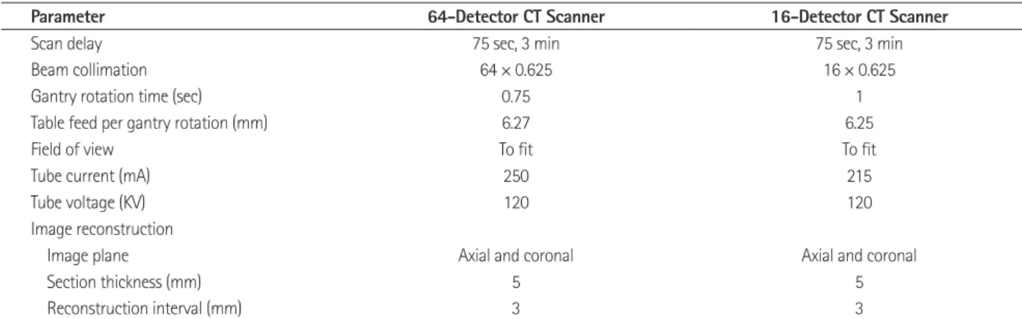
CT 소장조영술의 소장의 적절한 팽대정도를 분석한 결과는 다음과 같았다(Table 2). 소장 팽대정도가 90% 이상 되어 4점 을 받은 경우가 6예(9.1%), 70~90%(3점)가 30예(45.5%), 40~69%(2점)가 28예(42.4%), 40% 미만(1점)이 2예 촬영조건은 64채널 다중검출기 CT의 경우 120 kVp, 250
mAs, 절편두께 5 mm였으며, 16채널 다중검출기 CT의 경우 120 kVp, 215 mAs, 절편두께 5 mm였다(Table 1).
소장의 연동운동을 감소시키기 위하여 scopolamine butyl- bromide (Buscopan, Boehringer Ingerheim Korea, Seoul, Ko- rea) 1 amp (1 mL, 20 mg/1 mL)를 정맥주사 하였으며, 환자 몸무게 kg당 2~3 mL의 요오드제제 정맥 조영제(PamirayⓇ, Dong Kook Pharm., Seoul, Korea)를 2.5 mL/sec 속도로 정 맥 주입하였고, 15~20 mL 생리식염수를 2 mL/sec의 속도로 강력주사기(power injector)를 사용하여 전주와 정맥으로 주사 하였다. 조영제 주입 후 75초, 3분 후에 영상을 얻었다.
영상분석
CT 영상은 임상적 질병 활성도, 수술 혹은 내시경적 소견에 대한 정보 없이 두 명의 영상의학과 의사에 의해 후향적으로 분 석되었고, 이견이 있는 경우 합의에 의해 판정하였다. 정맥기 영 상을 분석하였으며, Picture Archiving and Communication System (Infinitt, Seoul, Korea)상에 저장되어 있던 절편두께 5 mm의 횡단영상과 절편두께 3 mm의 관상영상을 분석하였다.
두 영상의 분석은 1개월의 간격을 두었다.
소장의 적절한 팽대 정도를 평가하였으며, 소장의 팽대 정도 가 90% 이상 잘 되어 있을 때 4점, 70~90% 팽대되어 있을 때 3점, 40~69% 정도 팽대되어 있을 때 2점, 40% 미만으로 팽 대되어 있을 때 1점으로 정의하였다(4).
CT 소견은 급성기 소견, 만성기 소견, 합병증의 동반여부를 분석하였고, 급성기 소견으로는 점막의 과다조영증강, 장벽 비 후(장벽두께 > 3 mm), 소장벽의 층별화와 장간막 혈관분포 증가, 장간막 지방 침착이 있는지 조사하였고, 만성기 소견으로 는 점막하층의 지방 침윤, 가성수포, 주변 지방섬유 증식, 섬유 Table 1. Scan Parameters for CT Enterography
Parameter 64-Detector CT Scanner 16-Detector CT Scanner
Scan delay 75 sec, 3 min 75 sec, 3 min
Beam collimation 64 × 0.625 16 × 0.625
Gantry rotation time (sec) 0.75 1
Table feed per gantry rotation (mm) 6.27 6.25
Field of view To fit To fit
Tube current (mA) 250 215
Tube voltage (KV) 120 120
Image reconstruction
Image plane Axial and coronal Axial and coronal
Section thickness (mm) 5 5
Reconstruction interval (mm) 3 3
Note.-16-detector CT scanner = Light speed, GE Healthcare, Milwaukee, WI, USA, 64-detector CT scanner = Brilliance 64, Philips Medical System, Cleve- land, OH, USA
침착은 횡단영상에서 28예(42.4%), 관상영상에서 25예 (37.9%)에서 관찰되었고, 횡단영상에서 3예(4.5%) 더 관찰되 었으나 두 군 간에 유의한 통계학적 차이를 보이지 않았다(Fig.
3). 소장벽의 층별화와 장간막 혈관분포 증가는 횡단영상과 관상영상에서 모두 16예(24.2%)에서 관찰되었으며 두 군 간 에 차이를 보이지 않았다.
Crohn병의 만성기 소견을 횡단영상과 관상영상에서 나누어 분석한 결과는 다음과 같았다(Table 3). 가성수포는 횡단영상에 서는 관찰되지 않고, 관상영상에서만 4예(6.1%)에서 관찰되었 으며, 이는 통계학적으로 유의한 차이가 있었다(p = 0.0423) (3.0%)에서 있었으며, 97%의 대부분의 환자에서 2점 이상의
소장의 적절한 팽대를 유도할 수 있었다.
Crohn병의 급성기 소견을 횡단영상과 관상영상에서 나누어 분석한 결과는 다음과 같았다(Table 3). 점막의 과다조영증강 은 횡단영상에서 52예(78.8%), 관상영상에서 49예(74.2%)에 서 관찰되어 횡단영상에서 3예(4.5%) 더 관찰되었으나 두 군 간에 유의한 통계학적 차이를 보이지 않았다. 장벽 비후도 횡단 영상에서 53예(80.3%), 관상영상에서 50예(75.6%)에서 관 찰되어 횡단영상에서 3예(4.5%) 더 관찰되었으나 두 군 간에 유의한 통계학적 차이를 보이지 않았다(Fig. 2). 장간막 지방 Table 2. Each Numbers of Bowel Distention Score
Score 4 (Optimal): > 90% Score 3 (Good): 70–90% Score 2 (Moderate): 40–69% Score 1 (Mild): < 40%
6 (9.1%) 30 (45.5%) 28 (42.4%) 2 (3.0%)
Data in parentheses are percentages.
Fig. 2. A 19-year-old man with Crohn disease at the ileum. CT enterogram shows concentric wall thickening and mucosal hyperenhancement (arrows) at the ileum on axial image (A), which were missed on coronal image (B).
A B
B C D
A
Fig. 3. A 24-year-old man with active Crohn disease at the ascending colon and ileum.
A, B. CT enterogram shows concentric wall thickening and mucosal hyperenhancement (arrows) at the ileum on axial image. The thickened wall has a stratified appearance. CT enterography also shows increased attenuation of the mesenteric fat on axial image, which was not shown on coronal image.
C. Endoscopy image shows erythematous mucosal change at the ileocecal valve.
D. Image from barium study demonstrates transient luminal narrowing, mucosal fold widening and thickening on the proximal ascending colon and the terminal ileum.
의한 통계학적 차이는 없었다(Fig. 7). 염증성 농양은 횡단영상 과 관상영상에서 모두 4예(6.1%)에서 관찰되었으며 두 군 간 의 차이가 없었다. 봉소직염은 횡단영상에서는 관찰되지 않았 으나 관상영상에서는 1예(1.5%)에서 관찰되었고, 동이 관찰된 증례는 없었다.
대장, 항문 주위 침범여부도 조사하였으며, 대장 침범을 동반 한 경우는 횡단영상과 관상영상에서 모두 39예(59.1%)에서 관 찰되었으며 두 군 간의 차이가 없었고, 항문 주위 침범을 동반 한 경우는 횡단영상과 관상영상에서 모두 32예(48.5%)에서 관찰되었으며 차이가 없었다(Table 7).
또한 사전에 분석하기로 한 CT 소견은 아니었으나, 횡단영상 에서는 십이지장 침범여부를 간과하였으나, 관상영상에서 발견 (Table 4, Fig. 4). 섬유성 협착은 횡단영상에서 4예(6.1%), 관
상영상에서 12예(18.2%)에서 관찰되었고, 관상영상에서 8예 (12.1%) 더 관찰되었으며 이는 통계학적으로 유의한 차이가 있 었다(p = 0.0329)(Figs. 5, 6, Table 5). 반면, 점막하층의 지 방 침윤은 횡단영상과 관상영상에서 모두 8예(12.1%)에서 관 찰되어 두 군 간의 차이가 없었으며 주변 지방섬유 증식도 횡단 영상과 관상영상에서 모두 4예(6.1%)에서 관찰되어 두 군 간 의 차이가 없었다.
Crohn병의 합병증 동반여부 소견을 횡단영상과 관상영상에 서 나누어 분석한 결과는 다음과 같았다(Table 6). 누공은 횡단 영상에서 7예(10.6%), 관상영상에서 8예(12.1%)에서 관찰되 었고, 관상영상에서 1예(1.5%) 더 관찰되었으나 두 군 간의 유
Table 3. Each Numbers of Active Findings of Crohn Disease at Axial and Coronal Images
Mural Hyper-Enhancement Wall Thickening (> 3 mm) Mural Stratification with a
Prominent Vasa Recta (Comb Sign) Mesenteric Fat Stranding
Axl 52 (78.8%) 53 (80.3%) 16 (24.2%) 28 (42.4%)
Cor 49 (74.2%) 50 (75.6%) 16 (24.2%) 25 (37.9%)
Data in parentheses are percentages.
Note.-Axl = axial, Cor = coronal
Table 4. Each Numbers of Chronic Findings of Crohn Disease at Axial and Coronal Images
Submucosal Fat Deposition Pseudosacculation Surrounding Fibrofatty Proliferation Fibrotic Strictures
Axl 8 (12.1%) 0 4 (6.1%) 4 (6.1%)
Cor 8 (12.1%) 4 (6.1%) 4 (6.1%) 12 (18.2%)
Data in parentheses are percentages.
Note.-Axl = axial, Cor = coronal
Table 5. Statistical Analysis and Significantly More Detected on Coronal Image
Axial Coronal p-Value
Pseudosacculation 0 4 (6.1%) 0.0423
Fibrotic strictures 4 (6.1%) 12 (18.2%) 0.0329 Data in parentheses are percentages, p-value less than or equal to 0.05 was considered to indicate a statistically significant difference.
Table 6. Each Numbers of Complication Findings of Crohn Disease at Axial and Coronal Images
Fistulas Sinus Tracts Abscesses Phlegmons
Axl 7 (10.6%) 0 4 (6.1%) 0
Cor 8 (12.1%) 0 4 (6.1%) 1 (1.5%)
Data in parentheses are percentages.
Note.-Axl = axial, Cor = coronal
Table 7. Each Numbers of Large Bowel and Perianal Involvement Findings of Crohn Disease at Axial and Coronal Images
Large Bowel Perianal Lesion
Axl 39 (59.1%) 32 (48.5%)
Cor 39 (59.1%) 32 (48.5%)
Data in parentheses are percentages.
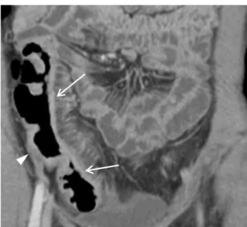
Note.-Axl = axial, Cor = coronal Fig. 4. A 31-year-old man with long-standing Crohn disease at the il-
eum. CT enterogram shows typical straightening of the mesenteric boder (arrows), a finding that indicates linear ulceration or ulcer scar and pseudosacculation of the antimesenteric border (arrowhead) at the ileum on coronal image.
점이 있다(10). 또한 CT 소장조영술은 고위관장법(enterocly- sis)에 비해 비침습적이고, 실행하기 쉽다는 장점이 있으며(11), 섬유성 협착이 의심되는 상황에서 캡슐내시경이 소장 폐색을 유발할 수 있기 때문에 금기로 알려져 있는 반면, CT 소장조영 술은 실행이 가능하다(12). MR 소장조영술도 최근에 사용이 증가되고 있으나 시간이 오래 걸리고, 가격이 비싸 영상의 질을 좋게 하는 데 CT 소장조영술보다 더 많은 고려를 해야 하며, 특히 숨을 참기 힘든 환자의 경우에는 MR보다 CT가 더 좋은 영상의 질을 얻을 수 있다(13, 14).
2011년에 발표된 ACR에 따르면 CT 소장조영술은 Crohn병 을 진단받은 환자에서 급성 악화나 합병증이 의심되는 상황에 서 평가하는데 현재까지 알려진 영상의학검사 중 가장 좋은 검 사로 알려져 있고, Crohn병 치료의 성공여부는 질병의 활성도 나 침범 정도에 달려 있다. 그러나 현재까지 보고된 문헌 중 Crohn병 환자에서 CT 소장조영술의 횡단영상과 관상영상에 대한 비교연구는 없었다.
본 연구에서 횡단영상에 비해 유의하게 관상영상에서 잘 관찰 된 소견은 가성수포(p = 0.0423)와 섬유성 협착(p = 0.0329)이 된 증례가 2예(3.0%) 있었고, 이들 증례는 추후 내시경 소견에
서 십이지장의 침범여부가 확인되었다.
고찰
최근 다중절편 CT는 해상도가 향상되고, 빠른 속도로 얇은 영상을 얻을 수 있어, 등방성(isotropic)의 관상면과, 시상면의 재구성 영상을 재현할 수 있게 되었다. 최근 연구에 따르면 이러 한 관상면의 재구성 영상을 횡단영상과 함께 평가할 경우, 위장 관 평가에 도움이 된다고 하였고, Horton과 Fishman (9)에 의 하면 특히 소장과 장간막 혈관의 평가에 도움이 된다고 하였다.
CT 소장조영술은 다량의 경구조영제를 투여하여 소장 내강 을 확장 시킴으로써, CT보다 소장의 내강과 소장벽에 대한 더 좋은 영상을 얻을 수 있다. 따라서 CT 소장조영술은 이전에 전 통적인 영상의학적 검사 방법으로 여겨져 왔던 경구소장바륨조 영술에 비해, 서로 중복되어 있는 골반내의 소장을 평가할 수 있고, 소장의 장벽이나, 소장 주변의 장간막, 고형 장기, 주변의 섬유성 협착이나 농양 등의 합병증도 함께 평가할 수 있다는 장
B C D
A
Fig. 5. A 27-year-old man with fibrotic stricture in Crohn disease.
A, B. CT enterogram shows fibrotic stricture (arrows) at the pelvic ileal loop on coronal image, which was missed on axial image.
C. Image from barium study also shows typical fibrotic stricture (arrows) at the pelvic ileal loop.
Fig. 6. A 27-year-old woman with fibrotic stricture in Crohn disease.
A-C. CT enterogram shows fibrotic stricture (arrows) at the ileocecal (IC) valve with dilatation of terminal ileum on serial coronal images, which was missed on axial image.
D. Endoscopy image shows numerous pseudopolyp and cecal deformity. Scope couldn’t be passed of stenotic portion of IC valve.
A B C
때문이라고 생각된다. 그러나, 누공과 봉소직염은 횡단영상과 관상영상에서 관찰되는 빈도가 유의한 차이를 보이지는 않았 는데, 이는 본 연구에 포함된 Crohn병 환자군 중 누공이나 봉 소직염 합병증을 보인 증례 자체가 적기 때문이며, 이 부분에 대해서는 향후 좀더 많은 수의 환자를 대상으로 한 연구가 이 루어져야 할 것으로 생각된다.
또한 본 연구에서 관상영상에 비해 횡단영상에서 점막의 과 다조영증강, 장벽 비후, 장간막 지방 침착이 잘 관찰되는 경향 을 보였으나 이들 소견은 통계학적으로 유의한 차이는 없었다.
본 연구의 제한점으로는 후향적으로 이루어졌으며, CT 소장 조영술에서 관찰된 Crohn병의 소장 병변을 확인하기 위한 방법 으로 캡슐내시경 검사를 이용하지 않았다. 캡슐내시경은 소장 의 점막을 평가하는 데 CT 소장조영술보다 더 민감한 것으로 알려져 있으나, 섬유성 협착이 의심되는 상황에서는 캡슐내시경 이 소장 폐색을 유발할 수 있어 금기로 알려져 있기 때문이다 (11). 본 연구에서는 Crohn병의 소장 병변 확인을 위하여, 대장 내시경을 통하여 원위부 회장에 대한 검사를 함께 시행하였으 나 그 역시 소장 병변을 모두 확인하지는 못하여 내시경 소견과 CT 소장조영술 소견의 직접적인 비교를 하지 못한 제한점이 있 다. 또한 각각의 CT 소장조영술 소견과 부위별 염증의 정도 및 었으며, 이는 통계학적으로 유의한 차이가 있었고 모두 Crohn병의
만성기에 나타나는 소견이었다. 가성수포는 Crohn병이 중기로 진행되면서, 아프타성 궤양이 길게 합쳐져 만들어지는 종축의 선형 궤양이 장간막쪽에 생기고 주변 장간막과 장벽의 비후 및 경화를 일으킴으로써, 상대적으로 장간막 반대쪽의 벽은 길이 가 남아 형성하게 된다.
따라서 가성수포는 주로 종축에 평행하게 형성되고, 관상영 상에서는 한 단면에서 가성수포 전체를 평가할 수 있기 때문에 관상영상에서 좀더 잘 관찰되었던 결과를 얻었다고 생각된다.
또한, 소장의 섬유성 협착이 있는지 여부를 평가하기 위해서는 소장의 내경 감소가 있는지 여부를 확인해야 하며, 전체 소장의 주행을 평가하는 것이 중요하다. 섬유성 협착이 관상영상에서 통계학적으로 유의하게 더 잘 관찰된 이유는 관상영상이 횡단 영상에 비해 소장의 전체 주행을 더 쉽게 평가할 수 있기 때문 이라고 생각된다.
횡단영상에 비해 관상영상에서 Crohn병의 합병증 소견에 해 당하는 누공이나 봉소직염이 잘 관찰되는 경향을 보였다. 이는 관상영상에서는 대부분의 대장이 한 단면에서 관찰되고, 소장 의 주행경로도 쉽게 평가되며, 횡단영상에서는 봉소직염이 정 상적인 장의 내강이나 장간막의 지방침착 등과 혼돈될 수 있기 Fig. 7. A 23-year-old woman with ileo-colonic fistula in Crohn disease.
A-E. CT enterography shows ileo-colonic fistula on serial coronal images, which was missed on axial image. Fistula extends from the distal ileum (arrowheads) to the adjacent distal sigmoid colon (open arrows).
F. Image from barium study obtained after 4 hours, demonstrates ileocolonic fistula on sagittal image (arrow). Sigmoid colon (open arrow) and rectum are seen.
D A
E B
F C
terobserver agreement for enteric phase CT enterography.
Radiology 2006;241:787-795
7. Goldberg HI, Gore RM, Margulis AR, Moss AA, Baker EL.
Computed tomography in the evaluation of Crohn disease.
AJR Am J Roentgenol 1983;140:277-282
8. Lee SS, Ha HK, Yang SK, Kim AY, Kim TK, Kim PN, et al. CT of prominent pericolic or perienteric vasculature in patients with Crohn’s disease: correlation with clinical disease activi- ty and findings on barium studies. AJR Am J Roentgenol 2002;179:1029-1036
9. Horton KM, Fishman EK. The current status of multidetec- tor row CT and three-dimensional imaging of the small bowel. Radiol Clin North Am 2003;41:199-212
10. Paulsen SR, Huprich JE, Fletcher JG, Booya F, Young BM, Fidler JL, et al. CT enterography as a diagnostic tool in eval- uating small bowel disorders: review of clinical experience with over 700 cases. Radiographics 2006;26:641-657; dis- cussion 657-662
11. Maglinte DD, Sandrasegaran K, Lappas JC, Chiorean M. CT Enteroclysis. Radiology 2007;245:661-671
12. Hara AK, Leighton JA, Heigh RI, Sharma VK, Silva AC, De Petris G, et al. Crohn disease of the small bowel: prelimi- nary comparison among CT enterography, capsule endos- copy, small-bowel follow-through, and ileoscopy. Radiolo- gy 2006;238:128-134
13. Tolan DJ, Greenhalgh R, Zealley IA, Halligan S, Taylor SA.
MR enterographic manifestations of small bowel Crohn disease. Radiographics 2010;30:367-384
14. Schmidt S, Lepori D, Meuwly JY, Duvoisin B, Meuli R, Mi- chetti P, et al. Prospective comparison of MR enteroclysis with multidetector spiral-CT enteroclysis: interobserver agreement and sensitivity by means of “sign-by-sign” cor- relation. Eur Radiol 2003;13:1303-1311
15. Bodily KD, Fletcher JG, Solem CA, Johnson CD, Fidler JL, Barlow JM, et al. Crohn Disease: mural attenuation and thickness at contrast-enhanced CT Enterography--correla- tion with endoscopic and histologic findings of inflamma- tion. Radiology 2006;238:505-516
16. Hara AK, Alam S, Heigh RI, Gurudu SR, Hentz JG, Leighton JA. Using CT enterography to monitor Crohn’s disease activ- ity: a preliminary study. AJR Am J Roentgenol 2008;190:
1512-1516 병리소견과의 관련성은 검증하지 못하였다는 제한점이 있다.
그러나 이전 연구에서, CT 소장조영술의 Crohn병 소견은 내 시경 소견과 매우 일치되며, 특히 급성기 Crohn병 환자에서 CT 소장조영술의 점막의 과다조영증강 소견, 장벽 비후 소견 은 내시경이나 병리소견과 통계학적으로 유의하게 상관관계를 보인다고 보고된 바가 있어 내시경 소견이나 병리소견을 CT 소 장조영술과 직접적인 비교를 하지 못하였다는 본 연구의 제한 점을 극복할 수 있다고 생각된다(15, 16).
또한 본 연구는 절편두께 5 mm의 횡단영상과 절편두께 3 mm의 관상영상을 분석하였는데 황단영상과 관상영상의 절편 두께가 다른 제한점이 있다.
결론적으로 Crohn병 환자에 있어서 CT 소장조영술의 소견 은 횡단영상과 관상영상에서 대부분 유의한 차이를 보이지 않 았으나, 만성기 소견인 가성수포와 섬유성 협착은 관상영상에 서 유의하게 높은 빈도로 관찰되었다. 이러한 점을 숙지하고, 만성기 Crohn병 환자의 CT 소장조영술 분석을 할 경우, 가성 수포와 섬유성 협착이 의심된다면, 관상영상을 보다 주의깊게 살펴, 정확한 진단과 치료계획 수립에 도움을 줄 수 있을 것으 로 생각된다.
참고문헌
1. Furukawa A, Saotome T, Yamasaki M, Maeda K, Nitta N, Takahashi M, et al. Cross-sectional imaging in Crohn dis- ease. Radiographics 2004;24:689-702
2. Jaffe TA, Martin LC, Thomas J, Adamson AR, DeLong DM, Paulson EK. Small-bowel obstruction: coronal reforma- tions from isotropic voxels at 16-section multi-detector row CT. Radiology 2006;238:135-142
3. Jaffe TA, Martin LC, Miller CM, Franklin KM, Merkle EM, Thompson WM, et al. Abdominal pain: coronal reformations from isotropic voxels with 16-section CT--reader lesion de- tection and interpretation time. Radiology 2007;242:175-181 4. Wold PB, Fletcher JG, Johnson CD, Sandborn WJ. Assess- ment of small bowel Crohn disease: noninvasive peroral CT enterography compared with other imaging methods and endoscopy--feasibility study. Radiology 2003;229:275-281 5. Elsayes KM, Al-Hawary MM, Jagdish J, Ganesh HS, Platt JF.
CT enterography: principles, trends, and interpretation of findings. Radiographics 2010;30:1955-1970
6. Booya F, Fletcher JG, Huprich JE, Barlow JM, Johnson CD, Fidler JL, et al. Active Crohn disease: CT findings and in-
Crohn병의 진단과 질병 활성도, 합병증 평가에 있어서 CT 소장조영술의 횡단영상과 관상영상의 유용성 비교
1안성은
1· 박성진
1· 문성경
1· 임주원
1· 이동호
1· 고영태
1· 김효종
2목적: Crohn병 환자에서 CT 소장조영술의 횡단영상과 관상영상을 비교하여, 질환의 진단과 합병증 평가에 있어서 차이 점이 있는지 알아보고자 하였다.
대상과 방법: 소장의 Crohn병이 의심되어 CT 소장조영술을 시행한 환자 168명 중, Crohn병으로 진단된 66명의 환자를 대상으로 하였다(말단회장에서 내시경 생검: 12명, 소장이나 말단회장의 수술: 6명, 말단회장의 내시경 생검소견, 임상증 상, 영상의학적 소견을 종합하여 판단: 48명). 2명의 영상의학과 의사가, CT 소장조영술의 횡단영상과 관상영상을 각각 1개월의 간격을 두고 분석하였다. CT 소견은 Crohn병의 급성기 소견, 만성기 소견, 합병증 동반여부를 분석하였고, 이 중 횡단영상과 관상영상에서 더 잘 관찰되는 소견을 카이제곱 검정을 이용하여 서로 비교하였다.
결과: CT 소장조영술의 횡단영상에서는 점막의 과다조영증강, 장벽 비후, 장간막 지방 침착이 잘 관찰되는 경향을 보였 고, 관상영상에서는 거짓주머니, 섬유성 협착, 누공, 봉소직염이 잘 관찰되었다. 특히, 만성기 소견인 가성수포와 섬유성 협착은 관상영상에서 통계적으로 유의하게 더 잘 관찰되었다(p = 0.0423, p = 0.0329).
결론: Crohn병의 CT 소장조영술의 소견은 횡단영상과 관상영상에서 대부분 유의한 차이를 보이지 않았으나, 만성기 소 견인 가성수포와 섬유성 협착은 관상영상에서 유의하게 높은 빈도로 관찰되었다.
경희대학교병원 1영상의학과, 2소화기내과