A STUDY ON THE CHANGE OF IMPLANT STABILITY USING RESONANCE FREQUENCY ANALYSIS
Chan-Jin Park, D.D.S., M.S.D., Ph.D.*, Yung-Soo Kim, D.D.S., M.S.D., Ph.D., M.Sc.(O.S.U.)**, Chang-Whe Kim, D.D.S., M.S.D., Ph.D.**, Lee-Ra Cho, D.D.S., M.S.D., Ph.D.*,
Yang-Jin Yi, D.D.S., M.S.D., Ph.D.*
Department of Prosthodontics, College of Dentistry, Kangnung National University*
Department of Prosthodontics, Graduate School, Seoul National University**
Statement of problem :Resonance frequency analysis (RFA) has been increasingly served as a non-invasive and objective method for clinical monitoring of implant stability. Many clin- ical studies must be required for standardized baseline data using RFA.
Purpose : This study was performed to evaluate RFA value changes in two stage surgery group and one stage surgery group in patients.
Material and method :Forty-seven mandibles in consecutively implant installed patients were selected for this study and 141 fixtures were installed. Ninety-three fixtures were double thread- ed, machined surface design (Bra�nemark�MK III, Nobel Biocare AB, Go¨teborg, Sweden) and 48 fixtures were root form, threaded, HA-coated surface one ( ReplaceTM, Steri-Oss/Nobel Biocare AB, USA). Among those, each 10 fixture was installed in one stage group patients. ISQ values were measured using OsstellTM(Integration Diagnostics Ltd. Sweden) during fixture installation, at healing abutment connection and in the loading period for two stage surgery group patients and during at each 4, 6, 8, 10, 12week and in the loading phase for one stage surgery group patients and evaluated the changes according to the time and fixture type.
Results :In two stage surgery group, mean and SD of ISQ values of machined surface implants were 76.85±3.74, 75.76±5.04, 75.73±4.41 and those of HA-coated surface implant were 75.05±6.23 , 77.58±5.23, 78.32±4.29 during fixtures installation, at healing abutment con- nection and in the loading period, respectively. In one-stage surgery group, the ISQ values of machined surface and HA-coated surface implants decreased until 4 or 6 week and maintained at plateau for 1-3 week and increased to the loading period.
Conclusions :Machined and HA-coated surface implants showed minimal ISQ changes with time if they were installed at the sites showing at least intact cortical plate and good bone qualities. And HA-coated implants had a tendency to show somewhat increased ISQ values with time.
Key Words
Implant stability, Resonance frequency analysis (RFA), Machined surface, HA-coated surface, Two stage surgery, One stage surgery
J Korean Acad Prosthodont : Volume 41, Number 3, 2003
F
or successful implant therapy, it is essential to achieve well-maintaining osseointegration.Osseointegration is a continuing structural and functional coexistance, possibly in a symbiotic man- ner, between differenciated, adequately remodeling, biologic tissues and strictly defined and controlled synthetic components, providing long lasting, spe- cific clinical functions without rejection mechanics.
The factors controlling osseointegration have been established as material compatibility, surface macrostructure, surface microstructure, status of implant bed, surgical trauma of installation and prosthetic loading.1The above factors eventually affect implant stability from fixture installation to long-last- ing maintenance of bioactive interface thereafter.
Protocols incorporating considerations of these fundamentals were well established.2
Clinical follow-up studies have shown that the risk for implant failure is higher in soft bone qualities and when using short implants, which implies that the degree of stability is important.3 The concept of implant survival and success are quite different, sur- vival is commonly relating to the retention of an im- plant within the jaws of the patient and success requires acceptable its criteria. Comments on bone quality, overall stability and load bearing capacity are often not discussed in clinical reports. Albrektsson et al.4have discussed the concepts of survival and suc- cess for oral implants in some detail. There is a clear need for quantitative techniques to enable the success criteria for endosseous implants to be more clearly defined.
Nowadays, resonance frequency analysis (RFA) technique has been increasingly served as a sensi- tive and objective tool for clinical monitoring of implant stability.5,6However, for the precise evaluation of individual implant stability or comparison with other implants in various clinical conditions, stan- dardized baseline data using RFA are urgently re- quired.7Therefore, more clinical studies are needed to elucidate resultant implant stabilities in such specific conditions as identification of implant sta- tus at risk for implant failure, individualization of healing periods after implant placement and so on. In present study, it was planned and performed that RFA value changes with time would be observed after implants placement in patients.
The aims of this study were to evaluate the RFA values of machined and HA-coated surface im- plants in patients immediately after implant place- ment, at healing abutment connection and in the load- ing phase of two stage surgery group and to evaluate the relationships between RFA values and other parameters such as bone quality, fixture length, marginal bone level, and cover screw exposure in two stage surgery group and to evaluate the RFA value changes during healing period in one stage surgery group.
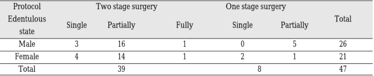
MATERIALS AND METHODS 1. Patients selection (Table I)
During the spring and summer of 2001 in the Implant Clinic of Seoul National University Dental
Table Ⅰ.Overview of patients distribution
Protocol Two stage surgery One stage surgery
Edentulous
Single Partially Fully Single Partially Total
state
Male 3 16 1 0 5 26
Female 4 14 1 2 1 21
Total 39 8 47
Hospital (Seoul, Korea), 47 mandibles in consecutively implant installed patients were selected for this study. They were 21 females and 26 males and their age range was 17-68 years (mean age 49.8 years). Twelve patients were smokers, who were on- ly included in two stage surgery group that was con- sisted of 39 patients. In one stage surgery group, all 8 patients were nonsmokers. All patients were con- sidered to be a good general condition. The cases re- quired bone grafting material or showed no intact cortical bone plate such as wound healing site were ruled out. Edentulous states of patients were 9 sin- gle tooth loss, 36 partially edentulous and 2 fully eden- tulous states. Opposing teeth conditions were nat- ural teeth or fixed bridges except 3 patients who were removable partial denture wearers.
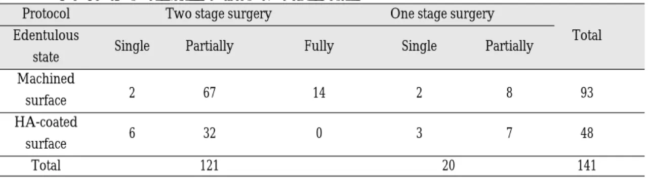
2. Installed fixtures (Table II)
Total 141 fixtures were installed. Ninety-three fixtures were double threaded, machined surface im- plants (Bra�nemark�MK III, Nobel Biocare AB, Go¨ teborg, Sweden) and 48 fixtures were root form, threaded, HA-coated surface implants (ReplaceTM, Steri-Oss/Nobel Biocare AB, USA). The diameter of fixtures was 3.75 or 4.0 mm for Bra�nemark� and 4.3 mm for ReplaceTMfixtures. Each 10 fixture was in- stalled in one stage surgery group and 121 fixtures were installed in two stage surgery group.
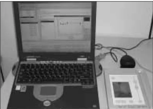
3. Resonance frequency analyser
OsstellTM(Integration Diagnostics Ltd. Sweden, Fig.
1) was used for implant stability measurements in this study. It is composed of OsstellTMinstrument (a LCD graphical display, Fig. 2), transducers (Fig.
3), data manager (Fig. 4, 5). The fixture level trans- ducer (Type F1) was used for Bra�nemark�RP and ReplaceTMand abutment level transducers were used for Bra�nemark�Standard, EstheiCone, Multi- Unit abutments (Type A1, A2 and A3, respectively).
The transducers were orientated perpendicular to the alveolar ridge with lingual position of the upright part of the beam and were tightened by hand.
Results were displayed graphically and represent- ed as an ISQ (Implant Stability Quotient,1-100).8 Initially, ISQs were obtained three times at each fixture and averaged. But because data repro- ducibility of OsstellTMinstrument was very excellent, ISQs were gained once then.
4. Bone quality and quantity assessment during initial drilling
During the initial drilling with a guide drill (Nobel Biocare AB, Go¨teborg, Sweden), bone qual- ities were assessed. For the details of the assessment, the status of cortical bone was decided from its thickness and feelings classfied as very hard, hard, less hard, soft and very soft senses, and that of
Table Ⅱ.Overview of installed fixtures distribution
Protocol Two stage surgery One stage surgery
Edentulous
Single Partially Fully Single Partially Total
state Machined
surface 2 67 14 2 8 93
HA-coated
surface 6 32 0 3 7 48
Total 121 20 141
Fig. 3.A transducer(Type A1) connection with a stan- dard abutment.
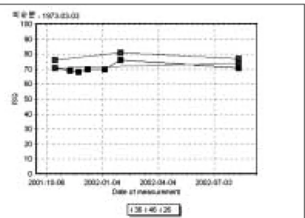
Fig. 4.A software-viewing example of one patient record form in OsstellTMdata manager.
Fig. 1.Setup of OsstellTMinstrument, data manager and supplied infrared link.
Fig. 2.A LCD graphical display of the OsstellTMinstru- ment.
cancellous bone was done from the feelings classi- fied as compact, coarse, loose and very loose sens- es. With bearing the combinations of these feel- ings in mind, bone qualities were scored (Grade 1- 4) by Lekholm and Zarb bone classification.9With periapical and panoramic radiographic images, bone quantities were also decided by that index and confirmed intraorally (Grade A-E).
5. Clinical primary stability acquisition during installation
For the installation of Bra�nemark�MK III fix- tures, recommended drill series were used. In some cases, tapping and countersinking procedures were not or minimally done for obtaining adequate fixtures stability. For ReplaceTMsystem, corresponding final size drills to the fixture were used at the terminal stage of high speed drilling and tapping procedure was minimally used. But counterbore drill was not used due to its characteristics of final size drill shape and fixture design. Resultant clinical primary stabilities were fair or excellent, and confirmed with resonance frequency analysis using OsstellTMinstrument. Most of all implants were inserted that their tops of fixture
flanges were slightly located at the subcrestal bone level aimed at the level of the external hex of the fix- tures with bony crest.
6. Stability measurement schedules and cover screw exposure evaluation and marginal bone level assessment
6.1. Two stage surgery group
ISQ values and periapical films were obtained right after implant insertion, at healing abutment con- nection, and in the loading phase of implants. The loading durations of prosthesis on each fixture in two stage surgery group were 5-10 months. All prostheses were fixed type restorations. Before second stage un- covering operation, the patterns of spontaneous early exposures of cover screw were assessed and clas- sified. Tal H. classified the patterns of early exposures of cover screw.10-12Because there was no statisti- cally significant difference between class 0 and class 1 in his classification, modification of classes was done and used in this study (Table III).
6.2. One stage surgery group
For this work, all RepalceTMfixtures were imme- diately connected with healing abutments at implant insertion stage after RFA measurements. 1mm height EsthetiCone�abutments (Noblel Biocare AB, Sweden) were connected to all Bra�nemark� fixtures in this group and healing caps were placed after RFA measurement. Transducer for EsthetiCone� abutment (type A2) was used for this measure- ment. Further ISQ values were measured at 4, 6, 8, 10, 12 weeks and in the loading phase, respective- ly. Patients were instructed to maintain good oral hy- giene and to take soft diet. Periapical radiographs were taken at implant insertion, 12 weeks and in the loading phase. The loading durations of prosthesis on fixtures in one-stage surgery group were 6- 9months.
Fig. 5. An example of the ISQ changes of 3 implants in one patient according to the time lapse (1 implant for one stage surgery, 2 implants for two stage surgery).
7. Periapical radiograph taking and reading point
To evaluate the marginal bone level of implants, periapical radiographs were taken with long cone par- alleling technique. Periapical radiographs taking was done such that the films were positioned with the axis of implant and the aperture of long cone as parallel as possible. Because RFA values were known to be sensitive to the marginal bone level sur- rounding the implants13and were known to vary 2- 3 ISQ according to the 1mm bone level change8, the marginal bone levels of implants were determined and scored through the findings as to the level of thread of installed implant with x 5.5 magnifying lens (Table IV). Scored levels were evaluated with ISQ val- ues at each period.
8. Statistical analysis
SPSS ver.10.0 package for Windows was used. For evaluations of RFA changes with time factor and op- eration method, repeated measure ANOVA test and Scheffe's post-hoc test were used (p<0.05). The Pearson correlation test (p<0.01) was used for sta- tistical analysis of the correlation between fixture length and RFA values at implant installation as well as for the correlation between marginal bone levels and RFA values from the installation to the loading period. The correlation between cover screw ex- posure and marginal bone level changes was also an- alyzed.
Table IV.Assessed marginal bone level score of implant fixture in periapical radiograph. Matched scores and the lowest bone contact levels in each mesial and distal side of implant were as followings
Score Radiographically assessed points
0 above the flange bottom of implant fixture 1 at or above the 1st thread end of implant fixture 2 at or above the 2nd thread end of implant fixture 3 at or above the 3rd thread end of implant fixture
Table III. Classification of early cover screw exposure before second stage surgery
Class Clinically assessed points
0 No exposure or breach in the mucosa covering the implant is observed.
1
The mucosa above the cover screw is fenestrated. The cover screw is visible. The boders of the perforation's aperture do not reach or overlap the borders of the cover screw at any point.
2 Cover screw is visible. In some parts, the borders of the perforation aperture overlap the borders of the cover screw.
3 Cover screw is completely exposed.
RESULTS
Among 141 fixtures, 2 fixtures failed and were con- firmed with OsstellTM. Their ISQ values were 78 and 81 at the installation, but showed 51 and 45 at the healing abutment connection and then de- creased to 43 and 40, respectively, 2 weeks later. They were removed and ruled out from statistical analy- sis. One (78-51-43 ISQ value change) of the two fixtures was not showed clinical mobility at the healing abutment connection. Even though this was short-term observation , other implants had no problems in the loading period and showed good clin- ical performances and ISQ values. Therefore, over- all survival rate was 98.6%. The highest ISQ value was 92, it was recorded on one fixture at the healing abutment connection and the lowest value was 62, it was recorded on two fixtures at the installation.
1. Bone quality and bone quantity (Table V)
In bone qualities , Grade 2 or 3 was mainly assessed in both groups. They were 87.6% in two stage surgery group and 95% in one stage surgery group.
Grade B was mainly assessed in bone quantities of
both groups. It was 77.7% in two stage surgery group and 100% in one stage surgery group.
2. Mean ISQ values of two stage surgery group at fixture installation, healing abutment con- nection and in the loading period
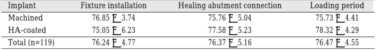
Mean and SD of ISQ values of machined sur- face implants were 76.85 ± 3.74, 75.76 ± 5.04, 75.73 ± 4.41 and those of HA-coated surface implant were 75.05 ± 6.23 , 77.58 ± 5.23, 78.32 ± 4.29 at each period in two stage group (Table VI). It showed small mean ISQ value change after fixture installation, so implants of that group were seemed to be stable.
Machined surface implants showed slightly lower ISQ values at healing abutment connection and maintained their values in the loading period. ISQ values of HA-coated surface implants showed high- er value tendency than those of machined ones (Fig. 6).
3. ISQ changes of implants in two stage surgery group according to the bone qualities Grade 4 group was ruled out in statistical analy-
Table V. Distribution of assessed bone qualities and quantities by Lekholm and Zarb Classification9)
Group Bone quality Bone quantity
1 2 3 4 A B C
Two stage 12 56 50 3 19 94 8
One stage 1 10 9 0 0 20 0
Table VI. Mean and SD of ISQ in two stage surgery group
Implant Fixture installation Healing abutment connection Loading period
Machined 76.85 ± 3.74 75.76 ± 5.04 75.73 ± 4.41
HA-coated 75.05 ± 6.23 77.58 ± 5.23 78.32 ± 4.29
Total (n=119) 76.24 ± 4.77 76.37 ± 5.16 76.47 ± 4.55
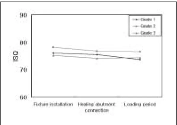
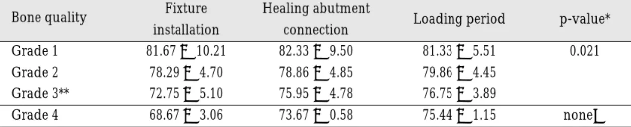
sis due to small sample size. Grade 3 group showed statistically significant value changes from those of other groups (p<0.05) in both fixture designs (Table VII, VIII). In machined surface implant, Grade 2 group showed the highest mean value in all periods (Table VII). In HA-coated surface implant, ISQ values were increased with time except Grade 1 group(Table VIII). In spite of small sample size, grade 4 group showed the lowest mean value at the fixture in- stallation but caught up the values of other groups gradually (Fig. 8).
4. Correlation between fixture lengths and ISQ values at fixture installation
10mm or 13mm fixture length was mainly in- stalled (31.9%, 45.4%, respectively). Pearson's cor- relation coefficient(r) between fixture lengths(Table IX) and corresponding ISQ values at fixture instal- lation was 0.038(p<0.01). Therefore, there was no cor- relation between two data (Fig. 9).
Fig. 6. The change of mean ISQ value in two stage surgery group.
Fig. 7.Mean ISQ value changes of machined surface fix- tures in two stage surgery group according to the bone qualities.
Table VII. Mean and SD of ISQ values of machined surface fixtures in two stage group ac- cording to classified bone qualities
Bone quality Fixture Healing abutment
Loading period p-value*
installation connection
Grade 1 76.11 ± 2.71 75.56 ± 3.97 73.78 ± 4.52 0.017
Grade 2 78.18 ± 3.44 76.90 ± 5.41 76.68 ± 4.32
Grade 3** 75.30 ± 3.82 74.30 ± 4.56 74.53 ± 4.21
* Repeated Measure ANOVA ( between subject effects)
** denotes that differences of each mean were statistically significant (p<0.05) by Scheffe's post-hoc test.
5. Mean ISQ values of one stage surgery group
To the 4-6 weeks after fixtures installation, mean ISQ values decreased(Table X). Decreasing ten- dency was more frequent in machined surface im- plant but the values of those increased with time.
Nevertheless, after some decreasing, HA-coated surface implants reached at plateau and showed in- creased values earlier than machined surface ones (Fig. 10).
6. Two stage surgery group vs one stage surgery group
The mean ISQ value changes according to time lapse between two stage and one stage surgery groups were likely minimal (Fig. 11). Mean ISQ values of one stage surgery group at 3 months after installation and in the loading period were larger.
Smaller sample size than two stage surgery group and meticulous case selection and controlled special care during study in one stage surgery group might affect that results.
7. Correlation between ISQ value changes and marginal bone level score changes in two stage surgery group.
Marginal bone level scores were increased with time (Table XI). That means that there were marginal bone resorptions after fixture installation. High score change from fixture installation to healing abut- ment connection means that mild complication such as cover screw exposure was existed. On the con- trary, low score change from healing abutment connection to the loading period means that marginal Fig. 8. Mean ISQ value changes of HA-coated surface
fixtures in two stage surgery group according to the bone qualities.
Table VIII. Mean and SD of ISQ values of HA-coated surface fixtures in two stage group ac- cording to classified bone qualities
Bone quality Fixture Healing abutment
Loading period p-value*
installation connection
Grade 1 81.67 ± 10.21 82.33 ± 9.50 81.33 ± 5.51 0.021
Grade 2 78.29 ± 4.70 78.86 ± 4.85 79.86 ± 4.45
Grade 3** 72.75 ± 5.10 75.95 ± 4.78 76.75 ± 3.89
Grade 4 68.67 ± 3.06 73.67 ± 0.58 75.44 ± 1.15 none�
* Repeated Measure ANOVA (between subject effects)
** denotes that difference of each mean were statistically significant (p<0.05) by Scheffe's post-hoc test.
� denotes that statistical analysis was not performed due to small sample size.
bone resorption process was decreased and marginal bone level had a tendency to be stable. Pearson's cor- relation coefficient(r) between ISQ value changes and marginal bone level score changes from fixture in- stallation to healing abutment connection period was -0.520 (p<0.01). That indicates a moderate to good negative relationship between two groups (Fig.
12). That r-value from healing abutment connection period to the loading period was -0.505 and indicates the same previous interpretation (Fig. 13).
8. Cover screw exposure observation before uncov- ering flap operation in two stage surgery group
In Table XII and Fig. 14, Class 0 (no exposure) was mainly observed before open flap surgery (75.6%).
Class 1 was the most frequent perforation (9.2%), fol- lowed by Class 2 and 3 (7.6%). Cover screw exposure means mucosal perforation which can cause in- flammatory reaction. The ISQ values of fixtures involved in classes showed perforation were all likely to be decreased at healing abutment connec- tion. That means that mucosal perforation caused mar- ginal bone resorption, which showed decreased ISQ values.
9. Correlation between cover screw exposure and marginal bone level score change from fixture in- stallation to healing abutment connection
Pearson's correlation coefficient(r) between cov- er screw exposure and marginal bone level score was Fig. 9.Plot of ISQ values against fixture lengths for implants at fixture installation. r=0.038(p<0.01)
Table X. Mean and SD of ISQ values in one stage surgery group according to time lapse
Measurement time Machined HA-coated Total (n=20)
0 week 76.60 ± 3.57 76.60 ± 3.84 76.60 ± 3.60
4 weeks 73.30 ± 4.72 74.60 ± 4.20 73.95 ± 4.39
6 weeks 72.40 ± 4.74 74.40 ± 4.01 73.40 ± 4.39
8 weeks 72.90 ± 4.65 75.90 ± 4.12 74.40 ± 4.55
10 weeks 73.70 ± 4.14 76.90 ± 3.67 75.30 ± 4.14
12 weeks 75.10 ± 4.25 78.00 ± 3.20 76.55 ± 3.95
Loading period 76.50 ± 2.92 79.30 ± 2.87 77.90 ± 3.16
Table IX. Installed fixture lengths Fixture length (mm) No. of fixtures
8.5 3
10 45
11.5 10
13 64
15 17
16 2
100
90
80
70
60
0 10 12 14 16 10
Length
Rsq=0.0015
ISQ
Fig. 10.Mean ISQ changes in one stage surgery group. Fig. 11.Mean ISQ value changes of two and one stage surgery groups.
Fig. 12.Plot of ISQ value changes against marginal bone level score changes from fixture installation to healing abutment connection period. r= -0.520 (p<0.01).
Fig. 13. Plot of ISQ value changes against marginal bone level score changes from healing abutment connec- tion to loading period. r= -0.505 (p<0.01).
Table XI. Mean and SD of marginal bone level scores at each period and scores change in two stage surgery group
Assessed time and scores change Mean ± SD
Fixture installation(1) 0.00
Healing abutment connection(2) 0.79 ± 0.84
Loading period(3) 1.08 ± 0.72
Scores change (2-1) 0.79 ± 0.84
Scores change (3-2) 0.29 ± 0.52
20
10
0 -10
-20
-30
-5 0.0 5 1.0 1.5 2.0 2.5 3.0 3.5 marginal bone level score change
Rsq=0.2706 Rsq=0.2551
ISQ value change
10
0
-10
-20
-5 0.0 5 1.0 1.5 2.0 2.5
marginal bone level score change
ISQ value change
0.618 (p<0.01). That indicates a moderate to good pos- itive relationship between two groups (Fig. 15).
Therefore, from the above result, cover screw ex- posure may cause to decrease ISQ value and result in lowering the implant stability.
DISCUSSION
In patients selection, patients with intact cortical bone plate were selected as possible as can. Among 73 patients who were consecutively received 232 fix- tures, only 47 patients were selected. One-stage surgery group patients were very meticulously se- lected and controlled, so they showed excellent initial fixture fixation, sufficient interocclusal distance, good oral hygiene, no smoking and cooperative passion. The diameters of selected fixtures were
3.75, 4.0 and 4.3mm. That size difference was not worth while to consider because matched trans- ducer was previously calibrated by manufactur- er.8
The resonance frequency value obtained from the OsstellTMtransducer is automatically translated into an index called the Implant Stablilty Quotient (ISQ), which runs from 1 to 100. Extensive clinical test- ing has made it possible to establish this scale for eval- uating the clinical stability of implants. The rela- tionship between the ISQ value and the resonance frequency value is close to linear, and factors such as individual differences between transducers are al- so taken into account. In this study, 4 types of transducers were used. As each transducer has its own calibration values, it is possible directly to compare ISQ values that originate from different Fig. 14.Graph of the number of each fixture according
to the classification of cover screws exposure.
Fig. 15. Plot of marginal bone level score changes against cover screws exposure or not (1.0:no expo- sure, 2.0: exposure). r= 0.618 (p<0.01).
3.5
2.5
1.5
5
-5
0.0 1.0 2.0
cover screw exposure or not
Rsq=0.3825 Bra�nemark Replace Total
marginal bone level score change
Table XII. Classified cover screw exposures and corresponding number of each fixture Class Bra�nemark�fixture ReplaceTM fixture Total(no./percentage)
(no./percentage) N=79 (no./percentage) N=40 N=119
0 61 77.2% 29 72.5% 90 75.6%
1 6 7.6% 5 12.5% 11 9.2%
2 5 6.3% 4 10% 9 7.6%
3 7 8.8% 2 5% 9 7.6%
transducers. A direct translation to ISQ from fre- quencies presented for early transducers is difficult, but when articles14-16presenting RF, the ISQ value can roughly be derived from : 1 ISQ per 50Hz.
On the failed 2 implants in this study, one showed largely decreased ISQ value change (-27 ISQ) with- out clinical mobility during second stage surgery. It presented an example of the effectiveness of RFA tech- nique.
Table VI showed mean ISQ value changes with time in two stage surgery group. Overall mean values of machined surface implants were maintained with small changes. Sennerby et al.17reported sim- ilar results that 6 months after loading and about 10- 11 months after implant placement did not show any changes compared with stability after placement and abutment connection in mandible. Al-Nawas et al.18also reported the experimental study results in dog. They evaluated RFA during implant inser- tion, at abutment connection and in the loading phase of dental implants and concluded that reso- nance freqeuncy at insertion was declining dur- ing the healing period regardless of the surface or screw design and during the loading phase no sig- nificant change was seen. From the result of stability measurement on the machined surface implants of mandibular molar area in functioning successfully during at least 12 months, mean ISQ values were maintained at the 71-73 level.19However, HA-coat- ed surface implants showed somewhat increased ISQ values tendency with time in spite of high value af- ter placement. It was seemed to be affected by rapid bone healing capacity of HA-coating.
In bone quality and bone quantity assessments, Lekholm and Zarb classification9was used in this study. Because primary stabilities were almostly very good or excellent, relatively high ISQ values were recorded at fixtures installation. Also, macrostruc- tures of implants such as double threaded or thread- ed root form and surgical techniques such as no or minimal usage of tap and countersink drill con- tributed to that results even in Grade 3 bone qual-
ity. In Table VII, the ISQ values of machined surface implants had a tendency to decrease with time in Grade 1 and 2 bone qualities, however, they main- tained their values with time in Grade 3 bone qual- ity. This pattern was not similar with those of HA- coated surface implants. In Table VIII, the ISQ val- ues of HA-coated surface ones showed maintained or increased pattern with time according to bone qual- ities. That difference of value change pattern between fixture surface types seems to be caused by interfacial bone response of HA-coating. Although bone qual- ity was scored with care on the initial drilling, there was a limitation of objectiveness. One of the ob- jective methods assessing bone quality is cutting re- sistance measurement.20-23Torque was determined by the current drawn by an electric motor while cut- ting a thread. Friberg et al.24 noted that there was a correlation between resonance frequency mea- surements at implant placement and thread cutting forces for the most coronal third of the implant site. This finding is significant as it is likely that it is the most coronal part of the implant bed which plays the most significant role in implant stability and the support of the implant under load. Also, they showed that the stability of implants placed in soft- er bone seemed to catch up over time with more dense bone sites. Table VIII and Fig. 8 showed sim- ilar patterns of increasing value like above with time in the poor bone quality in spite of small sam- ple size.
Table IX showed a correlation between fixture lengths and ISQ values at fixture installation. There was no correlation between both data, which coin- cided with other reports.14,15,19That means that the RFA value of each implant represents its stability status regardless of its length, so it is possible to com- pare each other and assess the relative stability.
The original idea of using a two stage surgery pro- cedure was to avoid preloading of the implant and to allow for bone formation and integration of the implant to occur.9Thus, it was anticipated that, with a healing period, an increased implant sta-
bility would be achieved as compared to the day of implant placement. Successful use of one stage, nonsubmerged ITI implants inserted in mandibles has, however, been reported by Buser et al.25,26 Deviations from the original protocol, i.e. to perform one instead of two surgical procedures, have been executed for both partially dentate7and edentu- lous patients using the Bra�nemark system.20,28,29The authors concluded that no clinical differences were seen between the two groups and the radiograph- ic bone level was similar with or without a trans- mucosal junction. The one stage implant result also compared well with those reported for the traditional two stage technique. Friberg et al.30showed that the RFA values slightly decreased for the majority of the implants during the study period (15 weeks) inde- pendent of design. Consequently, the implants were as stable at time of placement as when measured 3-4 months post-surgery, i.e when the prostheses were attached.
In relation with above study, similar decreased ISQ values of machined surface implants were shown un- til 6 weeks period in this study. But its values reached plateau at 6-8 weeks or 10 weeks, and in- creased to the loading period. This result is differ- ent from that of Friberg et al.30It seems to be a pos- sibility that no use of relined denture in this study affected that difference. In Table X, HA-coated im- plant showed more increased values to the loading period from after 6 weeks than installation, which is in contrast to the finding of machined surface im- plant. The ISQ values of machined surface im- plants showed recovery patterns of its own val- ues shown at the installation. All patients healed with- out any severe problems and all implants were found to be stable at the end of the study period. The one-stage surgery protocol did not present any major adverse tissue reactions or implant compli- cations postoperatively.
Effective implant length (EIL) means the sum of marginal bone height and the length of the abutment.
There is a strong negative correlation between the
EIL and resonance frequency.14,15,31Therefore, in- creased marginal bone resorption allow ISQ value to decrease. In present study, marginal bone re- sorptions were scored. That assessment method was somewhat rough, however, that was meaningful. The result showed that there was good negative corre- lation between marginal bone resorption and ISQ val- ue changes, so resonance frequency measurements were related to the effective length of an implant above the level of the bone.
It is believed that during the osseointegration phase of submerged dental implants, complete mucosal coverage and isolation of the implant from the oral cavity avoids trauma and infection and establishes favorble conditions for osseointegra- tion. Spontaneous early exposure is one of the com- plications that could adversely affect osseointe- gration of implants. Adell et al.2in a 15-year study of osseointegrated implants in the jaws, observed ear- ly perforations in the mucosa in 4.6% of their treat- ed patients in spite of careful surgical protocol and postsurgical care. They believed that sealing the communication between the implant and the oral cav- ity was crutial to the success of osseointegraton. Early perforation and partial exposure of the implant's cov- ering device are a focus for plaque accumulation which, if left untreated, may result in inflammation, damage to the peri-implant mucosa, and possible peri- implant loss.10,32
In present study, it was found that cover screw ex- posure before second stage surgery forced mar- ginal bone level to decrease, consequently, which low- ered ISQ values. That results were supported by the fact that correlation between cover screws exposures and marginal bone level score was strong. In com- parison with other studies,2,10relativley high per- centage of cover screw exposures was found and that might be caused 2 fully edentulous patients wear- ing relined denture during healing time. They showed mainly Class 3 mucosal perforations.
Without them, the percentages of Classes except class 0 would be a quite decreased. Tal et al.11,12suggest-
ed that different factors might result in the forma- tion of spontaneous early perforation, most of which were associated with mechanical trauma to the mucosa or tension in the tissue flaps covering the implants. And they noted that bone debris pro- duced during the osteotomy could act as an additional predisposing factor; these are sequestrated and ac- companied by chronic inflammatory cell infiltration as well as epithelial-covering reaction. In cases of cov- er screw exposure, more marginal bone resorption may be produced in HA-coated surface implant than machined surface implant due to its roughness character, which may play a role in accelerating the propagation of inflammatory reaction. Toljanic et al.32observed with HA- coated surface, press fit im- plants and reported that patients with exposed sites demonstrated a likelihood of bone loss 3.9 times greater than patients with nonexposed sites.
Although the present study included HA-coated sur- face ones, it was rarely found to show rapid and de- structive marginal bone resorption.
From the above results in this study, using reso- nance frequency analysis in two stage surgery group, implants stabilities in mandible with at least intact cortical bone plate and Grade 3 bone quality showed minimal ISQ changes with time if there was no complication such as mucosal perforation re- sulted in marginal bone resorption. One stage surgery group revealed different stability changes modes between machined and HA-coated surface implants with time. But because it has been be- lieved that bone remodelling is continuous process at least until 18 months,9more observations will be needed in further studies in related with RFA tech- nique.
CONCLUSIONS
Using the resonance frequency analysis, stabilities of mandibular implants in patients were measured from installation to the loading in two stage and one stage surgery groups and additional observations
were performed during healing period. The fol- lowings were concluded that :
1. In two stage surgery group, mean and SD of ISQ values of machined surface implants were 76.85 ± 3.74, 75.76 ± 5.04, 75.73 ± 4.41 and those of HA-coated surface implant were 75.05 ± 6.23 , 77.58 ± 5.23, 78.32 ± 4.29 immediately af- ter fixtures installation, at healing abutment con- nection and in the loading period, respectively.
2. In two stage surgery group, machined surface and HA-coated surface implants showed minimal ISQ changes with time if they were installed at the sites with intact cortical bone plate and good bone qualities.
3. In two stage surgery group, HA-coated implants had a tendency to show increased ISQ values with time.
4. In one stage surgery group, the ISQ values of ma- chined surface and HA-coated surface implants decreased until 4 or 6 weeks and maintained at plateau for 1-3 weeks and increased to the load- ing period.
5. In one stage surgery group, the ISQ values of HA- coated surface implants were likely to show shorter decreased and plateau period than ma- chined surface ones.
6. There was no correlation between installed fixture length and ISQ value at fixture installation (r=0.038).
7. There was a moderate to good negative correla- tion between ISQ changes and marginal bone lev- el score changes in two stage group (r=-0.520, -0.505).
8. There was a moderate to good negative correla- tion between marginal bone level scores and cover screw exposure in two stage group (r=0.618).
REFERENCES
1. Albrektsson T, Bra°nemark PI, Hansson H, Lindstr-
¨m J. Osseointegrated titanium implants.o Requirements for ensuring a long lasting direct bone- to-implant anchorage in man. Acta Orthop Scan
1981;52:155-170.
2. Adell R, Lekholm U, Rockler B, Bra°nemark PI. A 15-year study of osseointegrated implants in the treatment of the edentulous jaw. Int J Oral Surg 1981;10:387-416.
3. Sennerby L, Roos J. Surgical determinants of clin- ical success of osseointegrated implants. A re- view of the literature. Int J Prosthodont 1998;11:408- 420.
4. Albrektsson T, Sennerby L. State of the art in oral implants. J Clin Periodontol 1991;18:474-481.
5. Meredith N. Assessment of implant stability as a prognotic determinant. Int J Prosthodont 1998;11:491- 501.
6. Sennerby L, Meredith N. Resonance Frequency Analysis : Measuring implant stability and os- seointegration. Compendium 1998;19:493-502.
7. Park C. A study of the measurement of the implant stability using resonance requency analysis. PhD thesis, 2000, Dankook University, Korea.
8. OsstellTM resonance frequency analyser. Clinical Manual. Integration Diagnostics Ltd. Sa¨vedalen, Sweden. 2002.
9. Lekholm U, Zarb GA. Patient selection and prepa- ration, in Bra°nemark PI, Zarb GA, Albrektsson T.(eds): Tissue-Integrated Prostheses : Osseointegration in Clinical Dentistry.
Chicago:Quintessence, 1985:199-209.
10. Tal H. Spontaneous early exposure of submerged implants : I. Classification and clinical observations.
J Periodontol 1999;70:213-219.
11. Tal H, Dayan D. Spontaneous early exposure of sub- merged implants : II. Histopathology and histo- morphometry of non-perforated mucosa covering submerged implants. J Periodontol 2000;71:1224- 1230.
12. Tal H, Dayan D. Spontaneous early exposure of sub- merged implants : III. Histopathology of perforated mucosa covering submerged implants. J Periodontol 2000;71:1231-1235.
13. Meredith N. On the clinical measurement of implant stability and osseointegration. PhD thesis, 1997, University of Gothenburg, Sweden.
14. Meredith N, Alleyne D, Cawley P. Quantitative de- termination of the stability of the implant-tissue in- terface using resonance frequency analysis. Clin Oral Impl Res 1996;7:261-267.
15. Meredith N, Book K, Friberg B, Jemt T, Sennerby L. Resonance frequency measurements of implant stability in-vivo : A cross sectional and longitudinal study of resonance frequency measurements on im- plants in the edentulous and partially dentate maxilla. Clin Oral Impl Res 1997;8:226-233.
16. Meredith N, Sagaldi F, Alleyne D, Sennerby L, Cawley P. The application of resonance frequen- cy measurement to study the stability of titanium implants during healing in the rabbit tibia. Clin Oral Impl Res 1997;8:234-243.
17. Sennerby L, Friberg B, Linden B, Jemt T, Meredith
N. A comparison of implant stability in mandibu- lar and maxillary bone using RFA. European Commission Demonstration Project. Resonance Frequency Analysis Symposium. 2000, Gothenburg, Sweden.
18. Al-Nawas B, Groetz KA, Brahm R, Wagner W.
Resonance-frequency-analysis in the healing and loading period of dental implants. IADR abstracts 2002;1027.
19. Park CJ. Stability measurement on the implants of mandibular molar area using OsstellTM. J Korean Academy of Stomatognathic Function and Occlusion 2001;17:205-211.
20. Friberg B. On bone quality and implant stability measurements. PhD thesis. 1994, University of Gothenburg, Sweden.
21. Friberg B, Sennerby L, Roos J, Johansson P, Strid CG, Lekholm U. Evaluation of bone density using cutting resistance measurements and microradi- ography. : An in vitro study in pig ribs. Clin Oral Impl Res 1995;6:164-171.
22. Friberg B, Sennerby L, Roos J, Lekholm U.
Identification of bone quality in conjunction with insertion of titanium implants. Clin Oral Impl Res 1995;6:213-219.
23. Johansson P, Strid KG. Assessment of bone qual- ity from cutting resistance during implant surgery.
Int J Oral Maxillofac Impl 1994;9:279-288.
24. Friberg B, Sennerby L, Meredith N, Lekholm U. A comparison between cutting torque and reso- nance frequency measurements of maxillary im- plants. A 20-month clinical study. Int J Oral Maxillofacial Surg 1999a;28:297-303.
25. Buser D, Mericske-Stern R, Bernard JP. Long- term evaluation of non-submerged ITI implants.
Part 1:8-year life table analysis of a prospective mul- ticenter study with 2359 implants. Clin Oral Impl Res 1997;8:161-172.
26. Buser D, Weber HP, Bragger U, Balsiger C. Tissue integration of one-stage ITI implants :3-year results of a longitudinal study with hollow-cylinder and hollow-screw implants. Int J Oral Maxillofac Impl 1991;6:405-412.
27. Becker W, Becker B, Isarelson H. One-step surgi- cal placement of Branemark implants : A prospec- tive multicenter clinical study. Int J Oral Maxillofac Impl 1997;12:454-462.
28. Ericsson I, Rainbow K, Glantz PO, Lindhe J, Nilner K. Clinical and radiographical features of sub- merged and nonsubmerged titanium implants.
Clin Oral Impl Res 1994;5:185-189.
29. Ericsson I, Rainbow K, Nilner K, Petersson A.
Some clinical and radiographical features of sub- merged and non-submerged titanium implants. A 5-year follow-up study. Clin Oral Impl Res 1997;8:422-426.
30. Friberg B, Sennerby L, Linde′n B, Gro¨ndahl K, Lekholm U. Stability measurements of one-stage Bra°nemark implants during healing in mandibles.
A clinical resonance frequency study. Int J Oral Maxillofac Surg 1999b;28:266-272.
31. Park CJ. In vitro comparative study between ISQ and Peritest�values on the implant stability mea- surements according to the increased effective implant length. J Korean Academy of Prosthodontics 2001;39:625-635.
32. Toljanic J, Banakis M, Willes L, Graham L. Soft tis- sue exposure of endosseous implants between stage I and stage II surgery as a potential indica-
tor of early crestal bone loss. Int J Oral Maxillofac Impl 1999;14:436-441.
Reprint request to:
DR.YUNG-SOOKIM , D.D.S., Ph.D.
DEPT. OFPROSTHODONTICS,GRADUATESCHOOL,
SEOUL NATIONAL UNIV.
28-1 YEONGUN-DONG, CHONGNO-GU, 110-749, SEOUL KOREA [email protected]