103
J. Appl. Biol. Chem. 52(3), 103-108 (2009)
Article
지방세포배양액이 유방암세포주에서 GITRL의 발현을 유도
백아미·박미영·박정수·한정혜·양 영*
숙명여자대학교 생명과학부
Adipocyte Culture Medium Stimulates GITRL Expression in MDA-MB-231 Cells
Ahmi Baek, Miyoung Park, Jeongsu Park, Jeonghye Han, and Young Yang
*Department of Life Science, Sookmyung Women’s University, Seoul 140-742, Korea Received April 11, 2009; Accepted September 10, 2009
Little is known about roles of adipose tissue, although obesity is one of the potential risk factors in causing breast cancer and adipose tissue surrounding breast ductal cells is the largest organ. To identify the genes that are regulated by factors secreted from adipocytes in breast cancer cells, MDA-MB-231 cells were treated with the culture medium of adipocytes. In present study glucocor- ticoid-induced TNF receptor-related protein ligand (GITRL) gene was studied among the induced genes. It was found that GITRL was significantly increased by the culture medium of adipocytes.
Proteinase K-treated adipocyte culture supernatants failed to induce GITRL expression. These find- ings indicate that unknown protein factors are responsible for the induction of GITRL expression.
The expression of GITRL did not affect the invasive ability of MDA-MB-231 cells, but coculture with NK92 expressing GITR suppressed the invasiveness of MDA-MB-231 cells.
Key words: adipocytes, breast cancer, immune system
서 론
비만은유방암의전이를촉진하는위험요소로서잘 알려져 있고[Josefson, 2001; McCann, 2001; Bianchini 등, 2002], 지
방세포와유방암세포의상호작용에대하여연구된논문이많지 는 않지만몇몇의 연구가그 중요성에대하여 말해주고있다.
쥐에유방암 세포주를주입할때, 지방조직과 떨어진곳에주 입한경우보다지방조직에직접유방암세포를주입한경우암 의성장이 촉진되는것이알려졌고[Elliott 등, 1992], 미분화된 지방세포와분화된지방세포를 각각유방암 세포주와섞어쥐 에 주입했을때, 분화된 지방세포와 함께주입한경우에서암 세포의성장이증가됨이알려졌다[Johnston 등, 1992]. 또한유 방암세포주인 MCF-7에지방세포의 배양액을첨가하여 MCF- 7에서 변화하는 종양관련 유전자들을 연구한 그룹도 있다
[Iyengar 등, 2003].
지방조직은아디포넥틴, 렙틴과같은대사조절호르몬과 TNF-
α, IL-6, MCP-1 등과 같은 각종 사이토카인들을 생산하는데
[Fantuzzi, 2005] 이들을통칭하여아디포카인이라부른다. 아디 포카인들은 당대사나 지방대사, 면역 항상성을유지하는데 관
여하며 비만, 심혈관계질병, 인슐린저항성이나암과같은질 병에서는정상인경우와 비교해보면아디포카인의 발현정도 가 달라져 있다[Goodfriend 등, 1998; Grundy, 2000; Steppan
등, 2001; Baillargeon 등, 2006; Mauro 등, 2007; Schaffler 등, 2007; Spyridopoulos 등, 2007]. 예를들어비만환자에게서렙 틴의 양은정상의경우와비교해서증가되어져있으며그와반 대로 아디포넥틴의경우에있어서는비만환자에게서감소 경향 을 보인다. 또한유방암에있어서 렙틴의경우암세포의증식 능력을 증가시키나 아디포넥틴의경우반대로 증식억제기능 을 보인다[Baillargeon 등, 2006; Dos 등, 2008]. 이는 비만에 의하여감소되는아디포넥틴과증가하는렙틴이서로작용하여 비만한유방암환자의유방암세포증식을촉진할수있음을뜻한 다.
Glucocorticoid-induced tumor necrosis factor receptor(GITR)
은생쥐 hybridoma T 세포주에서 glucocorticoid에반응하는유
전자로 처음확인되었다[Nocentini 등, 1997]. GITR은 사람의 자연살해세포에서낮은레벨로발현하고있으며, T 세포, 대식
세포, B 세포 등에서도 발현되고 있다. Toll-like receptor ligand나자연살해세포성장인자인 IL-15들에 의해발현이증가 되며, 주로 T 세포를 자극시키며 regulatory T cell의 면역억제
*Corresponding author
Phone: +82-2-710-9590; Fax: +82-2-2077-7322 E-mail: [email protected]
doi:10.3839/jabc.2009.018
기능을약화시킨다고 알려져 있다[McHugh 등, 2002; Shimizu
등, 2002; Kim 등, 2003; Ji 등, 2004; Shevach and Stephens, 2006]. GITR의리간드인 GITRL은 주로 내피세포와항원제시 세포에서발현되며, 근래에는많은 암세포에서 발현된다고보
고되고 있다. 현재까지 GITR-GITRL system의 기능에 대해서 는 T 세포에 초점이 맞춰져 있었다. 하지만 최근 human plasmacytoid DC(pDC)에 발현되는 GITRL은 자연살해세포를 활성화시키는역할을하고있으며[Hanabuchi 등, 2006], 암세포
에서발현하는 GITRL은자연살해세포의 IFN-γ생산과세포살 상능력을낮춘다는 연구보고가있다[Baltz 등, 2007]. 본연구 에서는지방세포가유방암세포에미치는작용을알아보기위하 여 지방세포배양액을 MDA-MB-231 유방암세포주에처리하여 변화하는유전자중에 GITRL의 발현증가를확인하고암세포의
전이에미치는기능을조사하였다. 재료 및 방법
세포 배양. 인간유방암 세포주인 MDA-MB-231 세포와생 쥐 미분화 지방세포 OP9 세포주는 American Type Culture Collection(ATCC)로부터 구입하였다. 인간 유방암세포주인
MDA-MB-231세포는 2 mM L-glutamine, 항생제 그리고 10%
fetal bovine serum(FBS)가 포함된 Dulbecco’s modified Eagle’s
medium(DMEM) 배지를사용하여배양하였고생쥐골수유래기
질세포주인 OP9세포는 2 mM L-glutamine, 항생제그리고 20%
FBS가 포함된 Alpha minimum essential medium(alpha-MEM)
를사용하여 37oC, 5% CO2배양기에서 배양하였다. 세포배양
에 사용된 DMEM, Trypsin-EDTA, Antibiotic-Antimycotics, FBS는 Invitrogen(Carlsbad, CA)에서구입하였다.
지방세포 배양액 준비. 미분화지방세포 배양액은 OP9 세포 를 100 mm dish에 5×10⁶ cells/mL로 분주하고 2-3일 동안 배
양한다. 80% 정도의 confluent가 될 때, 1X PBS 두 차례세 척후 10% FBS가포함된 DMEM 배지로바꾸어 24시간동안 배양하고회수하였다.
지방분화 배양액은 OP9 세포주를 100 mm dish에 5×10⁶ cells/mL로 분주하고 2-3일 동안 배양한 후, 0.5 M isobutylmethlxanthine, 2µM dexamethasone, 1.7µM insulin을 첨가하여분화를 유도하였다. 분화유도배지는 24시간후에제
거해주었으며 배지는 20% FBS가포함된 α-MEM 배지를이 틀마다 교체하였다. 약 99%의 세포에 lipid droplet이 찼을때 10% FBS가들어간 DMEM 배지로바꿔준후 24시간배양하고 그배양액을회수하여실험에 사용하였다.
RNA의 분리 방법. MDA-MB-231 세포를 1X PBS를이용하 여 2회 세척한후 RNAzolB reagent 1 mL을 이용하여세포막 을용해시켰다. Chloroform 100µL를첨가하고잘섞은후원 심분리하여단백질층과 phenol을제외한상등액을얻었다. 이 상층액에 동일량의 isopropanol을 넣고 얼음에서 20분간 보관 후 13,000 rpm에서 20분간원심분리하여 MDA-MB-231 세포 의 전체 RNA를 얻었다. 얻어진 RNA 침전은 80% ethanol/
diethylenepyrocarbonate(DEPC)로 세척하였다. 세척된 RNA는
DEPC가첨가된멸균수에녹이고 260 nm의흡광도에서정량하
였다.
역전사-중합효소 연쇄 반응. 준비된 전체 RNA중 5µg를 70oC에서 10분간 반응시켜 denaturation시킨 후 6µL 10 mM dNTP mixture, 8µL 5X RT buffer, 1µL random primer (0.5µg/µL)와 함께 1µL의 M-MLV RTase(200 U/µL)(Promega, Madison, WI)를혼합하여 40µL의부피로 37oC 항온기에서 1
시간 동안역전사 반응시킨 후 80oC에서 5분간가열하여 M- MuLV RTase의 활성을 제거하고 얼음에 냉각 시켜 single- stranded cDNA를 얻었다. 얻어진 cDNA중 1µL를 1µL의
primer(10 pmol/µL), 10µL의 i-maxⅡ PCR premix, 8µL의
D.W.가함유된 총 20µL의반응혼합물을만들었다. 반응조건 은 94oC에서 denaturation 30초, 55oC에서 annealing 30초, 72oC에서 extention 30초를 1cycle로 하여 30 cycle을반복하였
고 추가적으로 72oC에서 post extension을 10분을 더 주었다.
이와 같은반응은 PTC-500 thermocycler(MJ Research, Waltham, MA)를 이용하여수행하였다.
Amplification을위해 PCR이수행되었고, 이때사용한 primer pair는(GITRL forward 5'-CGA ATT CAT GTG TTT GAG CCA C-3', reverse 5'-GGA GCT CGA TCC TCT AGT TAA C-3', GITR forward 5'-ATG GCA CAG CAC GGG GCG ATG-3', reverse 5'-ACT GAA TTT CCC CTG GGA CT -3', CXCL2 forward 5'-ACC GAA GTC ATA GCC ACA-3' reverse 5'-AAT GGG AGA GTG TGC AAG-3', CXCL3 forward 5'-TAC TGA ACA AGG GGA GCA-3' reverse 5'-CCC CAC CCT GTC ATT TAT-3', Selectin E forward 5'-CTG GCA GTT TCC GTT ATG-3' reverse 5'- CAC TGC AGC TCA TGT TGA-3', b-actin forward 5'-CTC GGA CGA CAT GGA GAA-3' reverse 5'-GGA TCT TCA TGA GGT AGT C-3'이다
인간 GITRL, GITR 유전자의 발현벡터 제작. 인간 GITRL
의 stable cell line구축을위하여 reverse transcription을 통하여 얻어진 인간 MDA-MB-231 세포의 cDNA를 주형으로 하여 pfu-PCR을 수행하였다. 이때 제작된 primer는 5'-CGA ATTCATGTGTTTGAGCCAC와 3'-GTTAAGTAGAGGATCGA GCTCC를사용하였다. Primer의 양쪽말단에는 EcoR I과 Xho I을 삽입하여 제작하였다. PCR을 통하여 인간 GITRL 560 bp
를 얻었으며 얻어진 PCR 산물은 염기 서열 분석을 통하여
mutation이없음을확인한후 EcoR I과 Xho I의제한효소를 이용하여 37oC에서 24시간 동안 반응 시킨 후 정제하여 pcDNA3.1 벡터에 클로닝하였다.
암세포 전이 측정 방법. NK92 세포와 GITRL을 과발현 하 고있는 MDA-MB-231세포가만났을때, MDA-MB-231세포의
invasion 능력을 측정하였다. 두 세포주 모두 6-well plate에서 각각 transfection 되었고, 다음 날 matrigel-coated filters 24- well Boyden chamber(Corning Coastar; 8-µm pore size)에 각 각의 세포를 넣었다. Transwell의 upper chamber에 합하여
5×10⁴ 개가되도록 1:1의 비율로 MDA-MB-231 세포와 NK92
세포를 넣었다. Lower chamber에는 10% FBS가 함유된 DMEM 을 50µL 넣었다. 37oC, 5% CO2 incubator에서 48시 간 동안 배양 후 lower chamber로 invaded 된 세포는 1×
지방세포배양액이유방암세포주에서 GITRL의발현을유도 105
PBS로 2차례씻어 NK92 세포를제거한후, MDA-MB-231 세 포만을 Calcein-AM reagent(Molecular probes, Carlsbad, CA)
가 포함된 disassociation buffer를 300µL 넣어chamber 에서 분리한다. 그 후 Wallac 1420 Victor3 plate reader(Perkin Elmer, Waltham, MA)를 사용하여 excitation 485±10 nm, emission 520±10 nm의흡광도로세포의 invasion을측정하였다.
통계처리. 실험군당 triplicates로 실험을 수행하고 결과는 student t-test 분석했으며 p값이 0.05보다크면기각하였다.
결 과
지방세포 배양액에 의하여 MDA-MB-231세포에서 발현이 증 가하는 유전자. 이전의 연구에서인간유방암 세포주중 하나
인 MDA-MB-231세포에지방세포배양액을처리하고변화하는
유전자를 microarray 방법으로분석하였다[Kim 등, 2008]. 이때 발현차이를 보인 유전자중에 CXCL2, CXCL3 케모카인과
selectin E 그리고 GITRL의 발현이지방세포배양액의처리에 의하여증가 하였는데이를재확인하기위하여인간유방암세
포주인 MDA-MB-231 세포에미분화 지방세포배양액과분화
된 지방세포 배양액을 6시간 동안 각각 처리하였다. 그 후 RNA를 isolation한후 RT-PCR을수행하여각각의유전자발현 변화를측정하였다. 그결과이들유전자가미분화된지방세포 배양액에의해서는변화하지않지만분화된지방세포배양액을 처리한경우그발현정도가증가됨을확인하였다(Fig. 1).
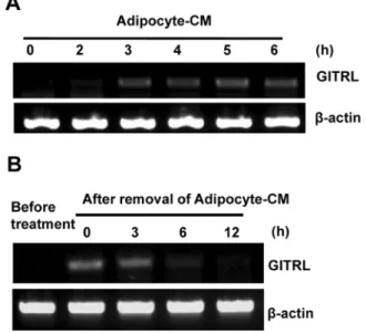
GITRL 발현이 지방세포 배양액에 의하여 증가. 지방세포배 양액에의해 MDA-MB-231 세포에서 GITRL의발현이증가됨 을 다시 한 번 확인하기위해, MDA-MB-231 세포를 6 well
plate에 배양하고다음날 준비된지방세포배양액을처리한후
에시간에따라전체 RNA를분리하였다. 그후 RT-PCR을수
행하여 GITRL의 mRNA level을 측정하였다. 그 결과 GITRL mRNA level은지방세포 배양액처리후 3시간부터 충분히증
가되었으며 6시간까지그발현이 유지되었다(Fig. 2A). 다음으
로지방세포배양액에의해증가된 GITRL 발현이지방세포배
양액 제거 후 얼마나 오래 유지되는지를 알아보았다. MDA- MB-231 세포에 6시간동안지방세포배양액을처리한후 10%
FBS를포함한 DMEM으로바꾸어주었다. 지방세포배양액제 거 후 12시간까지 GITRL 발현변화추이를 살펴보았는데 지 방세포의 제거후에 MDA-MB-231세포의 GITRL 발현이 6시 간째부터 사라짐을 알았다. 유방세포에서 발현되는 GITRL이 기능을하기위해서는지방세포의배양액이지속적으로필요함 을 나타낸다(Fig. 2B).
지방세포에 존재하는 단백질이 GITRL의 발현을 증가시킴.
지방세포는 유리지방산뿐만아니라렙틴, 레지스틴, 아디포넥틴 을포함하는아디포카인 등을분비한다고 알려져있다. 어떤인
자가 MDA-MB-231 세포에서 GITRL mRNA 발현을 증가시키 는지 알아보기위해, 우리는지방세포배양액에 proteinase K를 처리하여 단백질인자들을 제거하였다. 그 후단백질이제거된
지방세포배양액을이전실험과동일하게 MDA-MB-231 세포
Fig. 1. MDA-MB-231 cells were incubated with preadipocyte-CM or adipocyte-CM for 6 h. Total RNA was isolated and subjected to
RT-PCR analysis. Fig. 2. (A) MDA-MB-231 cells were treated with adipocyte-CM. At
the indicated time points, cells were harvested and total RNA was isolated. Then RT-PCR analysis was performed using GITRL primer.
(B) MDA-MB-231 cells were treated with adipocyte-CM for 6 h and adipocyte-CM was removed. The total RNA was isolated at the indicated time points after removal of the adipocyte-CM, and then subjected to RT-PCR analysis.
Fig. 3. MDA-MB-231 cells were treated with adipocyte-CM or proteinase K-treated adipocyte CM, for 6 h. Total RNA was isolated and subjected to RT-PCR analysis.
에 6시간동안 처리하여 GITRL 발현을 살펴보았다. 그 결과
단백질이제거된 지방세포 배양액은 GITRL의 발현을 올리지
못하였다. 즉, 지방세포가분화되면서증가되는단백질인자중
하나가 유방암 세포주인 MDA-MB-231 세포에서 GITRL
mRNA 발현을증가시킴을말한다(Fig. 3).
GITR-GITRL의 상호작용이 MDA-MB-231 세포의 전이에 미치는 영향. GITR-GITRL 상호작용의영향을알아보기위해, MDA-MB-231 세포에 GITR과 GITRL을 각각과발현시켰다. MDA-MB-231 세포가 GITR, GITRL을각각발현하는지알아 보기위해 transfection 후, RT-PCR로 각각의 발현을확인하였 다(Fig. 4A). 그 후 세포의 invasion 능력을 알아보기 위해
GITR을 발현하는 MDA-MB-231 세포와 GITRL을 발현하는
MDA-MB-231 세포를 1:1의비율로섞어 48시간 동안전이정 도를 관찰하였다. GITR을 발현하는 MDA-MB-231 세포와
GITRL을발현하는 MDA-MB-231 세포그리고각각을발현하 는 세포주들에서 전이능력의 차이를 관찰 할 수 없었다(Fig.
4B).
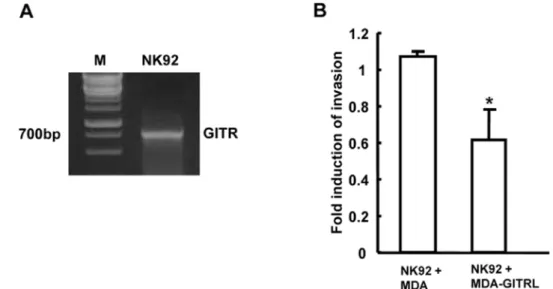
NK세포와 MDA-MB-231세포의 GITR-GITRL interaction.
이번에는 GITRL을 발현하고있는 유방암 세포가 GITR을 발 현하고있는면역세포에의해어떤영향을받는지살펴보았다.
먼저 자연살해 세포주인 NK92 세포를 사용하여 우선적으로
NK92 세포에서 GITR이발현되는지확인하였다. GITR primer
를 이용하여 RT-PCR을 수행한결과 700bp 정도크기의밴드 가증폭되는것으로보아 NK92 세포주가 GITR을발현하는것
Fig. 4. (A) MDA-MB-231 cells were transfected with human GITRL and GITR expression plasmids, respectively. Expression was examined using RT-PCR. (B) GITRL or GITR expressing MDA-MB-231 cells were mixed and then incubated in upper chamber of transwell for 48 h. After incubationfor 48h, the cells that invaded to lower chamber were disassociated with 500 L of disassociation buffer containing Calcein-AM reagents.
Invaded cells were detected using Wallac 1420 Victor3 plate reader. The experiment was performed at the triplicates. Data represent means±SEM.
Fig. 5. (A) GITR expression was determined in NK92 cells. (B) GITRL expressing MDA-MB-231 cells and GITR expressing NK92 cells were mixed and then incubated in upper chamber of transwell for 48 h. After incubationfor 48 h, the cells that invaded to lower chamber were disassociated with 500 L of disassociation buffer containing Calcein-AM reagents. Invaded cells were detected using Wallac 1420 Victor3 plate reader. The experiment was performed at the triplicates. Data represent means±SEM. *p<0.05 versus MDA-MB-231 cells+NK92 cells.
지방세포배양액이 유방암세포주에서 GITRL의 발현을 유도 107 을 알 수 있었다(Fig. 5A). GITRL이 과발현 된 MDA-MB-
231 세포와 GITR을 발현하는 NK92 세포를 1:1의 비율로섞
어 24 시간동안 배양하면서전이능력을조사하였다. GITRL을 과발현한 MDA-MB-231 세포와 GITR을발현하는 NK92 세포
를함께 넣은 chamber에서 MDA-MB-231 세포의 invasion 능 력이감소됨을확인하였다(Fig 5B). 결론적으로유방암 세포인 MDA-MB-231 세포는 주변의 microenvironment 변화에 의해
GITRL 발현이증가될수있으며, 이때 GITR을발현하는 NK
세포에의해그 invasion 능력이감소하게될수있다.
고 찰
암의성장과전이는그 주변환경에 의해많은영향을받는 다. 암세포를둘러싸고있는기질세포와암세포는서로여러물 질들을분비하며직간접적으로상호작용한다. 어떤인자들은암
의침윤과전이등을조절하고, 또다른인자들은암세포의생 존에도영향을미친다. 지방세포는유방을둘러싸는 가장흔한
세포로써이들간의상호작용은유방암의생성과, 증식, 전이에 중요한 영향을 미친다. 이를 뒷받침하는 증거로 지방세포인 3T3-L1 세포 배양액을 유방암 세포주인 MCF-7 세포에 처리
하였을때 MCF-7 세포의 생존과 전이가 촉진됨이 보고되었다
[Iyengar 등, 2003]. 본연구에서는지방세포의배양액을 MDA-
MB-231 유방암세포주에처리하여변화하는여러유전자들중
에 GITRL을 연구하였다. 미분화 지방세포 배양액과 더불어
NIH3T3 섬유아세포주의 배양액을 처리하면 GITRL의 발현이 증가하지못하지만지방세포배양액은 GITRL의유전자 발현을
증가시키었다.
지방세포의분비 물질이 암세포에긍정적인 영향을주는지,
부정적인영향을주는지는인체의상황에따라다르게 나타난 다. 미분화된 지방세포 배양액은 MCF-7, MDA-MB-231, MDA-MB-435 세포의증식을유도하는 반면에, 분화된지방세 포배양액은오히려유방암 세포주의증식을 억제한다는보고 도있다[Chamras 등, 1998]. 지방세포분비물질인아디포카인 의하나인 아디포넥틴은유방암 세포의증식을억제시키는역 할을하는반면[Kang 등, 2005] 렙틴은유방암세포의 증식을
촉진시키는 역할을 한다[Mauro 등, 2007]. 이들아디포카인의 발현은비만의정도에따라변화하기때문에비만으로인한아 디포카인의발현차이가 유방암세포의 증식과전이를 조절하는 조건에대한 연구가매우중요하다. 또한지금까지밝혀진아
디포카인들외에 새로운유방암세포조절 아디포카인의 발굴과 이들아디포카인에의하여유방암세포가 어떻게영향을받는지 연구하는것 또한중요한하나의과제이다.
TNF family의 member들은면역체계와 암세포 사이의 상호 작용에서중요한역할을한다[Locksley 등, 2001]. 본 연구에서 는지방세포배양액에의하여유방암에서발현이증가하는 TNF family, GITRL과 수용체인 GITR의 밝혀지지 않은역할에 대 해초점을맞추어연구하였다. 지방세포배양액에의한 GITRL
의발현은지방세포배양액에존재하는단백질인자임을알수 있었고(Fig. 3) GITRL의발현이유방암세포주의전이에는영향 을미치는않음을알 수있었다(Fig. 4B). 이전보고에따르면,
GITRL을 발현하는 plasmacytoid predendritic 세포와 GITR을 발현하는자연살해세포의 coculture는자연살해세포의 cytotoxicity
를 증가시킨다고한다[Hanabuchi 등, 2006]. 하지만 반대로암 세포에 GITRL이과발현되면자연살해세포의 IFN-γ의분비를
억제시켜서 암세포가면역회피를 하게돕는다고한다[Baltz 등,
2007]. 본 연구에서는 지방세포가 분비하는특정단백질 인자
가유방암세포주인 MDA-MB-231 세포에서 GITRL의발현을 증가시켜, 자연살해세포의 감시로부터벗어나게 할 수 있다는
사실과다른한편으로는 GITRL이과발현된 MDA-MB-231 세 포는 자연살해세포주 NK92 세포의 GITR과 상호작용을 통해 전이가 감소될 수있음을 알았다. 이런상반된결과는 앞으로 더 많은실험을 수행하여야해석이가능할것이다.
암세포의경우언제나그주변의 미세환경의변화에민감하 게 반응한다. 그 중 유방암은주위를둘러싸는가장 대표적인 기질세포인 지방세포와의 상호작용이매우 중요할 수밖에 없 다. 정상인경우와비만의경우지방세포가분비하는물질의종 류나 그것들의 비율이 달라지며 이것은 직간접적으로 유방암
세포의 변화에 영향을 미치게 되는 것이다. GITRL 이외에도
많은유전자들이 지방세포배양액에 의하여증감되는데 이들의 연구 또한유방암과 비만사이의 관계를 규명하는 기초가 될 것이다.
초 록
비만은유방암을일으키는잠재적위험요소중하나이며현 재 이들의 상관관계를밝히려는 많은연구들이 진행중에 있 다. 지방세포는 유방조직을이루는 기질세포중 가장높은비 중을 차지하며, 이들이 분비하는 물질인 아디포카인이 유방암
의 성장과 전이에영향을 줄 것으로예상되어지고 있다. 지방 조직이 생산하는아디포카인들 중렙틴은 유방암세포의증식 능력을 증가시키고 아디포넥틴의경우반대로 증식억제기능 을 보인다는연구결과가 있다. 또한정상의 경우와 비교해서 비만환자에게서렙틴의 양은증가되어져있으며그와반대로 아디포넥틴은 비만환자에게서감소 경향을 보인다. 이는비만 에 의하여변화되는아디포카인들이서로작용하여비만한유 방암환자의유방암세포증식을촉진할수 있음을뜻한다. 본연 구에서는지방세포의분비 물질인아디포카인들에의해조절되 는 유방암세포의 유전자들을 조사하기 위해 유방암세포주인
MDA-MB-231 세포주에 지방세포배양액을 처리하였으며, 이
때 증가되는 유전자 중 glucocorticoid-induced TNF receptor- related protein ligand(GITRL)를선택 연구하였다. GITRL은지
방세포 배양액을처리한 MDA-MB-231 세포주에서 높게발현
되었으며, proteinase-K를 처리한 지방세포 배양액에 의해서는 발현정도에변화가없었다. 이는지방세포가분비하는단백질
인자 중하나가 유방암 세포의 GITRL 발현을증가시킴을 말
한다. 또한 유방암 세포주 MDA-MB-231 세포에서 증가된
GITRL은 그 자체의 전이 능력에는 영향을 주지 못하였으나,
glucocorticoid-induced TNF receptor-related protein(GITR)을
발현하는 자연살해세포주인 NK92 세포와의상호작용 때에는
전이능력을감소시켰다.
Key words: adipocytes, breast cancer, immune system 감사의 글
이 연구는 2008년 숙명여자대학교 교비에 의하여 지원받았다.
참고문헌
Baillargeon J, Platz EA, Rose DP, Pollock BH, Ankerst DP, Haffner S, Higgins B, Lokshin A, Troyer D, Hernandez J, Lynch S, Leach RJ, and Thompson IM (2006) Obesity, adipokines, and prostate cancer in a prospective population- based study. Cancer Epidemiol Biomarkers Prev 15, 1331- 1335.
Baltz KM, Krusch M, Bringmann A, Brossart P, Mayer F, Kloss M, Baessler T, Kumbier I, Peterfi A, Kupka S, Kroeber S, Menzel D, Radsak MP, Rammensee HG, and Salih HR (2007) Cancer immunoediting by GITR (glucocorticoid-induced TNF- related protein) ligand in humans: NK cell/tumor cell interactions. FASEB J21, 2442-2454.
Bianchini F, Kaaks R, and Vainio H (2002) Overweight, obesity, and cancer risk. Lancet Oncol3, 565-574.
Chamras H, Bagga D, Elstner E, Setoodeh K, Koeffler HP, and Heber D (1998) Preadipocytes stimulate breast cancer cell growth. Nutr Cancer32, 59-63.
Dos Santos E, Benaitreau D, Dieudonne MN, Leneveu MC, Serazin V, Giudicelli Y, and Pecquery R (2008) Adiponectin mediates an antiproliferative response in human MDA-MB 231 breast cancer cells. Oncol Rep20, 971-977.
Elliott BE, Tam SP, Dexter D, and Chen ZQ (1992) Capacity of adipose tissue to promote growth and metastasis of a murine mammary carcinoma: effect of estrogen and progesterone. Int J Cancer 51, 416-424.
Fantuzzi G (2005) Adipose tissue, adipokines, and inflammation. J Allergy Clin Immunol 115, 911-919 (quiz 920).
Goodfriend TL, Egan BM, and Kelley DE (1998) Aldosterone in obesity. Endocr Res24, 789-796.
Grundy SM (2000) Metabolic complications of obesity. Endocrine
13, 155-165.
Hanabuchi S, Watanabe N, Wang YH, Wang YH, Ito T, Shaw J, Cao W, Qin FX, and Liu YJ (2006) Human plasmacytoid predendritic cells activate NK cells through glucocorticoid- induced tumor necrosis factor receptor-ligand (GITRL). Blood
107, 3617-3623.
Iyengar P, Combs TP, Shah SJ, Gouon-Evans V, Pollard JW, Albanese C, Flanagan L, Tenniswood MP, Guha C, Lisanti MP, Pestell RG, and Scherer PE (2003) Adipocyte-secreted factors synergistically promote mammary tumorigenesis through induction of anti-apoptotic transcriptional programs and proto- oncogene stabilization. Oncogene 22, 6408-6423.
Ji HB, Liao G, Faubion WA, Abadia-Molina AC, Cozzo C, Laroux FS, Caton A, and Terhorst C (2004) Cutting edge: the natural ligand for glucocorticoid-induced TNF receptor-related protein abrogates regulatory T cell suppression. J Immunol172, 5823- 5827.
Johnston PG, Rondinone CM, Voeller D, and Allegra CJ (1992)
Identification of a protein factor secreted by 3T3-L1 preadipocytes inhibitory for the human MCF-7 breast cancer cell line. Cancer Res52, 6860-6865.
Josefson D (2001) Obesity and inactivity fuel global cancer epidemic. Br Med J 322, 945.
Kang JH, Lee YY, Yu BY, Yang BS, Cho KH, Yoon DK, and Roh YK (2005) Adiponectin induces growth arrest and apoptosis of MDA-MB-231 breast cancer cell. Arch Pharm Res 28, 1263- 1269.
Kim JD, Choi BK, Bae JS, Lee UH, Han IS, Lee HW, Youn BS, Vinay DS, and Kwon BS (2003) Cloning and characterization of GITR ligand. Genes Immun4, 564-569.
Kim JH, Kim KY, Jeon JH, Lee SH, Hwang JE, Lee JH, Kim KK, Lim JS, Kim KI, Moon EY, Lee HG, Ryu JH, and Yang Y (2008) Adipocyte culture medium stimulates production of macrophage inhibitory cytokine 1 in MDA-MB-231 cells.
Cancer Lett261, 253-262.
Locksley RM, Killeen N, and Lenardo MJ (2001) The TNF and TNF receptor superfamilies: integrating mammalian biology.
Cell104, 487-501.
Mauro L, Catalano S, Bossi G, Pellegrino M, Barone I, Morales S, Giordano C, Bartella V, Casaburi I, and Ando S (2007) Evidences that leptin up-regulates E-cadherin expression in breast cancer: effects on tumor growth and progression. Cancer Res67, 3412-3421.
McCann J (2001) Obesity, cancer links prompt new recommendations.
J Natl Cancer Inst93, 901-902.
McHugh RS, Whitters MJ, Piccirillo CA, Young DA, Shevach EM, Collins M, and Byrne MC (2002) CD4(+)CD25(+) immunoregulatory T cells: gene expression analysis reveals a functional role for the glucocorticoid-induced TNF receptor.
Immunity16, 311-323.
Nocentini G, Giunchi L, Ronchetti S, Krausz LT, Bartoli A, Moraca R, Migliorati G, and Riccardi C (1997) A new member of the tumor necrosis factor/nerve growth factor receptor family inhibits T cell receptor-induced apoptosis. Proc Natl Acad Sci
94, 6216-6221.
Schaffler A, Scholmerich J, and Buechler C (2007) Mechanisms of disease: adipokines and breast cancer-endocrine and paracrine mechanisms that connect adiposity and breast cancer. Nat Clin Pract Endocrinol Metab3, 345-354.
Shevach EM and Stephens GL (2006) The GITR-GITRL interaction: co-stimulation or contrasuppression of regulatory activity? Nat Rev Immunol6, 613-618.
Shimizu J, Yamazaki S, Takahashi T, Ishida Y, and Sakaguchi S (2002) Stimulation of CD25(+)CD4(+) regulatory T cells through GITR breaks immunological self-tolerance. Nat Immunol3, 135-142.
Spyridopoulos TN, Petridou ET, Skalkidou A, Dessypris N, Chrousos GP, Mantzoros CS, and the Obesity and Cancer Oncology Group (2007) Low adiponectin levels are associated with renal cell carcinoma: a case-control study. Int J Cancer
120, 1573-1578.
Steppan CM, Bailey ST, Bhat S, Brown EJ, Banerjee RR, Wright CM, Patel HR, Ahima RS, and Lazar MA (2001) The hormone resistin links obesity to diabetes. Nature 409, 307- 312.