• 검색 결과가 없습니다.

A Promising Treatment for Broncholith Removal Using Cryotherapy during Flexible Bronchosopy: Two Case Reports

N/A
N/A
Protected

Academic year: 2022

Share "A Promising Treatment for Broncholith Removal Using Cryotherapy during Flexible Bronchosopy: Two Case Reports"

Copied!
6
0
0

로드 중.... (전체 텍스트 보기)

전체 글

(1)

Tuberc Respir Dis 2012;73:282-287

CopyrightⒸ2012. The Korean Academy of Tuberculosis and Respiratory Diseases. All rights reserved.

A Promising Treatment for Broncholith Removal Using Cryotherapy during Flexible Bronchosopy: Two Case Reports

Jong Hwan Lee, M.D., Joong Hyun Ahn, M.D., Ah Young Shin, M.D., Sung Jin Kim, M.D., Sung Jun Kim, M.D., Gu-Min Cho, M.D., Hyun Jin Oh, M.D., In Ho Kim, M.D., Ju Sang Kim, M.D.

Division of Pulmonology, Critical Care Medicine, Department of Internal Medicine, Incheon St. Mary's Hospital, The Catholic University of Korea College of Medicine, Incheon, Korea

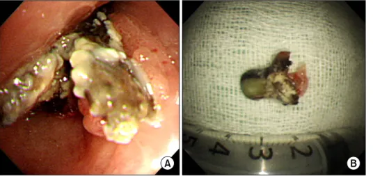
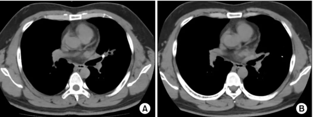
Broncholiths are defined as calcified materials that occur in a tracheobronchial tree or in a cavity communicating with that. Broncholith has variable clinical features. The therapeutic options to remove broncholiths are so variable that clinicians need to select the most safe and effective methods by mass size, mobility, and location. As yet, there is no consistent guideline removing a broncholith. We report 2 successful cases of removing a fixed broncholith by flexible bronchoscopy guided cryoadhesion. With repeated technique of thawing and freezing with cryoprobe, we could extract the fixed broncholith safely. This method is promising as a way to remove broncholith in the future.

Key Words: Bronchial Diseases; Calculi; Cryotherapy; Bronchoscopy

Address for correspondence: Ju Sang Kim, M.D.

Department of Internal Medicine, Incheon St. Mary's Hospital, The Catholic University of Korea College of Medicine, 665, Bupyeong 6-dong, Bupyeong-gu, Incheon 403-720, Korea

Phone: 82-32-280-5866, Fax: 82-280-5190 E-mail: [email protected] Received: Aug. 16, 2012

Revised: Sep. 18, 2012 Accepted: Oct. 25, 2012

CCIt is identical to the Creative Commons Attribution Non-Commercial License (http://creativecommons.org/licenses/by-nc/3.0/).

Introduction

Broncholith is calcified material within tracheobron- chial tree. The therapeutic options are so variable that clinicians should choose the proper way to remove it considering by mass size, mobility, location, patient's symptoms. As yet, for removing broncholith, flexible bronchoscopy has been applied only in limited cases.

We report here on 2 cases of successful removal of broncholith using cryotherapy-assisted flexible broncho- scopy.

Case Report 1. Case 1

A 32-year-old female was admitted on the chief com- plaint of blood-tinged sputum and fever for 3 weeks.

The patient had an episode of hemoptysis 3 years ago at which she was treated with for pulmonary tuber- culosis.

The patient complained of fever, myalgia and sweat- ing for 3 weeks and physical examination revealed de- creased lung sounds and crackles on right upper lung filed.

Chest radiographs showed multifocal ill defined pat- chy, nodular densities in the right upper lobe; therefore, a chest computed tomography (CT) scan was perform- ed. Chest CT suggested a mass-like calcified endobron- chial lesion and post-obstructive pneumopathy (Figure 1A).

Bronchoscopy was performed to examine the endo-

bronchial lesion. On bronchoscopy, a fixed broncholith

with sharp and speculated margins was seen (Figure

2A). At the opening of right upper lobe bronchus, it

was difficult to approach the orifice due to nearly total

(2)

Figure 1. A 32-year-old woman's computed tomography (CT) scan image. (A) Chest CT scan image (mediastinal setting) showing calcifications present within the right upper lobe bronchus and resulting pneumonia. (B) Follow-up CT scan image (mediastinal setting) after bronchoscopic broncholith removal showing a patent airway and recovered pneumonic infiltrations.

Figure 2. A 32-year-old woman's bronchoscopy image and extracted bron- choliths. (A) The broncho- scopic view showing the sharp marginated bron- cholith which attached at the opening of the right upper lobe bronchus. (B) The extracted broncholith which was about 1.8 cm sized.

obstruction with the impacted mass. Immediate cry- otherapy was performed to extract the broncholith. Just 5 seconds freezing without thawing, we could fInd the cryoprobe stucked to the broncholith. We forced the probe which was attached to the mass backward. There was a small amount of bleeding. A bleeding control with argon plasma coagulation (APC) was successfully done without major complications. The extracted bron- cholith was 1.8 cm sized and attached bronchial mucosa with irregular margin (Figure 2B).

Tuberculosis study including both acid fast bacilli stain and tuberculosis polymerase chain reaction were negative and no pathogen was identified on cultures.

Obstructive pneumonia improved (Figure 1B) and the patient was followed on out-patient department on rou- tine basis without anti-tuberculosis medication.

2. Case 2

A 32-year-old man visited for management of he- moptysis for 5-days. He has a history of 10 pack-year smoking and incomplete treatment of pulmonary tuber- culosis 17 years ago. This was the second episode of massive hemoptysis.

At his first admission, the hemoptysis resolved with conservative management and thus he was discharged without any invasive management for removing the broncholith.

Chest CT on this admission showed ill defined patchy consolidation and ground glass opacities surrounding lingular segment and a calcified density within Lt. upper lobar bronchus suggesting broncholith with pulmonary hemorrhage (Figure 3A).

Bronchoscopy showed 1.5 cm sized yellowish mass

(3)

Figure 3. A 32-year-old man's computed tomography (CT) scan image. (A) Chest CT scan image (mediastinal setting) showing patchy opacities surrounding lingular segment and a calcified density within left upper lobe bronchus suggesting broncholith with pulmonary hemorrhage. (B) Follow-up CT scan image (mediastinal setting) after bronchoscopic broncho- lith removal showing no endobronchial mass.

Figure 4. A 32-year-old man's bronchoscopy im- age and extracted bron- choliths. (A) The broncho- scopic view showing the impacted broncholith in the left upper lobe lingular divi- sion orifice. (B) The extra- cted broncholith was about 1.6 cm sized tooth-like yel- lowish mass.

(probably broncholith) at left upper lobe lingular divi- sion (Figure 4A).

Follow-up bronchoscopy and cryoadhesion were performed. We performed the freezing procedure about 5 seconds. The cryoprobe stucked to the mass, but we could not force the probe backward because the bron- cholith was tightly fastened to the bronchial wall. For thawing procedure, we waited for another 10 seconds, which made the linkage between the probe and the broncholith weakened. By repeating freezing-thawing techinique 5 times, the stucked mass became movable due to the damaged bronchial wall. So we could extract the total broncholith completely (Figure 4B) and addi- tional APC was done for bleeding control. After 1 week, follow-up chest CT shows patent airway without bron- cholith and hemorrhage resorption (Figure 3B).

Discussion

Broncholiths are endogenous calcified material within

tracheobronchial tree. Its pathogenesis can be explained

in three mechanisms. First, inhaled materials in the

bronchus mixed up with calcium develop into bron-

cholith. Second, bronchial cartilage is either calcified or

ossified which then breaks apart in the endobronchial

spaces. Third, the most common cause, lung paren-

chymal infection progresses and brings about calcium

deposits in lymph nodes under alkalic environment

1

.

Major diseases that cause such symptoms as men-

tioned above include tuberculosis in Korea and Europe,

while in US it is histoplasma. Occasionally they are also

caused by fungal infection, lung abscess, pulmonary in-

farction, silicosis or inhalation of foreign materials

2

.

(4)

Depending on the location of the broncholith and the extent of bronchial erosion, clinical manifestations range from asymptomatic, mild coughing and sputum to he- moptysis, chest pain, fever, secondary pneumopathy, bronchiectasis and atelectasis. Whereas conservative care is adequate in patient with cough, hemoptysis and obstructive pneumopathy, surgical intervention such as lobectomy, segmentectomy or even pneumonectomy are rarely needed in some cases of refractory hemopt- ysis or obstructive pneumopathy suggestive of lung can- cer

1

.

In clinical settings, broncholiths are often detected during evaluating symptoms of complications such as hemoptysis or recurrent pulmonary infections. If com- plications develop, both symptomatic care and the re- moval of broncholiths are often needed.

Even if a proper symptomatic care was done, re- current symptoms would be often develop without the removal of broncholiths. But, some cases of these are studded tightly to bronchial mucosa granulation tissue and obstructed completely, it is not easy to remove the entire material by bronchoscopy without major com- plications. The therapeutic options to remove broncho- liths are so variable, and it should be determined by mass size, mobility, location and patient's symptoms.

In general, a symptomatic, large and fixed broncho- lith were likely to be removed by a rigid bronchoscopy or if unsuccessful, thoracotomy. Success rates using flexible bronchoscopy for the removal of broncholiths reported to be approximately 30%

3,4

.

Rigid bronchoscopy is also employed for the removal of broncholiths with success rates ranging from 67 to 87%

5

. Olson et al.

4

reported that 100% of their patients with loose (free in the airway) broncholiths were under- went flexible and rigid bronchoscopic extraction to at- tempts without severe complications. But, these results are only explained in easy cases resulting from loose broncholiths.

In many cases, a rigid bronchoscopy is preferred than a flexible bronchoscopy. Because, once the rigid bron- choscope was performed, jet ventilation is established that enabling the inspector more room to work through

the end of the scope concomitantly while still oxygen- ating the patient

6

. But, rigid bronchoscopy needs gen- eral endotracheal anesthesia which sometimes induce respiratory depression or hemodynamic instability.

Therefore, many pulmonologist have been trying var- ious ways to remove broncholiths by a flexible bron- choscopy. However as yet, there is no consistent guide- lines for broncholithectomy or broncholithotomy using flexible bronchoscopy. As pulling with the forceps, or using a balloon catheters

3

, and YAG laser incision to broncholithotomy

7

, a variety of methods have been performed. Moreover, surgical treatment should be con- sidered over bronchoscopic removal when patients present with hemoptysis, the broncholith is firmly at- tached to the bronchial wall, or bronchoscopic excision is not available or feasible

6

. Preferred operative proce- dures include segmentectomy, lobectomy, and pneumo- nectomy.

In both cases mentioned above, we used a flexible bronchoscopy guided cryoadhesion to remove the bron- choliths which were firmly attached to the bronchial wall.

In the first case, the patient presented with ob- structive pneumopathy caused by a fixed broncholith.

Bronchoscopy was performed to identify the broncho- lith and the tip of cryoprobe was attached to the mass to freeze and it was removed with pull-out method. The procedure was successfully performed despite minor bleeding that was controlled by APC.

In the second case, the patient complained of mas- sive recurrent hemoptysis that was revealed to be caused by a fixed broncholith. The same procedure was performed untill cryoprobe attached to the mass to freeze but then pulling out was risky as it may concur massive bleeding. This time, pulling out was retained until repeated freezing and thawing eased separation of broncholith and bronchial mucosa without any exertion.

Bleeding was minimal.

Bronchoscopic cryotherapy is most often employed

as a palliative therapy for airway obstruction due to a

malignancy, usually in patients who cannot tolerate

lung resection due to poor respiratory function or who

(5)

are deemed inoperable due to the proximity of the tu- mor to the carina. Cryoadhesion can be used to extract foreign bodies, mucous plugs, or blood clots. During cryoadhesion, the cryoprobe freezes the target material, causing the material to adhere to the probe

8-10

. In 2007, there was a first attempt to remove the broncholith by using a cryoprobe with flexible bronchoscopy

11

. A conventional method to remove broncholiths is to pull them out with a forcep. This method is applicable when the broncholith is not firmly attached to the bron- chial wall. When dealing with the fixed masses how- ever, which is the subject of this study, it is unlikely to remove them with forcep as it may tear the tissue surrounding which may result in major bleeding while pulling them out.

Another method applicable to remove broncholiths is to grind them with YAG laser

7

. Having to be performed in a very narrow space, it could also inflict damages on the surrounding tissue and entail subsequent bleed- ing. In particular, if the mass is adjacent to the vessel, it can trigger massive life-threatening hemoptysis secon- dary to fistula or rupture of aorta or pulmonary arteries and systemic air embolism

12

.

Meanwhile, cryoadhesion does not inflict any direct damages on the bronchial wall. While it freezes bron- cholith, the cold stimuli is gradually transmitted from the objective to the surrounding tissue. Tissue affected by cold stimuli will lose its density making it easier for the mass to fall apart

13,14

. All the procedures can take place without making the patient unconcious. Cryoad- hesion obviously provides us with safer method to re- move broncholith and foreign body in a limited space

14

. There are two ways to perform the cryoadhesion de- pending on the conditions of the patients: pull-out method and repeated thawing-freezing method

14,15

. Pull-out method means to forcep it out after rapid freezing of the broncholith. It has the merit of removing the broncholith with a single attempt but in such a case where surrounding tissue is not fully loosened, it could inflict damages on the tissue causing subsequent com- plications

14

. It is therefore applicable only to not firmly attached, movable endobronchial lesion or foreign

body.

Repeated thawing-freezing method is suggested when the broncholith is deemed not easily removable by the pull-out method. The repeated thawing and freezing of the objective will reduce the resistance of the surround- ing tissue ensuring safer removal of the fixed mass.

Repeated thawing-freezing method obviously is more time consuming than pull-out method. Pull-out method as part of the cryoadhesion has greater risk of bleeding than repeated thawing-freezing method in which only a small amount of bleeding is noticed when removing the broncholith

14

. Repeated thawing-freezing method is recommendable to the patients with high risk of com- plications.

Our cases report successful removal of broncholith with cryoadhesion on patient of semi-sedative state. The procedure introduces safer method of repeated freezing and thawing that loosens the connection between the soft tissue and the broncholith unlike the classical way of attaching and pulling out. In this way, bleeding risk is notably reduced and manipulation much easier in small spaces and thus flexible bronchoscopy guided cry- oadhesion can be considered as a primary therapy for removal of broncholith whether movable or fixed even though the patien's main symptom is hemoptysis.

Thus, introduction of bronchoscopic cryoadhesion method can spare unnecessary general anesthesia or lung surgery.

References

1. Kwak SM, Ahn CM, Kim HJ, Lee JS, Oh SH. A case of middle lobe syndrome due to broncholithiasis.

Korean J Med 1988;34:834-9.

2. Lee HL, Kim SK, Chang J, Kim SK, Lee WY, Chung KY. A study on broncholithiasis. Korean J Med 1995;

48:353-60.

3. Yi KY, Lee HK, Park SJ, Lee YC, Rhee YK, Lee HB.

Two cases of broncholith removal under the guidance of flexible bronchoscopy. Korean J Intern Med 2005;

20:90-1.

4. Olson EJ, Utz JP, Prakash UB. Therapeutic broncho-

scopy in broncholithiasis. Am J Respir Crit Care Med

1999;160:766-70.

(6)

5. Ferguson JS, Rippentrop JM, Fallon B, Ross AF, McLennan G. Management of obstructing pulmonary broncholithiasis with three-dimensional imaging and holmium laser lithotripsy. Chest 2006;130:909-12.

6. Cerfolio RJ, Bryant AS, Maniscalco L. Rigid broncho- scopy and surgical resection for broncholithiasis and calcified mediastinal lymph nodes. J Thorac Cardiovasc Surg 2008;136:186-90.

7. Miks VM, Kvale PA, Riddle JM, Lewis JW Jr. Bronchol- ith removal using the YAG laser. Chest 1986;90:295-7.

8. Bolliger CT, Mathur PN, Beamis JF, Becker HD, Cavaliere S, Colt H, et al. ERS/ATS statement on inter- ventional pulmonology. European Respiratory Society/

American Thoracic Society. Eur Respir J 2002;19:356- 73.

9. Ernst A, Feller-Kopman D, Becker HD, Mehta AC.

Central airway obstruction. Am J Respir Crit Care Med 2004;169:1278-97.

10. Asimakopoulos G, Beeson J, Evans J, Maiwand MO.

Cryosurgery for malignant endobronchial tumors: anal- ysis of outcome. Chest 2005;127:2007-14.

11. Reddy AJ, Govert JA, Sporn TA, Wahidi MM. Bronchol- ith removal using cryotherapy during flexible broncho- scopy: a case report. Chest 2007;132:1661-3.

12. Tellides G, Ugurlu BS, Kim RW, Hammond GL. Path- ogenesis of systemic air embolism during broncho- scopic Nd:YAG laser operations. Ann Thorac Surg 1998;65:930-4.

13. Kim HO, Kim SJ, Lee SH, Lee EJ, Hur GY, Lee SY, et al. A case of atypical bronchial carcinoid treated by cryotherapy. Tuberc Respir Dis 2011;70:521-5.

14. Lyu J, Song JW, Hong SB, Oh YM, Shim TS, Lim CM, et al. Bronchoscopic cryotherapy in patients with cen- tral airway obstruction. Tuberc Respir Dis 2010;68:6-9.

15. Jung JY, Lee SY, Kim DH, Lee KJ, Lee EJ, Kang EH,

et al. Clinical benefits and complications of cryothera-

py in advanced lung cancer with central airway ob-

struction. Tuberc Respir Dis 2008;64:272-7.

수치

Figure  2.  A  32-year-old  woman's  bronchoscopy  image and extracted  bron-choliths
Figure  3.  A  32-year-old  man's  computed  tomography  (CT)  scan  image.  (A)  Chest  CT  scan  image  (mediastinal  setting)  showing patchy opacities surrounding lingular segment and a calcified density within left upper lobe bronchus suggesting bronc

참조

관련 문서

 However, since the schools for catechumens used the same catechetical method, the term catechetical schools is often used in a more generic sense to include both

The index is calculated with the latest 5-year auction data of 400 selected Classic, Modern, and Contemporary Chinese painting artists from major auction houses..

The “Asset Allocation” portfolio assumes the following weights: 25% in the S&P 500, 10% in the Russell 2000, 15% in the MSCI EAFE, 5% in the MSCI EME, 25% in the

(Attached to a noun) This is used to indicate the topics, to compare two information or to emphasize the preceding noun in a sentence.. (Attached to a noun) This is used

(Attached to a verb) This is used to show that the succeeding action takes place right after the preceding action.. (Attached to a verb) This is used to indicate a

(Attached to a verb or an adjective) This is used to indicate a succeeding action or situation. This is used

(Attached to a verb or an adjective) This is used to reflect on a past situation. (Attached to an adjective) This is used to show another

(Attached to a noun) This is used to indicate an object has the characteristics and qualities of both a preceding and succeeding