INTRODUCTION
Osteosarcoma is the most widespread malignant bone tumor and a major cause of cancer death among adolescents and young adults, with a incidence of approximately 10 to 26 new cases per million worldwide annually.1,2 Due to high aggres- siveness and early systemic metastasis, the 5-year overall sur- vival rate of those diagnosed with metastatic osteosarcoma is
only about 20−30%.1,3 Although the combination of surgery re- section and neoadjuvant chemotherapy has become a stan- dard treatment strategy for osteosarcoma patients for more than 30 years, patients with recurrent or metastatic osteosar- coma still have an extremely poor outcome.4 Till now, the mo- lecular mechanisms involved in osteosarcoma origination, metastasis, and chemoresistance has remained largely un- known. Therefore, new prognostic biomarkers and treatment targets are expected to be identified to improve therapeutic efficacy for osteosarcoma patients.
MicroRNAs (miRNAs) are small non-coding RNAs with 18 to 25 nucleotides in length, which lead to translation inhibition or target mRNA degradation by interacting with the 3’-UTR or coding region of mRNA targets.5 Multiple miRNAs, such as miR-199a-3p, miR-125b, and miR-29, have been shown to be implicated in cell proliferation and migration and patient prog- nosis in osteosarcoma.6-8 miR-194 has been found to prevent metastasis and progression of various tumors, such as liver can- Received: April 27, 2017 Revised: July 26, 2017
Accepted: July 26, 2017
Corresponding author: Dr. Heping Wang, Department of Orthopedics, Zhoukou Central Hospital, East Section of Renmin Road, Zhoukou 466000, China.
Tel: 86-0394-8222893, Fax: 86-0394-8269016, E-mail: [email protected]
•The authors have no financial conflicts of interest.
© Copyright: Yonsei University College of Medicine 2017
This is an Open Access article distributed under the terms of the Creative Com- mons Attribution Non-Commercial License (http://creativecommons.org/licenses/
by-nc/4.0) which permits unrestricted non-commercial use, distribution, and repro- duction in any medium, provided the original work is properly cited.
Knockdown of Long Non-Coding RNA NEAT1 Inhibits Proliferation and Invasion and Induces Apoptosis of Osteosarcoma by Inhibiting miR-194 Expression
Heping Wang1, Yanzhang Yu2, Shuxin Fan1, and Leifeng Luo1
Departments of 1Orthopedics and 2Surgery, Zhoukou Central Hospital, Zhoukou, China.
Purpose: Long non-coding RNA (lncRNA) nuclear paraspeckle assembly transcript 1 (NEAT1) has been implicated as an onco- gene in the development and progression of osteosarcoma. This study aims to explore the mechanism of NEAT1 in osteosarcoma.
Materials and Methods: Expressions of NEAT1 and miR-194 in osteosarcoma tissues and cells were detected by quantitative real- time PCR. The effects of NEAT1 knockdown or miR-194 overexpression on cell proliferation, invasion, and apoptosis were deter- mined by 3-[4, 5-dimethylthiazol-2-yl]-2, 5 diphenyl tetrazolium bromide (MTT) assay, transwell invasive assay, and flow cytometry analysis, respectively. Luciferase reporter assay was performed to observe the possible interaction between NEAT1 and miR-194.
Results: NEAT1 was upregulated and miR-194 was downregulated in osteosarcoma tissues and cells. Knockdown of NEAT1 or overexpression of miR-194 suppressed proliferation and invasion and induced apoptosis of osteosarcoma cells in vitro. Luciferase reporter assay validated that NEAT1 could interact with miR-194 and negatively modulated its expression. Furthermore, inhibi- tion of miR-194 reversed the suppression of proliferation and invasion and the promotion of apoptosis induced by NEAT1 deple- tion in osteosarcoma cells.
Conclusion: Knockdown of NEAT1 suppressed proliferation and invasion and induced apoptosis in osteosarcoma cells by inhib- iting miR-194 expression.
Key Words: lncRNA, tumorigenesis, osteosarcoma, miR-194
pISSN: 0513-5796 · eISSN: 1976-2437 Yonsei Med J 2017 Nov;58(6):1092-1100
https://doi.org/10.3349/ymj.2017.58.6.1092
cer,9 endometrial cancer,10 and gastric cancer.11 Moreover, a pre- vious study showed that miR-194 partially inhibited the prolif- eration, migration, and invasion of osteosarcoma cells in vitro, as well as tumor growth and pulmonary metastasis of osteosar- coma cells in vivo.12 Moreover, there were statistically significant relationships between miR-194 expression and clinical stage, distant metastasis, and patient mortality. Downregulation of miR-194 has been shown to be associated with poor prognosis in osteosarcoma patients.12 However, how miR-194 is regulated in osteosarcoma is still unclear.
Long non-coding RNAs (lncRNAs) are a class of RNAs with length surpassing 200 nucleotides with little or no protein-cod- ing ability.13 LncRNAs are often dysregulated and are involved in the occurrence and development of human cancers,14 includ- ing osteosarcoma: for instance, lncRNA urothelial carcinoma associated 1 contributed to the initiation and progression of os- teosarcoma.15 LncRNA tumor suppressor candidate 7 hindered cell proliferation and served as a tumor suppressor in osteosar- coma.16 LncRNA taurine up-regulated gene 1 (TUG1) was over- expressed and improved cell proliferation in osteosarcoma.17 LncRNA modified frailty index 2 knockdown suppressed cell proliferation, migration, and invasion, and induced cell apop- tosis in osteosarcoma.18 Nuclear paraspeckle assembly tran- script 1 (NEAT1) has been identified as a nuclear-restricted ln- cRNA and could act as an oncogene in osteosarcoma progres- sion.19,20 However, the molecule mechanism through which NEAT1 exerts its role in osteosarcoma progression is still unde- fined. In the present study, we aimed to explore the function and molecule mechanism of lncRNA NEAT1 in osteosarcoma.
MATERIALS AND METHODS
Tissue specimens and cell culture
Fifteen osteosarcoma patient tissue samples and fifteen nor- mal tissue samples were obtained from Zhoukou Central Hos- pital. This study was approved by the local Ethic Review Com- mittees.
Human osteosarcoma cell lines (MG63 and U2OS) and the human normal osteoblastic cell line hFOB 1.19 were obtained from the American Type Culture Collection and cultured at 37°C in a humidified atmosphere containing 5% CO2. All cells were maintained in Dulbecco’s modified Eagle’s medium (In- vitrogen, Carlsbad, CA, USA) containing 10% fetal bovine se- rum (Invitrogen) and 1% penicillin/streptomycin (Invitrogen).
Quantitative real-time PCR (qRT-PCR)
Total RNA was extracted from cultured cells using TRIzol (In- vitrogen). The expression levels of NEAT1 and miR-194 were evaluated using the SYBR-Green PCR Master Mix Kit (Takala, Dalian, China) and an ABI7500 Real-Time PCR System (Ap- plied Biosystems, Foster City, CA, USA). The primer sequenc- es for β-actin were 5’-TGA GAG GGA AAT CGT GCG TGA C-3’
(forward primer) and 5’-AAG AAG GAA GGC TGG AAA AGA G-3’ (reverse primer). The primer sequences for NEAT1 were 5’-CTT CCT CCC TTT AAC TTA TCC ATT CAC-3’ (forward primer) and 5’-CTC TTC CTC CAC CAT TAC CAA CAA TAC- 3’ (reverse primer). The primer sequences for U6 were 5’-CTC GCT TCG GCA CA-3’ (forward primer) and 5’-AAC GCT TCA CGA ATT TGC GT-3’ (reverse primer). The primer sequences for miR-194 were 5’-ACA GCA ACT CCA TGT GG-3’ (forward primer) and 5’-GAA CAT GTC TGC GTA TCT C-3’ (reverse primer). β-actin and U6 were used as internal reference for NEAT1 and miR-194, respectively. The relative expression levels of NEAT1 and miR-194 were calculated and normalized using the 2-ΔΔCt method.21
Transfection
The scrambled control miR (miR-con), miR-194 mimics (UGUA ACAGCAACUCCAUGUGGA), scrambled control anti-miR (anti-miR-con), and anti-miR-194 (UCCACAUGGAGUUGCU GUUACA) were synthesized by Genepharma (Shanghai, Chi- na). NEAT1 (GenBank #EF177379.1) was amplified from the cDNA of MG63 and U2OS cells and cloned into the pcDNA3.1 plasmid, which was named pcDNA-NEAT1. The siRNA sequence targeting NEAT1 was 5’-GUGAGAAGUUGCUUAGAAACUU UCC-3’ (si-NEAT1). si-NEAT1 and negative control siRNA (si- con) were obtained from GenePharma. Cells were transfected with plasmid or nucleotide sequences using Lipofectamine 2000 (Invitrogen) according to the manufacturer’s instructions.
MTT assay
Transfected MG63 and U2OS cells (4×10³ cells per well) were seeded in 96-well plates. After cell incubation for 24, 48, 72, and 96 h, 10 µL of MTT (Sigma, St. Louis, MO, USA) was added to each well for another 4 h. Then, the cultural supernatant was discarded, and cells were treated with 150 µL of dimethyl sul- phoxide to resolve formazan crystals. Absorbance values at 490 nm were determined by a microplate reader (Molecular Devices, Sunnyvale, CA, USA).
Cell invasion assay
The invasion ability of MG63 and U2OS cells was determined by Transwell invasion assay (Bection Dickinson, Bedford, MA, USA). Cells were planted on the top side of the membrane pre- coated with 10 μg/mL of Matrigel and incubated for 24 h. Then, the invaded cells in the lower membrane were fixed and stained with 0.5% crystal violet solution, and counted under a micro- scope (Olympus, Tokyo, Japan).
Flow cytometry
Cell apoptosis was evaluated using Annexin V/fluorescein iso- thiocyanate (FITC) and propidium iodide (PI) apoptosis detec- tion kits (Becton Dickinson, Franklin Lakes, NJ, USA). Briefly, MG63 and U2OS cells were resuspended and stained with 5 μL of Annexin V/FITC and 5 μL of PI (Sigma) for 15 min at room
temperature in the dark. Flow cytometry (Becton Dickinson) was used to detect apoptosis of MG63 and U2OS cells.
Luciferase reporter assays
MG63 and U2OS cells (5×10⁴ cells per well) were cultured in a 24-well plate and co-transfected with the luciferase reporter plasmids pGL3 (Promega, Madison, WI, USA) containing wild- type or mutant NEAT1 putative sites at miR-194 binding do- main (Wt-NEAT1 or Mut-NEAT1) and miR-con or miR-194. The luciferase activities were measured using a dual-luciferase re- porter assay system (Promega) at 48 h post-transfection.
Statistical analysis
All data are presented as means±SD and analyzed using SPSS 19.0 software (SPSS Inc., Chicago, IL, USA). The significant dif- ferences among groups were calculated with Student’s t-test or one-way ANOVA. p<0.05 was considered statistically signifi- cant.
RESULTS
NEAT1 is upregulated and miR-194 is downregulated in osteosarcoma tissues and cells
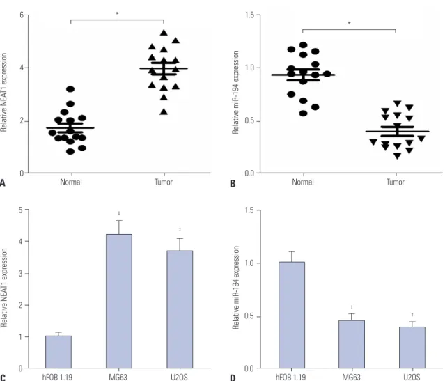
Real-time PCR assay was performed to detect lncRNA NEAT1 and miR-194 expression in osteosarcoma tissue samples (n=15) and normal tissue samples (n=15). The level of NEAT1 was markedly increased and the expression of miR-125a was sig- nificantly reduced in osteosarcoma tumor tissues, compared with normal tissues (Fig. 1A and B). Moreover, the levels of NEAT1 and miR-194 were further detected in osteosarcoma cell lines MG63 and U2OS or the human normal osteoblastic cell line hFOB 1.19. The results indicated that NEAT1 expres- sion was significantly elevated and miR-194 level was mark- edly decreased in MG63 and U2OS cells, compared with that in hFOB 1.19 cells (Fig. 1C and D). These results suggested that dysregulated expression of NEAT1 and miR-194 may be involved in the development of osteosarcoma.
6
4
2
0
1.5
1.0
0.5
0.0
Relative NEAT1 expression Relative miR-194 expression
Normal Tumor Normal Tumor
*
*
A B
5
4
3
2
1
0
1.5
1.0
0.5
0.0
Relative NEAT1 expression Relative miR-194 expression
hFOB 1.19 MG63 U2OS hFOB 1.19 MG63 U2OS
‡
‡
†
†
C D
Fig. 1. Expression of NEAT1 and miR-194 in osteosarcoma tissues and cells. (A and B) The expression levels of NEAT1 and miR-194 in osteosarcoma tissue samples (n=15) and normal tissue samples (n=15) by qRT-PCR analysis. (C and D) qRT-PCR analysis of NEAT1 and miR-194 in MG63 and U2OS cells. *p<0.05, †p<0.01, ‡p<0.001. NEAT1, nuclear paraspeckle assembly transcript 1; qRT-PCR, quantitative real-time PCR.
1.5
1.0
0.5
0.0
Relative NEAT1 expression levels
si-con si-NEAT1 si-con si-NEAT1
MG63 U2OS
‡ ‡
0.6
0.4
0.2
0.0
0.8
0.6 0.4 0.2
0.0
Absorbance Absorbance
Day 1 Day 2 Day 3 Day 4 Day 1 Day 2 Day 3 Day 4
MG63 U2OS
† †
† †
si-con si-NEAT1
si-con si-NEAT1
A
C
B
D 100
80 60 40 20 0
104 103 102 101 100
104 103 102 101 100
100 80 60 40 20 0
Number of invasive cells Number of invasive cells
si-con si-NEAT1 si-con si-NEAT1
MG63 U2OS
‡
‡
U2OS
30
20
10
0
Apoptotic cells (%)
si-con si-NEAT1
‡
MG63
PI
FITC
100 101 102 103 104 100 101 102 103 104
F
40
30
20
10
0
Apoptotic cells (%)
si-con si-NEAT1
‡
PI
FITC
Fig. 2. Knockdown of NEAT1 inhibits proliferation and invasion and promotes apoptosis of MG63 and U2OS cells. MG63 and U2OS cells were trans- fected with si-NEAT1 or si-con. (A) qRT-PCR analysis was performed to detect NEAT1 levels in MG63 and U2OS cells. (B) MTT assay was performed to detect viability of MG63 and U2OS cells at days 1, 2, 3, and 4 after transfection. (C and D) Transwell invasive assay was carried out to assess inva- sive ability of MG63 and U2OS cells 24 h post transfection. (E and F) Flow cytometry analysis was conducted to determine the apoptotic rate of MG63 and U2OS cells 24 h after transfection. †p<0.01, ‡p<0.001 vs. si-con. NEAT1, nuclear paraspeckle assembly transcript 1; qRT-PCR, quantitative real-time PCR; MTT, 3-[4, 5-dimethylthiazol-2-yl]-2, 5 diphenyl tetrazolium bromide; PI, propidium iodide; FITC, fluorescein isothiocyanate.
E
104 103 102 101 100
104 103 102 101 100
100 101 102 103 104 100 101 102 103 104
Knockdown of NEAT1 suppresses proliferation and invasion and promotes apoptosis of osteosarcoma cells To elucidate the regulatory roles of NEAT1 on osteosarcoma, MG63 and U2OS cells were transfected with si-NEAT1 or si- con. The results of qRT-PCR analysis revealed that si-NEAT1 significantly reduced NEAT1 expression in MG63 and U2OS cells, compared with si-con (Fig. 2A). MTT assay showed that downregulation of NEAT1 led to a significant reduction of via- bilities, compared with control group, in MG63 and U2OS cells (Fig. 2B). Transwell invasion assay indicated that the in- vasive ability of MG63 and U2OS cells was significantly sup- pressed by NEAT1 knockdown, compared with the si-con group (Fig. 2C and D). Flow cytometry analysis revealed that the apoptotic rate of MG63 and U2OS cells in si-NEAT1 transfec- tion group was significantly increased, compared to the si-con group (Fig. 2E and F). These findings suggested that silencing of NEAT1 inhibited proliferation and invasion and induced apo- ptosis of osteosarcoma cells.
miR-194 inhibits proliferation and invasion and induces apoptosis of osteosarcoma cells
To further assess the effects of miR-194 on proliferation, inva- sion, and apoptosis of osteosarcoma cells, MG63 and U2OS cells were transfected with miR-194 or miR-con. The results of qRT-PCR analysis indicated that miR-194 was successfully overexpressed in MG63 and U2OS cells (Fig. 3A). MTT assay revealed that miR-194 transfection dramatically repressed the cell viability of MG63 and U2OS cells (Fig. 3B). Consistently, the invasive capacity of MG63 and U2OS cells was markedly hindered by miR-194 upregulation (Fig. 3C and D). Moreover, enforced expression of miR-194 resulted in a remarkable en- hancement of apoptosis in MG63 and U2OS cells (Fig. 3E and F). Taken together, all these results demonstrated that overex- pression of miR-194 impeded proliferation and invasion and induced apoptosis of osteosarcoma cells.
C D
80
60 40 20
0
100 80 60 40 20 0
Number of invasive cells Number of invasive cells
miR-con miR-194 mimic miR-con miR-194 mimic
MG63 U2OS
‡ ‡
E F
40
30 20 10 0
40
30 20 10 0
Apoptotic cells (%) Apoptotic cells (%)
miR-con miR-194 mimic miR-con miR-194 mimic
MG63 U2OS
‡
‡
Fig. 3. miR-194 overexpression suppressed proliferation and invasion and induced apoptosis of MG63 and U2OS cells. MG63 and U2OS cells were transfected with miR-194 mimic or miR-control. (A) qRT-PCR analysis was performed to detect the miR-194 expression in MG63 and U2OS cells. (B) MTT assay of viability at days 1, 2, 3 and 4 in MG63 and U2OS cells. (C and D) Transwell invasive assay of invasive capacity 24 h post transfection in MG63 and U2OS cells. (E and F) Flow cytometry analysis of apoptosis of 24 h post transfection in MG63 and U2OS cells. †p<0.01, ‡p<0.001 vs. miR-con.
qRT-PCR, quantitative real-time PCR; MTT, 3-[4, 5-dimethylthiazol-2-yl]-2, 5 diphenyl tetrazolium bromide.
8
6 4 2
Relative miR-194 expression levels 0 miR-con miR-194
mimics miR-con miR-194 mimics
MG63 U2OS
‡
‡ 0.8
0.6 0.4 0.2
0.0
0.8
0.6 0.4 0.2
0.0
Absorbance Absorbance
Day 1 Day 2 Day 3 Day 4 Day 1 Day 2 Day 3 Day 4
MG63 U2OS
†
† † †
miR-con miR-194 mimic
miR-con miR-194 mimic
A B
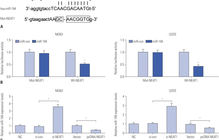
NEAT1 inhibits miR-194 expression in osteosarcoma cells Bioinformatics analysis by using starBase v2.0 showed that NEAT1 contains a binding site of miR-194 (Fig. 4A). To confirm the relationship between NEAT1 and miR-194, the luciferase reporter assay was performed. The results displayed that trans- fection of miR-194 significantly reduced the luciferase activity of Wt-NEAT1, but not with Mut-NEAT1, in MG63 and U2OS cells (Fig. 4B). To further confirm whether NEAT1 could indeed regulate miR-194 expression, MG63 and U2OS cells were transfected with si-NEAT1 or pcDNA-NEAT1. The results dem- onstrated that knockdown of NEAT1 improved the expression of miR-194 and that overexpression of NEAT1 suppressed the level of miR-194 in MG63 and U2OS cells (Fig. 4C). Together, these results clarified that NEAT1 functioned as a sponge to modulate miR-194 expression in osteosarcoma cells.
miR-194 inhibition reverses the suppression of prolif- eration and invasion and the induction of apoptosis triggered by NEAT1 knockdown in osteosarcoma cells To further explore whether NEAT1 exerted its function in os- teosarcoma cells through regulating miR-194 expression,
MG63 and U2OS cells were transfected with si-NEAT1 or co- transfected with si-NEAT1 and anti-miR-194. Then, MTT assay, Transwell invasion assay, and flow cytometry analysis were performed to determine the cell viability, invasive ability, and apoptosis of MG63 and U2OS cells, respectively. MTT assay revealed that NEAT1 knockdown resulted in prominent inhi- bition of cell viability in MG63 and U2OS cells, which was ab- rogated by miR-194 inhibition (Fig. 5A and B). Transwell inva- sion assay confirmed that downregulation of miR-194 reversed the inhibitory effect of NEAT1 silencing on the invasive ability of MG63 and U2OS cells (Fig. 5C and D). Flow cytometry analy- sis indicated that inhibition of miR-194 markedly attenuated NEAT1 depletion-induced apoptosis in MG63 and U2OS cells (Fig. 5E and F). Collectively, NEAT1 knockdown suppressed proliferation and invasion and the induced apoptosis of osteo- sarcoma cells through modulating miR-194 in osteosarcoma cells.
DISCUSSION
Growing reports have shown that many lncRNAs are dysregu-
Fig. 4. NEAT1 inhibited miR-194 expression in MG63 and U2OS cells. (A) The binding sequences of miR-194 in NEAT1 were marked. (B) Luciferase re- porter assay was performed in MG63 and U2OS cells co-transfected with Wt- or Mut-NEAT1 and miR-194 mimic or miR-con. (C) qRT-PCR analysis of miR- 194 expression in MG63 and U2OS cells transfected with si-NEAT1 or pcDNA-NEAT1. †p<0.01, ‡p<0.001 vs. controls. Wt-NEAT1, wild type-nuclear para- speckle assembly transcript 1; Mut-NEAT1, mutant-nuclear paraspeckle assembly transcript 1; NC, blank control; qRT-PCR, quantitative real-time PCR.
1.5
1.0
0.5
Relative Iuciferase activity 0.0
Mut-NEAT1 Wt-NEAT1
MG63
†
B
miR-con miR-194 1.5
1.0
0.5
Relative Iuciferase activity 0.0
Mut-NEAT1 Wt-NEAT1
U2OS
†
miR-con miR-194
5 4 3 2 1 0
4 3 2 1
Relative miR-194 expression levels NC si-con si-NEAT1 Vector pcDNA-NEAT1 Relative miR-194 expression levels 0 NC si-con si-NEAT1 Vector pcDNA-NEAT1
MG63
‡ ‡
† †
C
U2OS A
Wt-NEAT1
hsa-miR-194
Mut-NEAT1
lated in various cancers and exhibit important functions in cancer progression.14 Recently, numerous lncRNAs have been reported to be involved in osteosarcoma. For instance, lncRNA MALAT1 accelerated the proliferation and metastasis of os- teosarcoma cells via activation of the PI3K/Akt pathway.22 Ln- cRNA HOXA transcript at the distal tip enhanced chemoresis- tance of osteosarcoma cells by activating the Wnt/β-catenin pathway.23 Sun, et al.24 revealed that lncRNA highly upregulat- ed in liver cancer reflected poor prognosis and promoted os- teosarcoma cell metastasis. LncRNA HNF1A-AS1 was found to promote cell proliferation and metastasis in osteosarcoma by activating the Wnt/β-catenin signaling pathway.25 The present study focused on lncRNA NEAT1.
NEAT1 is a novel lncRNA localized specifically to nuclear paraspeckles.26 To date, many studies have reported the bio- logical function of NEAT1 and its potential mechanism in var-
ious cancers. For example, NEAT1 was illuminated to function as a competing endogenous lncRNA by sponging miR-98-5p to alleviate its suppression on copper transporter 1, subse- quently enhancing the sensitivity of non-small cell lung cancer cells to cisplatin.27 NEAT1 knockdown suppressed proliferation and invasion and induced apoptosis in laryngeal squamous cell carcinoma through upregulating miR-107 to repress CDK6 (a cyclin-dependent kinase) expression.28 NEAT1 pro- moted proliferation, migration, and invasion in esophageal squamous cell carcinoma.29 All these studies demonstrated that NEAT1 could function as an oncogene in cancers. In the present study, its oncogenic activities were also confirmed in osteosarcoma. Our study revealed that NEAT1 was overex- pressed in osteosarcoma tissues and cells, and knockdown of NEAT1 suppressed proliferation and invasion and promoted apoptosis of osteosarcoma cells. Consistent with our findings, Fig. 5. miR-194 inhibition overturned suppression of proliferation and invasion and the promotion of apoptosis elicited by NEAT1 knockdown in MG63 and U2OS cells. MG63 and U2OS cells were transfected with si-NEAT1 or co-transfected with si-NEAT1 and anti-miR-194. (A and B) MTT assay was used to test the viability of transfected MG63 and U2OS cells. (C and D) Transwell invasive assay was applied to measure the invasive ability of trans- fected MG63 and U2OS cells. (E and F) Flow cytometry analysis was conducted to observe the apoptotic rate of transfected MG63 and U2OS cells.
†p<0.01, ‡p<0.001 vs. controls. NEAT1, nuclear paraspeckle assembly transcript 1; MTT, 3-[4, 5-dimethylthiazol-2-yl]-2, 5 diphenyl tetrazolium bromide.
1.5
1.0
0.5
0.0
Relative cell viability
MG63
A si-con si-NEAT1 si-NEAT1+anti-
miR-con
si-NEAT1+anti- miR-194
†
‡ 2.0
1.5 1.0
0.5 0.0
Relative cell viability
U2OS
B si-con si-NEAT1 si-NEAT1+anti-
miR-con
si-NEAT1+anti- miR-194
†
‡
30
20
10
0
Apoptotic cells (%)
MG63
E si-con si-NEAT1 si-NEAT1+anti-
miR-con
si-NEAT1+anti- miR-194
‡ ‡ 40
30
20 10 0
Apoptotic cells (%)
U2OS
F si-con si-NEAT1 si-NEAT1+anti-
miR-con
si-NEAT1+anti- miR-194
‡ ‡
100 80 60 40 20 0
Number of invasive cells
MG63
C si-con si-NEAT1 si-NEAT1+anti-
miR-con
si-NEAT1+anti- miR-194
†
‡ 120
100 80 60 40 20 0
Number of invasive cells
U2OS
D si-con si-NEAT1 si-NEAT1+anti-
miR-con
si-NEAT1+anti- miR-194
†
‡
Zhao, et al.20 confirmed that NEAT1 was upregulated in osteo- sarcoma tissues, and silencing of NEAT1 inhibited osteosarco- ma cell proliferation, migration, and invasion.
Previous documents have implicated that miRNAs exert im- portant functions in regulating biological processes, including cell proliferation, metastasis, and apoptosis, in various can- cers.30 Specifically, many miRNAs involved in osteosarcoma carcinogenesis could be regulated by lncRNAs. For instance, lncRNA plasmacytoma variant translocation 1 promoted pro- liferation, migration, and invasion and suppressed apoptosis and cell cycle arrest in osteosarcoma cells by acting as a mo- lecular sponge to regulate miR-195.31 LncRNA TUG1 knock- down suppressed cell proliferation and colony formation, and induced G0/G1 cell cycle arrest and apoptosis by sponging miR-9-5p to alleviate its repression on POU class 2 homeobox 1 (POU2F1) expression in osteosarcoma.32 Previous studies discovered that miR-194 acted as a tumor suppressor through suppressing its targets, such as bone morphogenetic protein 1 and ras-related gene.33,34 Additionally, miR-194 has been shown to inhibit osteosarcoma cell proliferation, migration, and inva- sion in vitro through targeting Cadherin 2 and type 1 insulin- like growth factor receptor and to suppress tumor growth and metastasis of osteosarcoma in vivo.12 In agreement with the pre- vious study, our findings suggested that miR-194 repressed proliferation and invasion and induced apoptosis of osteosar- coma cells. Next, we applied luciferase reporter analysis and qRT- PCR analysis to conclude that NEAT1 functioned as a miR-194 sponge to regulate its expression. Further rescue experiments indicated that miR-194 downregulation abolished the NEAT1 knockdown-induced suppression on proliferation and inva- sion, as well as promotion of apoptosis in osteosarcoma cells, indicating that NEAT1 suppresses proliferation and invasion and induces apoptosis of osteosarcoma by acting as a sponge to modulate miR-194 expression.
In summary, our study showed that NEAT1 is upregulated in osteosarcoma cells. Functionally, knockdown of NEAT1 suppressed proliferation and invasion and induced apoptosis of osteosarcoma cells. Mechanistically, NEAT1 promoted os- teosarcoma progression by functioning as a miR-194 sponge to suppress its expression. Thus, NEAT1 could potentially serve as a novel therapy target for osteosarcoma.
REFERENCES
1. Ottaviani G, Jaffe N. The epidemiology of osteosarcoma. Cancer Treat Res 2009;152:3-13.
2. Stiller CA. International patterns of cancer incidence in adoles- cents. Cancer Treat Rev 2007;33:631-45.
3. Yang J, Zhang W. New molecular insights into osteosarcoma tar- geted therapy. Curr Opin Oncol 2013;25:398-406.
4. Luetke A, Meyers PA, Lewis I, Juergens H. Osteosarcoma treat- ment - where do we stand? A state of the art review. Cancer Treat Rev 2014;40:523-32.
5. Chen L, Wang X, Wang H, Li Y, Yan W, Han L, et al. miR-137 is fre-
quently down-regulated in glioblastoma and is a negative regula- tor of Cox-2. Eur J Cancer 2012;48:3104-11.
6. Duan Z, Choy E, Harmon D, Liu X, Susa M, Mankin H, et al. Mi- croRNA-199a-3p is downregulated in human osteosarcoma and regulates cell proliferation and migration. Mol Cancer Ther 2011;
10:1337-45.
7. Liu LH, Li H, Li JP, Zhong H, Zhang HC, Chen J, et al. miR-125b suppresses the proliferation and migration of osteosarcoma cells through down-regulation of STAT3. Biochem Biophys Res Com- mun 2011;416:31-8.
8. Hong Q, Fang J, Pang Y, Zheng J. Prognostic value of the microRNA-29 family in patients with primary osteosarcomas. Med Oncol 2014;
31:37.
9. Meng Z, Fu X, Chen X, Zeng S, Tian Y, Jove R, et al. miR-194 is a marker of hepatic epithelial cells and suppresses metastasis of liver cancer cells in mice. Hepatology 2010;52:2148-57.
10. Dong P, Kaneuchi M, Watari H, Hamada J, Sudo S, Ju J, et al. Mi- croRNA-194 inhibits epithelial to mesenchymal transition of en- dometrial cancer cells by targeting oncogene BMI-1. Mol Cancer 2011;10:99.
11. Song Y, Zhao F, Wang Z, Liu Z, Chiang Y, Xu Y, et al. Inverse asso- ciation between miR-194 expression and tumor invasion in gas- tric cancer. Ann Surg Oncol 2012;19 Suppl 3:S509-17.
12. Han K, Zhao T, Chen X, Bian N, Yang T, Ma Q, et al. microRNA-194 suppresses osteosarcoma cell proliferation and metastasis in vitro and in vivo by targeting CDH2 and IGF1R. Int J Oncol 2014;45:
1437-49.
13. Mercer TR, Dinger ME, Mattick JS. Long non-coding RNAs: in- sights into functions. Nat Rev Genet 2009;10:155-9.
14. Cheetham SW, Gruhl F, Mattick JS, Dinger ME. Long noncoding RNAs and the genetics of cancer. Br J Cancer 2013;108:2419-25.
15. Li W, Xie P, Ruan WH. Overexpression of lncRNA UCA1 promotes osteosarcoma progression and correlates with poor prognosis. J Bone Oncol 2016;5:80-5.
16. Cong M, Li J, Jing R, Li Z. Long non-coding RNA tumor suppres- sor candidate 7 functions as a tumor suppressor and inhibits pro- liferation in osteosarcoma. Tumour Biol 2016;37:9441-50.
17. Yun-Bo F, Xiao-Po L, Xiao-Li L, Guo-Long C, Pei Z, Fa-Ming T. Ln- cRNA TUG1 is upregulated and promotes cell proliferation in os- teosarcoma. Open Med (Wars) 2016;11:163-7.
18. Yin Z, Ding H, He E, Chen J, Li M. Overexpression of long non-cod- ing RNA MFI2 promotes cell proliferation and suppresses apop- tosis in human osteosarcoma. Oncol Rep 2016;36:2033-40.
19. Choudhry H, Albukhari A, Morotti M, Haider S, Moralli D, Smythies J, et al. Tumor hypoxia induces nuclear paraspeckle formation through HIF-2α dependent transcriptional activation of NEAT1 leading to cancer cell survival. Oncogene 2015;34:4482-90.
20. Zhao H, Zhao Y, Tao J, Ma C, Zhang J, Xu H, et al. Up-regulated ex- pression of lncRNA NEAT1 promotes progression of osteosarco- ma by regulating the activity of Wnt/β-catenin pathway. Int J Clin Exp Pathol 2016;9:11466-72.
21. Schmittgen TD, Livak KJ. Analyzing real-time PCR data by the comparative C(T) method. Nat Protoc 2008;3:1101-8.
22. Dong Y, Liang G, Yuan B, Yang C, Gao R, Zhou X. MALAT1 pro- motes the proliferation and metastasis of osteosarcoma cells by ac- tivating the PI3K/Akt pathway. Tumour Biol 2015;36:1477-86.
23. Li Z, Zhao L, Wang Q. Overexpression of long non-coding RNA HOTTIP increases chemoresistance of osteosarcoma cell by acti- vating the Wnt/β-catenin pathway. Am J Transl Res 2016;8:2385-93.
24. Sun XH, Yang LB, Geng XL, Wang R, Zhang ZC. Increased expres- sion of lncRNA HULC indicates a poor prognosis and promotes cell metastasis in osteosarcoma. Int J Clin Exp Pathol 2015;8:2994- 3000.
25. Zhao H, Hou W, Tao J, Zhao Y, Wan G, Ma C, et al. Upregulation of lncRNA HNF1A-AS1 promotes cell proliferation and metastasis in osteosarcoma through activation of the Wnt/β-catenin signaling pathway. Am J Transl Res 2016;8:3503-12.
26. Clemson CM, Hutchinson JN, Sara SA, Ensminger AW, Fox AH, Chess A, et al. An architectural role for a nuclear noncoding RNA:
NEAT1 RNA is essential for the structure of paraspeckles. Mol Cell 2009;33:717-26.
27. Jiang P, Wu X, Wang X, Huang W, Feng Q. NEAT1 upregulates EGCG-induced CTR1 to enhance cisplatin sensitivity in lung can- cer cells. Oncotarget 2016;7:43337-51.
28. Wang P, Wu T, Zhou H, Jin Q, He G, Yu H, et al. Long noncoding RNA NEAT1 promotes laryngeal squamous cell cancer through regulating miR-107/CDK6 pathway. J Exp Clin Cancer Res 2016;
35:22.
29. Chen X, Kong J, Ma Z, Gao S, Feng X. Up regulation of the long non- coding RNA NEAT1 promotes esophageal squamous cell carci- noma cell progression and correlates with poor prognosis. Am J
Cancer Res 2015;5:2808-15.
30. Stahlhut C, Slack FJ. MicroRNAs and the cancer phenotype: pro- filing, signatures and clinical implications. Genome Med 2013;5:
111.
31. Zhou Q, Chen F, Zhao J, Li B, Liang Y, Pan W, et al. Long non-cod- ing RNA PVT1 promotes osteosarcoma development by acting as a molecular sponge to regulate miR-195. Oncotarget 2016;7:82620-33.
32. Xie CH, Cao YM, Huang Y, Shi QW, Guo JH, Fan ZW, et al. Long non-coding RNA TUG1 contributes to tumorigenesis of human os- teosarcoma by sponging miR-9-5p and regulating POU2F1 expres- sion. Tumour Biol 2016;37:15031-41.
33. Wu X, Liu T, Fang O, Leach LJ, Hu X, Luo Z. miR-194 suppresses metastasis of non-small cell lung cancer through regulating ex- pression of BMP1 and p27(kip1). Oncogene 2014;33:1506-14.
34. Zhang M, Zhuang Q, Cui L. MiR-194 inhibits cell proliferation and invasion via repression of RAP2B in bladder cancer. Biomed Phar- macother 2016;80:268-75.