INTRODUCTION
Liver fibrosis is a chronic wound-healing response represent- ed by excessive extracellular matrix (ECM) deposition that oc- curs in most types of chronic liver diseases,
1,2including hepa- tocellular carcinoma and hepatitis B/C. Activation of hepatic stellate cells (HSCs), the major mesenchymal cells in the liver, are the critical cellular basis for liver fibrogenesis:
3,4activated HSCs are responsible for excess synthesis and deposition of ECM following a fibrotic stimulus.
5,6A variety of cytokines and growth factors induced in liver injury can activate HSCs to pro- duce α-smooth muscle actin (α-SMA) and cause liver fibro-
miR-140-3p Knockdown Suppresses Cell Proliferation and Fibrogenesis in Hepatic Stellate Cells
via PTEN-Mediated AKT/mTOR Signaling
Shi-Min Wu 1 *, Tian-Hong Li 2 *, Hao Yun 1 , Hong-Wu Ai 3 , and Ke-Hui Zhang 1
1
Wuhan Center for Clinical Laboratory, Wuhan Forth Hospital; Puai Hospital, Tongji Medical College, Huazhong University of Science and Technology, Wuhan;
Departments of
2Ophthalmology and
3Clinical Laboratory, Wuhan Children’s Hospital, Tongji Medical College, Huazhong University of Science and Technology, Wuhan, China.
Purpose: Liver fibrosis is a major cause of morbidity and mortality and the outcome of various chronic liver diseases. Activation of hepatic stellate cells (HSCs) is the key event in liver fibrosis. Studies have confirmed that miR-140-3p plays a potential regulato- ry effect on HSC activation. However, whether miR-140-3p mediates the liver fibrosis remains unknown.
Materials and Methods: Expression of miR-140-3p was detected by real-time quantitative PCR (qPCR). Cell proliferation was measured by MTT, while cell apoptosis rate was determined via flow cytometry. Western blot assay was used to detect the expres- sion of cleaved PARP. The fibrogenic effect was evaluated by expression of α-smooth muscle actin and desmin. Functional experi- ments were performed in transforming growth factor β1 (TGF-β1)-induced HSC-T6 cells with transfection of anti-miR-140-3p and/or siPTEN. Target binding between miR-140-3p and PTEN was predicted by the TargetScan database and identified using luciferase reporter assay and RNA immunoprecipitation.
Results: TGF-β1 induced the activation of HSC-T6 cells, and miR-140-3p expression varied according to HSC-T6 cell activation status. Knockdown of miR-140-3p reduced cell proliferation and the expressions of α-SMA and desmin, as well as increased apoptosis, in TGF-β1-induced HSC-T6 cells, which could be blocked by PTEN silencing. Additionally, inactivation of the AKT/
mTOR signaling pathway stimulated by miR-140-3p knockdown was abolished when silencing PTEN expression. PTEN was neg- atively regulated by miR-140-3p via direct binding in HSC-T6 cells.
Conclusion: miR-140-3p is an important mediator in HSC-T6 cell activation, and miR-140-3p knockdown suppresses cell prolif- eration and fibrogenesis in TGF-β1-induced HSC-T6 cells, indicating that miR-140-3p may be a potential novel molecular target for liver fibrosis.
Key Words: miR-140-3p, PTEN, liver fibrosis, TGF-β1, hepatic stellate cells (HSCs)
pISSN: 0513-5796 · eISSN: 1976-2437
Received: November 27, 2018 Revised: January 7, 2019 Accepted: February 14, 2019
Corresponding author: Ke-Hui Zhang, MB, Wuhan Center for Clinical Laboratory, Wuhan Forth Hospital; Puai Hospital, Tongji Medical College, Huazhong University of Science and Technology, 473 Hanzheng Street, Qiaokou, 430033, Wuhan, Hubei, China.
Tel: 86-027-68834898, E-mail: [email protected]
*Shi-Min Wu and Tian-Hong Li contributed equally to this work.
•The authors have no potential conflicts of interest to disclose.
© Copyright: Yonsei University College of Medicine 2019
This is an Open Access article distributed under the terms of the Creative Com- mons Attribution Non-Commercial License (https://creativecommons.org/licenses/
by-nc/4.0) which permits unrestricted non-commercial use, distribution, and repro- duction in any medium, provided the original work is properly cited.
Yonsei Med J 2019 Jun;60(6):561-569
https://doi.org/10.3349/ymj.2019.60.6.561
sis,
7,8later leading to cirrhosis. Transforming growth factor-β1 (TGF-β1) is considered the main pro-fibrogenic mediator of the transdifferentiation of fibroblasts to myofibroblast-like cells.
9Despite fundamental advances in understanding of the pathophysiology of liver fibrosis, detail mechanisms of liver fi- brosis remain elusive. Therefore, a comprehensive under- standing of the molecular mechanisms involved in HSC acti- vation and fibrogenesis will be helpful to providing novel therapeutic targets with which to inhibit, even mitigate, this devastating progression.
There has been an increasing number of studies on microR- NAs (miRNAs) as key regulators in the development of liver fi- brosis.
10miRNAs are endogenous, small, noncoding RNAs ranging in size from 20 to 25 nt, and deregulation of miRNAs affect various intracellular functional complexes,
11such as cell proliferation, apoptosis, and differentiation; ECM; cytoskele- ton; tyrosine kinase; and G-protein signaling. With the use of in vitro cell activation and miRNA microarray hybridization, many differentially expressed miRNAs, among which miR- 140 was upregulated, were identified in rat HSCs during acti- vation.
12Serum miR-138 and miR-140 were highly detected in early fibrosis and late fibrosis, compared to healthy patients.
13Moreover, increasing expression thereof during the develop- ment of fibrosis of the liver and progressive liver fibrosis have been posited in the late stages of various chronic liver diseas- es. Research has demonstrated that miR-140-3p has a pro-fi- brotic effect in the mammary glands
14and is deeply involved in liver disorders,
12,15,16including hepatic impact injury, non-al- coholic fatty liver disease, and hepatocellular carcinoma. Thus, we planned to investigate the role of miR-140-3p in HSC activa- tion and its molecular signaling pathway.
Phosphatase and tensin homolog deleted on chromosome 10 (PTEN), a tumor suppressor, is a dual phosphatase, and its major function is to dephosphorylate phosphatidylinositol 3, 4, 5-triphosphate (PIP3) to phosphatidylinositol 4, 5-bisphos- phate (PIP2), antagonizing PI3K/AKT signaling.
17Alteration of PTEN expression and activity has been recognized as being pervasive among tumor cells.
18Accumulating evidence has indicated that PTEN is dysregulated in liver diseases
19,20and has demonstrated decreased PTEN expression in fibrotic dis- eases of the lungs, kidneys, and skin.
21-23PTEN activity and ex- pression are controlled by several mechanisms, including phosphorylation, acetylation, oxidation, ubiquitination, non- coding RNAs, and DNA methylation.
24Here, we sought to deter- mine the role of miR-140-3p in HSC activation through PTEN.
In this work, we studied the fibrogenic role of miR-140-3p in rat hepatic stellate HSC-T6 cells and its downstream regulation.
MATERIALS AND METHODS
Cells and cell culture
This study was approved by the Institutional Review Board of
Puai Hospital, Tongji Medical College, Huazhong University of Science and Technology. The HSC-T6 cell line was obtained from the Cell Bank of Type Culture Collection (Chinese Acade- my of Sciences, Shanghai, China) and cultured in Dulbecco’s modified Eagle’s medium (DMEM, Hyclone, Logan, UT, USA), 10% (v/v) fetal bovine serum (FBS, Hyclone), 100 units/mL of penicillin, and 100 μg/mL of streptomycin in a humidified at- mosphere containing 5% (v/v) CO
2at 37°C.
Cell medium was refreshed every other day, and cells grown to subconfluence were pretreated with serum-free DMEM for 16 h. Then, cells were incubated with DMEM supplemented with platelet derived growth factor (PDGF)-BB (GF310; Merck;
MO, USA) and TGF-β1 (T7039; Merck) for 48 h.
Cell transfection
In six-well plates (Corning, NY, USA), 10 ng/mL of TGF-β1 treated HSC-T6 cells were seeded at a density of 2×10
5cells per well 24 h prior to the transfection. siRNA against PTEN (siP- TEN)/scramble, pre-miR-140-3p/NC, and anti-miR-140-3p/
NC were provided by GenePharma (Shanghai, China). Oligo- nucleotides were transfected into cells at a final concentration of 100 nM using Lipofectamine 2000 (Invitrogen, Shanghai, China) according to the manufacturer’s instructions. Samples were collected after 48 h of transfection for further studies, such as RNA isolation and protein extraction.
Cell proliferation assay
Cell proliferation assay was determined by standard 3-(4, 5-di- methylthiazol-2-yl)-2, 4-diphenyl-tetrazolium bromide (MTT, Sigma-Aldrich, Louis, MO, USA) assay. After transfection, cells were implanted at a density of 2×10
4cells per well in 96-well plates (Corning) for 24 h. Briefly, 5 mg/mL of MTT (Sigma-Al- drich) was added and incubated at 37°C for another 4 h; there- after, the medium was replaced and the formazan crystals were dissolved in 150 μL of dimethyl sulphoxide (DMSO, Dingguo, Beijing, China). The optical density was determined with a Thermomax microplate reader (Bio-Tek EL, VT, USA) at 490 nm wavelength. All experiments were performed in triplicate.
Cell apoptosis assay
Cell apoptosis assay was performed on flow cytometry using Cell Cycle and Apoptosis Analysis Kits (Beyotime, Shanghai, China). In brief, cells were collected and washed with cold phosphate buffer solution (PBS) three times after transfection.
Then, cells were fixed with 70% ethanol, stained with 0.5 mL
of propidium iodide (PI) staining buffer, which contained 200
mg/mL of RNase A and 50 μg/mL of PI, at 37°C for 30 min in
the dark. Analyses were performed on a BD LSR flow cytome-
ter (BD Biosciences, Jiangsu, China), and apoptotic cells were
calculated. The experiments were repeated three times.
Western blot assay
TGF-β1-treated HSC-T6 cells with translation or not were lysed in RIPA (Beyotime), and the protein concentration was determined by Bradford. 20 μg of sample were separated by sodium dodecyl sulfate-polyacrylamide gel electrophoresis and transferred onto PVDF membranes (Millipore, Billerica, MA, USA). Prior to incubation of primary antibodies overnight at 4°C with gentle shaking, the blots were blocked in 5% of nonfat milk. GAPDH on the same membrane was the loading control. After incubation of horseradish peroxidase conjugat- ed secondary antibodies correspondingly, proteins were visu- alized with ECL Plus (Solarbio, Beijing, China), and ImageJ (National Institutes of Health, Bethesda, MD, USA) was uti- lized to analyze the gray intensity of the bands. Primary anti- bodies were purchased from Cell Signaling Technology (Dan- vers, MA, USA) and Abcam (Cambridge, UK): α-SMA (#14968), COL1A1 (#84336), PCNA (#13110), cleaved caspase 3 (#9664), caspase 3 (#9665), p-AKT (#2965), p-mTOR (#2971), AKT (#9272), mTOR (#2972), GAPDH (#97166), and fibronectin (#6328).
Real-time quantitative PCR
Total RNA from cultured cells was isolated using TRIzol re- agent (Thermo, Waltham, MA, USA) following the protocol, and 300 ng of total RNA was reverse transcribed to cDNA us- ing Bestar QPCR RT Kit (DBI Bioscience, Ludwigshafen, Ger- many). Real-time quantitative PCR (qPCR) was performed with Bestar SYBR Green qPCR Master Mix (DBI Bioscience) on ABI 7500. The expression of miR-140-3p was calculated ac- cording to the comparative threshold cycle value (2
−ΔΔCt) meth- od, compared with U6 small nuclear RNA (U6). PCR primers were as follows: miR-140-3p, 5’-TCGGCAGGTAACACTG TCTGGT-3’ (sense) and 5’-CTCAACTGGTGTCGTGGA-3’ (an- tisense); U6, 5’-CTCGCTTCGGCAGCACA-3’ (sense) and 5’-AACGCTTCACGAATTTGCGT-3’ (antisense). All experi- ments were performed at least in triplicate.
Luciferase reporter assay
The potential binding sites of miR-140-3p on PTEN 3’UTR were mutated and cloned by PCR into plasmid pGL3-basic. Cells were plated in a 24-well plate (Corning) at 1×10
4cells/well, fol- lowed by co-transfection with 20 nM of miR-140-3p mimic/NC and 20 ng of either PTEN wt/mut 3’UTR in TGF-β1 treated HSC-T6 cells for 48 h. Cells were collected to measure the rela- tive luciferase using the Dual-luciferase Reporter Assay Sys- tem (Promega, Madison, WI, USA). The ratio of Firefly to Re- nilla luciferase activity was used as the relative luciferase activity, and all experiments were carried out in triplicate.
RNA immunoprecipitation
TGF-β1-treated HSC-T6 cells were transfected with miR-140- 3p/NC. RNA immunoprecipitation (RIP) was performed with cell extract, and Magna RIP
TMRNA-binding protein immuno-
precipitation kit (Millipore) was chosen to detect expression of PTEN from the samples bound to the Ago2 antibody or IgG. All procedures followed the standard protocol. Total RNA isolation was extracted from RIP-Ago2/IgG immunoprecipitation using TRIzol (Thermo). All operations were performed in triplicate.
Transwell assay
For migration assay, HSC-T6 cells with different processing were supplemented in 200 μL of serum-free medium and plat- ed in the upper chamber with a non-coated membrane (Corn- ing). The medium containing 10% FBS (Gibco) was used as a chemo-attractant and loaded in the lower chamber. The tran- swell system was maintained at 37°C for 48 h. The migratory cells into the lower chambers were stained with 0.5% crystal violet and quantitated under a light microscope (DM2500M, Leica, Wetzlar, Germany) separately, and the ability of migra- tion/invasion was assessed as the percentage of migratory/in- vasive cells compared with control group.
Statistical analysis
All statistical analyses were performed using SPSS 17.0 (SPSS Inc., Chicago, IL, USA). Values given represent mean±SEM, and the differences between two groups were determined by two- tailed Student’s t-test. p<0.05 was considered to be statistically different.
RESULTS
miR-140-3p is highly expressed upon HSC-T6 cell activation
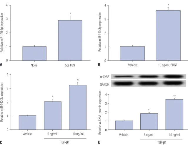
First and foremost, the expression of miR-140-5p was mea- sured in activated HSC-T6 cells and compared with that in quiescent cells. 5% FBS, PDGF-BB (PDGF), and TGF-β1 were selected to stimulate cells independently. As was shown in Fig. 1A-C, miR-140-3p expression was upregulated over three- fold in 5% FBS-, 10 ng/mL of PDGF-, and 10 ng/mL of TGF- β1-incubated HSC-T6 cells. Additionally, the levels of α-SMA, a marker of HSC activation, in TGF-β1-treated cells and relative α-SMA protein expression were elevated (Fig. 1D). To explore the role of miR-140-3p in HSC activation, HSC-T6 cells were exposed to 10 ng/mL of TGF-β1 for further experiments. This data indicated that miR-140-3p is highly expressed in activated HSCs.
miR-140-3p knockdown reduces cell fibrosis in HSC-T6 cells
The role of miR-140-3p knockdown in cell fibrosis were mea-
sured in TGF-β1-induced HSC-T6 cells. miR-140-3p knock-
down was obtained after transfection with anti-miR-140-3p
(Fig. 2A). Cell proliferation was impaired by anti-miR-140-3p
(Fig. 2B), and apoptosis rate and level of cleaved caspase 3
were higher in miR-140-3p knockdown cells (Fig. 2C and D);
meanwhile, expression of the proliferation marker proliferat- ing cell nuclear antigen (PCNA) was opposite to that of cleaved caspase 3. Additionally, migratory cells were decreased (Fig.
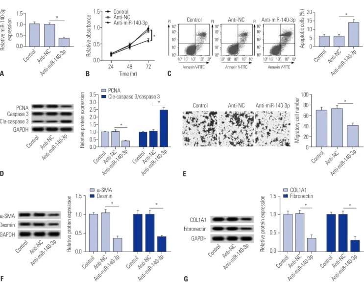
2E). Moreover, we observed that anti-miR-140-3p induced lower expression levels of markers of fibrosis α-SMA, desmin, COL1A1, and fibronectin (Fig. 2F and 2G). These results illu- minated a certain cytotoxicity and fibrosis suppressor role of miR-140-3p knockdown in TGF-β1-induced HSC-T6 cells.
PTEN is negatively regulated by miR-140-3p target binding in HSC-T6 cells
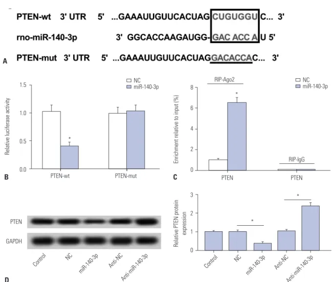
Potential target sites were predicted on TargetScan software (http://www.targetscan.org/) (Fig. 3A). The site on PTEN wide type (wt) 3’UTR was mutated (mut), and full-length PTEN-wt/
mut 3’UTR was cloned into pGL3 vector. Luciferase reporter assay (Fig. 3B) and RIP (Fig. 3C) were performed to identify target binding in TGF-β1 treated HSC-T6 cells. As shown, lucif- erase activity relatively declined after co-transfection with PTEN-wt 3’UTR and miR-140-3p mimic, compared with co- transfected with PTEN-mut 3’UTR and miR-140-3p mimic.
Besides, dramatically high expression of PTEN was obtained from Ago2 immunoprecipitation. We also observed that ex-
pression of PTEN protein was upregulated by anti-miR- 140-3p and downregulated by miR-140-3p mimic (Fig. 3D).
These outcomes indicated that PTEN was a downstream tar- get gene of miR-140-3p in HSC-T6 cells.
PTEN silencing blocks the effect of miR-140-3p knockdown in HSC-T6 cells
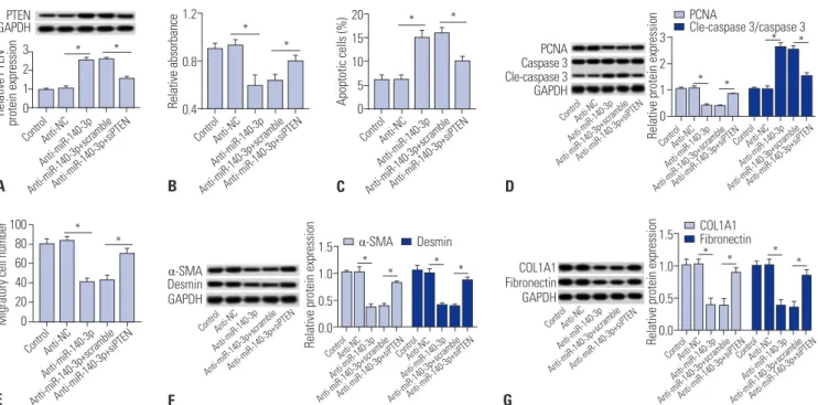
Further, the effects of PTEN silencing on miR-140-3p knock- down-induced inhibition of cell proliferation and fibrosis were measured in TGF-β1-induced HSC-T6 cells. Silencing expres- sion of PTEN was obtained after transfection of specific siRNA against PTEN (siPTEN). Expression levels of PTEN were up- regulated in cells transfected with anti-miR-140-3p and then reduced with siPTEN incubation (Fig. 4A). Cell proliferation was impaired by anti-miR-140-3p, which was improved by siPTEN (Fig. 4B). Apoptosis rate and levels of cleaved caspase 3 were higher in miR-140-3p knockdown cells, and were sub- sequently blocked with siPTEN incubation (Fig. 4C and 4D).
PCNA expression was opposite that of cleaved caspase 3. In addition, the number of migratory cells declined (Fig. 4E).
Next, we found that anti-miR-140-3p-induced lower expres- sion levels of α-SMA, desmin, COL1A1, and fibronectin were
4
3
2
1
0
Relative miR-140-3p expression
B
4 3 2 1 0
Vehicle 5 ng/mL 10 ng/mL
Relative α-SMA protein expression
D TGF- β1
α-SMA GAPDH
Vehicle 10 ng/mL PDGF
*
*
*
†4
3
2
1
0
None 5% FBS
Relative miR-140-3p expression
A
Vehicle 5 ng/mL 10 ng/mL 4
3
2
1
0
TGF- β1
Relative miR-140-3p expression
C
*
*
*
†Fig. 1. miR-140-3p expression according to HSC-T6 cell activation status. Expression of miR-140-5p was measured in HSC-T6 cells by qPCR. (A) Cells were
incubated with 5% FBS for 48 h. The control was FBS-free cells. (B) Cells were treated with 10 ng/mL of PDGF-BB (PDGF) for 48 h. The control was no treat-
ment cells. (C) Cells were exposed to TGF-β1 for 48 h. (D) Levels of the marker of hepatic stellate cell activation, α-SMA in TGF-β1 treated cells were detect-
ed using western blot assay. The control was no treatment cells. All experiments were performed in triplicate, and *p<0.05,
†p<0.01. PDGF, platelet derived
growth factor; α-SMA, α-smooth muscle actin; TGF-β1, transforming growth factor β1.
rescued with treatment of siPTEN (Fig. 4F and G). These re- sults showed that PTEN silencing blocks the inhibitory effects of miR-140-3p knockdown on cell proliferation and fibrosis.
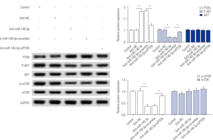
miR-140-3p knockdown inactivates AKT/mTOR signaling, which is abolished by PTEN silencing While we discovered that PTEN silencing blocks the effect of miR-140-3p knockdown in TGF-β1-induced HSC-T6 cells, the associated signaling pathway remains unknown. The effects of siPTEN on anti-miR-140-3p-inactivated signaling pathway were measured, and the expressions of p-AKT, p-mTOR, and PTEN were detected using Western blot assay (Fig. 5). As shown, anti-miR-140-3p significantly reduced the p-AKT and p-mTOR, while the expressions of total AKT and mTOR were not affect- ed. Co-expression of anti-miR-140-3p and siPTEN improved p-AKT and p-mTOR expression. These data indicated that miR-
140-3p knockdown inhibits AKT/mTOR signaling in HSC-T6 cells, which could be blocked by PTEN silencing.
DISCUSSION
In this study, we discovered that miR-140-3p is highly expressed in TGF-β1-induced HSC-T6 cells, indicating the regulatory role of miR-140-3p in HSCs activation and fibrogenesis. Knock- down of miR-140-3p reduced cell proliferation and increased cell apoptosis and fibrosis through upregulating its target PTEN, thus inhibiting p-AKT and p-mTOR. Our work suggests a novel pro-fibrotic factor, miR-140-3p, that affects PTEN ex- pression and PTEN-mediated AKT/mTOR signaling in rat HSCs.
miRNAs have become promising targets for liver fibrosis 20
15 10 5 Apoptotic cells (%) 0
C 1.5
1.0 0.5
0.0 24 48 72 Time (hr)
Relative absorbance
B 1.5
1.0 0.5 0.0
Control Anti-NC Anti-miR-140-3p Relative miR-140-3p expression
A
100 80 60 40 20 Migratory cell number 0
E 3.5
3.0 2.5 2.0 1.5 1.0 0.5 Relative protein expression 0.0
D
1.5
1.0
0.5
Relative protein expression 0.0
G 1.5
1.0
0.5
Relative protein expression 0.0
F
*
* Control
Anti-NC
Anti-miR-140-3p PI Control Anti-NC Anti-miR-140-3p
10
410
310
210
110
010
410
310
210
110
010
410
310
210
110
010
010
110
210
310
410
010
110
210
310
410
010
110
210
310
4Annexin V-FITC Annexin V-FITC Annexin V-FITC
*
* Control Anti-NC
Anti-miR-140-3p
Control
Control Control
Anti-NC
Anti-NC Anti-NC
Anti-miR-140-3p
Anti-miR-140-3p Anti-miR-140-3p
Control Anti-NC Anti-miR-140-3p Caspase 3 PCNA
Cle-caspase 3 GAPDH
PCNA
Cle-caspase 3/caspase 3
*
*
Control Anti-NC Anti-miR-140-3p
α-SMA Desmin GAPDH
Control Control
Control
Control
Control Control
Anti-NC Anti-NC
Anti-NC
Anti-NC
Anti-NC Anti-NC
Anti-miR-140-3p Anti-miR-140-3p Anti-miR-140-3p
Anti-miR-140-3p
Anti-miR-140-3p Anti-miR-140-3p
* *
COL1A1 Fibronectin GAPDH
* *
α-SMA
Desmin COL1A1
Fibronectin
Fig. 2. miR-140-3p knockdown reduces cell fibrosis in HSC-T6 cells. miR-140-3p knockdown was obtained after transfection of anti-miR-140-3p into TGF- β1-induced HSC-T6 cells. (A) Relative expression levels of miR-140-3p. (B) Cell proliferation was measured with cell proliferation assay. (C) Cell apoptosis was measured on flow cytometry, and apoptosis rate was calculated. (D) Expressions of PCNA, caspase 3, and cleaved caspase 3 (cle-caspase 3) were detected using Western blot assay, and the gray intensity was analyzed. (E) Cell migration was determined using transwell assay (0.5% crystal violet,
×100). (F and G) Expressions of α-SMA, desmin, COL1A1, and fibronectin were detected using Western blot assay, and the gray intensity was analyzed.
All experiments were compared with cells transfected of anti-NC and performed in triplicate, *p<0.05. TGF-β1, transforming growth factor β1; α-SMA, α-smooth muscle actin; anti-miR-140-3p, miR-140-3p inhibitor; PCNA, proliferating cell nuclear antigen; COL1A1, collagen type 1A1.
PI PI
therapy. Targeting specific molecules to combat liver fibrosis is a growing challenge in translational medicine. Though signif- icant progress has been made to elucidate the physiopathology related to liver fibrosis, there is still a lack of effectively estab- lished drugs for liver fibrosis prognosis and treatment. Also, with the central role of HSCs activation in the development of liver fibrosis,
4,25suppression of its activation is the most prom- ising therapeutic approach. Inhibition of cytokine-mediated ECM synthesis
26and promotion of HSCs apoptosis
27are the predominant strategies of liver fibrosis. In this study, we found that a noncoding RNA, namely miR-140-3p, universally af- fected TGF-β1-induced HSC-T6 cell proliferation, apoptosis, and activation. In other research, the miR-29 family exhibited dramatic decreases in the two models of liver fibrosis, com- pared with nonfibrotic liver, as well as in patients with liver fi- brosis, through inducing apoptosis of HSCs and reducing the accumulation of ECM.
28The miR-34 family
29has also been found to play promotion roles in liver fibrosis by target binding
to acyl-CoA synthetase long-chain family member 1 (ACSL1) and PPARγ to deposit ECM proteins and metabolize fatty acids in HSCs: the accumulation of vitamin A-containing lipid drop- lets is characteristic of quiescent HSCs.
miR-140-3p has a pro-fibrosis effect. It has been demon- strated that miR-140-3p has a pro-fibrotic effect in the mam- mary gland
14and is deeply involved in liver disorders,
13,15,16in- cluding hepatic impact injury, non-alcoholic fatty liver disease, and hepatocellular carcinoma. Progressive liver fibrosis is ob- served in the late stages of various chronic liver diseases, and miR-140 has been predicted to be upregulated in rat HSCs dur- ing activation.
12Additionally, serum miR-138 and miR-140 have been highly detected in early fibrosis and late fibrosis, com- pared to the healthy patients, with increasing expression in late stages of liver fibrosis.
13Thus, we planned to investigate the role of miR-140-3p in HSC activation and its molecular signaling pathway. Herein, we hypothesized and verified the pro-fibrotic properties of miR-140-3p in TGF-β1-induced HSC-T6 cells. Be- 1.5
1.0
0.5
0.0 PTEN-wt PTEN-mut
Relative luciferase activity
B A
8
6
4
2
0
Enrichment relative to input (%)
C 3
2
1
Relative PTEN protein expression 0
D
*
NC miR-140-3p
PTEN
GAPDH
Control Anti-NC Anti-NC
NC NC
Anti-miR-140-3p Anti-miR-140-3p
miR-140-3p miR-140-3p
PTEN PTEN
*
*
*
RIP-IgG
RIP-Ago2 NC
miR-140-3p
Control
Fig. 3. PTEN is negatively regulated by miR-140-3p target binding in HSC-T6 cells. (A) Potential target sites (red) on PTEN 3’UTR were predicted on Tar- getScan software and mutated. (B) Luciferase reporter assay was conducted to identify the binding sites. Relative luciferase activity was significantly reduced in cells transfected with PTEN wild type (wt) 3’UTR and miR-140-3p mimic (miR-140-3p). (C) RIP were performed. Dramatically high expres- sion of PTEN were obtained from Ago2, separately. (D) Effect of miR-140-3p on expression of PTEN in cells. Relative expression levels of PTEN were eval- uated using Western blotting, and the gray intensity was analyzed. All experiments were performed in triplicate, *p<0.05. RIP, RNA immunoprecipitation;
PTEN, phosphatase and tensin homolog deleted on chromosome.
cause our results were from in vitro experiments depending on rat immortalized HSC line, primary HSCs
30and rat liver fibrosis models
31should be established to further identify the pro-fibro- sis effect of miR-140-3p. Additionally, more investigations are desperately needed to provide evidence of the pro-fibrosis ef- fect of miR-140-3p in fibrotic diseases involving the lungs, heart, and skin.
Decreased PTEN controlled by miRNAs in the liver is a mech- anism of fibrogenesis. PI3K/AKT pathway is believed to regu- late liver fibrosis,
32,33Its upstream natural inhibitor PTEN exhibits a tumor suppressor role and is dysregulated in liver diseases.
19,34PTEN has recently been identified as a target/biomarker for pharmacological therapy of liver fibrosis,
35and approaches to decrease PTEN expression would be promising strategies in liver injury.
34,36A growing number of miRNAs have been found to directly/indirectly target PTEN in fibrogenesis: miR-21 me- diates PDGF-BB-induced LX-2 cell activation,
37and its target, PTEN-mediated PI3K/AKT pathway, mediates the fibrogenic effects of miR-21. Adiponectin-induced upregulation of miR- 29b
34can suppress transcription of the DNA methylation pro- tein DNMT3B in LX-2 cells, thus resulting in reduced methyla- tion of PTEN CpG islands and ultimately suppressing the PI3K/
AKT pathway. P53 and PTEN deficiency has been shown to be associated with chronic hepatitis B-induced liver fibrosis.
36PTEN regulates ECM protein expression, including collagen metabolism and α-SMA, as well as hepatic macrophage activa-
tion and function, in progression and reversal of liver fibrosis.
In this study, miR-140-3p-stimulated PTEN silencing im- proved cell proliferation and expression of α-SMA and des- min, accompanied by less apoptosis, through facilitating p- AKT and p-mTOR levels.
38Recently published studies have clarified the role of miR- NAs in liver fibrosis and miRNAs, and not surprisingly, both play an anti-fibrogenic and a pro-fibrogenic role, depending on the genes targeted and the nature of the stimulus. Here, we showed that miR-140-3p is upregulated according to HSC-T6 cell activation status. Moreover, miR-140-3p knockdown in- creases PTEN expression, while miR-140-3p overexpression decreases PTEN expression. PTEN silencing could partly block, even reverse, the effects of miR-140-3p knockdown on anti-proliferation, pro-apoptosis, and anti-fibrosis. Our data suggest the presence of a novel miR-140-3p/PTEN/AKT/mTOR signaling pathway underlying the pathogenesis of HSC activa- tion and fibrogenesis.
ACKNOWLEDGEMENTS
This work was supported by the Applied Basic Research Pro- gram of Wuhan Municipal Bureau of Science and Technology (Grant No. NO 2017060201010154).
3 2 1 Relative PTEN protein expression 0
A
1.2
0.8
Relative absorbance 0.4
B
20 15 10 5 Apoptotic cells (%) 0
C
3 2 1 0
COL1A1 Fibronectin GAPDH
Relative protein expression
D
1.5 1.0 0.5 Relative protein expression 0.0
G 1.5
1.0 0.5 0.0
Relative protein expression
Migratory cell number
F 100
80 60 40 20 0
E
Control
Control
Anti-NC
Anti-NC
Anti-miR-140-3p
Anti-miR-140-3p
Anti-miR-140-3p+scramble
Anti-miR-140-3p+scramble
Anti-miR-140-3p+siPTEN
Anti-miR-140-3p+siPTEN ControlAnti-NC ControlAnti-NC ControlAnti-NC ControlAnti-NC
Anti-miR-140-3p Anti-miR-140-3p Anti-miR-140-3p Anti-miR-140-3p
Anti-miR-140-3p+scrambleAnti-miR-140-3p+siPTENAnti-miR-140-3p+scrambleAnti-miR-140-3p+siPTEN Anti-miR-140-3p+scrambleAnti-miR-140-3p+siPTENAnti-miR-140-3p+scrambleAnti-miR-140-3p+siPTEN ControlAnti-NC
Anti-miR-140-3p Anti-miR-140-3p+scrambleAnti-miR-140-3p+siPTEN
α-SMA Desmin GAPDH
COL1A1 Fibronectin α-SMA Desmin
* * * *
* * *
*
* *
Control Control Control
Control
Anti-NC Anti-NC Anti-NC
Anti-NC
Anti-miR-140-3p Anti-miR-140-3p Anti-miR-140-3p
Anti-miR-140-3p
Anti-miR-140-3p+scramble Anti-miR-140-3p+scramble Anti-miR-140-3p+scramble
Anti-miR-140-3p+scramble
Anti-miR-140-3p+siPTEN Anti-miR-140-3p+siPTEN Anti-miR-140-3p+siPTEN
Anti-miR-140-3p+siPTEN
ControlAnti-NC ControlAnti-NC Anti-miR-140-3p Anti-miR-140-3p Anti-miR-140-3p+scramble Anti-miR-140-3p+scrambleAnti-miR-140-3p+siPTEN Anti-miR-140-3p+siPTEN