• 검색 결과가 없습니다.

Investigation of Standard Evaluation for the Quality Control of General X-ray Systems

N/A
N/A
Protected

Academic year: 2021

Share "Investigation of Standard Evaluation for the Quality Control of General X-ray Systems"

Copied!
9
0
0

로드 중.... (전체 텍스트 보기)

전체 글

(1)

◈ Original Article ◈

Investigation of Standard Evaluation for the Quality Control of General X-ray Systems

1)

Byung Sam Kang

1

ㆍJin Hyun Son

1

ㆍByoung Jun Kim

1

ㆍDeok Woo Park

1

ㆍ Byoung Hoon Jung

1

ㆍHyo Jin Lee

1

ㆍJi Young Hong

1

ㆍSeung Chul Kim

2

1Department of Radiological Technology, Shingu Universityㆍ

2Department of Radiological Technology, Songho College

Abstract

Thanks to the great development of technology in radiation, we are now able to reduce radiation exposure to the patients, and the radiographer and expenses in medical sector. We are also trying to produce ideal images which maintain useful information. These kinds of effort are increasing over the world. For that reason, we should get images which include necessary data of patients. Then it also can help to reduce radiation exposure to the patients. Therefore, we need to know the problems that cause a falling off in image’s quality and check on generator in case of their electronic and mechanical errors. And moreover, we should anticipate the possibility of devices errors and prevent them with regular quality control. This investigation was conducted in medical institutions, institute of educations and hospitals.

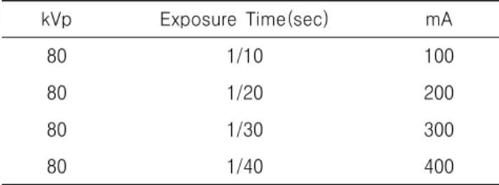
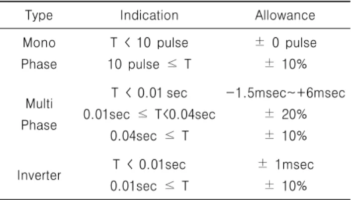
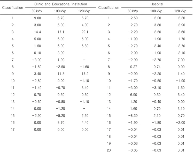
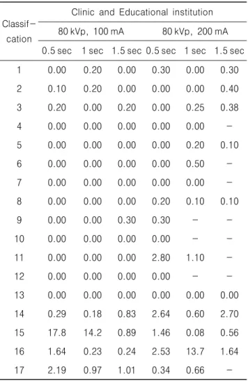
They are all in Seongnam-City. We used PMX-Ⅲ, kVp meter to implement kVp test, mR / mAs output test, light fiel / beam alignment test, Reproducibility of exposure dose, half value layer test, reproducibility of exposure time test. in the case of hospitals, they perceive the importance of regular quality control and organize the regular quality control team so they can be satisfied with the error standard in most experiments. On the other hand, when it comes to medical institutions and institute of educations, they perceive the importance of regular quality control less than hospitals do. Radiographer need to understand the importance of regular quality control and practice it so they can get the fine ideal image with the lower dose to the patient.

Key Words : Quality control, PMX-Ⅲ, kVp test, mR / mAs output test, Light field / beam alignment test

Ⅰ. Introduction

In modern medicine, As field of Radiation utilization is being expanded and the importance

Received August 24, 2010/ 1st Revised September 17, 2010/

Accepted for Publication October 20, 2010.

Corresponding Author: Byung Sam Kang

Department of Radiological Technology, Shingu University (462-743) 657, Gwangmyeong-ro, seongnam-si, Gyeonggi-do, Republic of Korea

Tel: 031) 740-1522 Fax: 031) 740-1589 E-mail: [email protected]

of this is growing, people try to reduce the

individual radiation exposure. And one of the

major things of those efforts is quality control

about general X-ray systems.

1

Thanks to the

great development of in radiation, we are now

able to reduce radiation exposure to patients and

workers and expense in medical sector. We are

also trying to produce ideal images which

maintain useful information. These kinds of effort

are increasing over the world.

2

We use the

standard evaluation research and the actual

(2)

condition investigation which are reported by 3 person except Jin-Soo Kim

3

, for developing the importance of quality control of General X-ray systems as the base of the investment. In this investment, we were targeted at medical institutions, institute of educations and general hospitals in Seongnam-City. first, then we researched on the actual condition of quality control and carried out the evaluation based on method of a safety management rule of the diagnosis radiation system, the diagnosis radiation system and QC, quality management for Radiog- raphic imaging, Korea Food&Drug Administration.

4~7

Ⅱ. Object and Method

1. Object

We were targeted at the General X-ray systems of medical institutions, institute of educations and hospitals in Seongnam-City. then we implemented many experiments of the safety supervision, such as kVp test, light field / beam alignment test, half value layer test, reproducibility of exposure dose, exposure time test. We substituted mAs test for and used the approximate value instead mA test. if the original one cannot be possibly used because of the equipment’s limited condition.

Lastly, we sent all the results to medical institutions, institute of educations and hospitals.

2. Method

1) kVp test

Voltage neighboring a X-ray tube determines X-ray volume as well as the energy generated in the X-ray tube. The purpose is to maintain contrast and photographic density of X-ray images consistently by maintaining kVp accurately and to reduce its exposure dose to patients.

Let a tube and PMX-Ⅲ warm up before test, and then place PMX-Ⅲ on the imaging stand. SID

section of PMX-Ⅲ, and then collimate. kVp measuring ranges are changed to 80, 100 and 120 kVp and each kVp are measured 5 times, 0.1 sec is fixed at the test. The test results are recorded on the measured value and have checked whether any abnormality is. The average error percentage of kVp(Percent Average Error : PAE) should be within ± 10% of the set point range. Use the following formula to check abnormality(Eq. 1).

  

 

    

×  (1)

PAE(Percent Average Error) Xp : Set value

 : Average of the set value

2) mR / mAs output test

It is designed to check if the same exposure always takes place by using mAs and kVp regardless of dose duration and mA combination. Place a Apron on the imaging stand that reduces back scattering, then puts PMX-Ⅲ on the table. Tests are conducted in the same procedure as kVp test and measured under the following conditions Table 1. mR / mAs ratios are calculated and recorded its value. After that, calculate changes of reproducibility. Reproducibility of mR / mAs output test must be within ± 10%.

3) Light field / beam alignment test

It is designed as accuracy of light field and beam alignment reduces unnecessary exposure dose and improves contrast of images. SID is fixed as 100 cm and a X-ray tube is located vertically onthe imaging stand. A collimator template is placed on t h e i m a g i n g s t a n d a n d t h e h o l e s i n t h e

Table 1. mR / mAs output test

kVp Exposure Time(sec) mA

80 1/10 100

80 1/20 200

(3)

template are identical to the right shoulder of a patient. Then, adjust beam alignment to match the rectangular exterior line of the template and place the beam alignment test tool in the center of collimator template. Shot condition irradiates in the hand exposure conditions. Images after taking images are measured and the results are recorded.

Surrounding error of beam alignment and light field must be within ± 2% of SID.

4) Reproducibility of exposure dose

The purpose is to assess quality and reliability of medical radiographic diagnostic devices. The measured value should be the same at every measurement when kVp, mAs, dose duration and imaging distance are set the same. It closely involves with the fact that picture density is the same at each imaging. Tests are conducted in the same procedure as mA test. measuring conditions are fixed at 80 kVp, 100 mA and 100 kVp 200 mA and dose duration is changed to 0.5, 1.0 and 1.5 sec. Each duration are irradiated 3 times respectively and the results are recorded. Lastly, calculate base on formula CV(Co-efficiency of Variation) using the results(Eq. 2). CV for the exposure dose of diagnostic radiation generating devices must be less than 0.05.

  

 (2)

5) Half value layer test

When X-rays are generated, They come from a tube as a number of energy beams and consist of various pulses and frequencies and soft lines and hard lines.

Soft lines have low energy and are absorbed in to soft tissues, thus, increase exposure dose to a patient. It is designed to check the tube settings to keep a proper level of exposure of a patient to

a minimum by using filters. For test, place a Apron on the imaging stand and place PMX-Ⅲ, half value layer test equipment and an X-ray tube. The distance between X-ray tube and the table is adjusted to be 100 cm and the condition is fixed at 80 kVp, 10 mA, 0.1 sec. The results of 2 times are recorded which decreases thickness of filters, opposite from thickness X-ray filters. And lastly, calculate the average of the above recorded and then generate the half value thickness by drawing an attenuated curve. At 80 kVp, half value layer must be more than equivalent to 2.3 mmAl.

6) Reproducibility of exposure time test The exposure time is one of the factor that depend on controlling exposure dose and affects to make an image. When we get a superior image to diagnose, we need to maintain stable exposure time. Place the PMX-Ⅲ on the imaging stand before test. Controlling light field, it matches the detecting area. The factor(80 kVp, 100 mA) is fixed and the time(0.05, 0.2, 0.4 sec) is changed. At each factor, we conduct the test for 3 times and write the result on the sheet. Averaging the result of test, we check the reproducibility of exposure time. Allowable error is the same as Table 2.

Table 2. Allowable error of Reproducibility of exposure time test

Type Indication Allowance

Mono Phase

T < 10 pulse 10 pulse ≤ T

± 0 pulse

± 10%

Multi Phase

T < 0.01 sec 0.01sec ≤ T<0.04sec

0.04sec ≤ T

-1.5msec~+6msec

± 20%

± 10%

Inverter T < 0.01sec 0.01sec ≤ T

± 1msec

± 10%

(4)

Table 3. kVp test of Clinic and Educational institution, Hospital (unit : %) Classification

Clinic and Educational institution

Classification

Hospital 

80 kVp 100 kVp 120 kVp 80 kVp 100 kVp 120 kVp

1 9.00 6.70 6.70 1 -2.50 -2.20 -2.30

2 3.00 5.00 4.00 2 -2.70 -3.80 -2.90

3 14.4 17.1 22.1 3 -2.20 -2.50 -2.60

4 5.00 6.00 5.00 4 -1.90 -1.90 -1.70

5 1.50 6.00 6.80 5 -2.70 -2.40 -2.70

6 0.10 3.00 - 6 -2.00 -1.90 -2.10

7 -3.00 1.00 - 7 -2.90 -2.70 7.00

8 -1.50 -2.50 -1.60 8 0.27 0.74 0.00

9 3.40 11.5 17.2 9 -2.90 -2.20 1.40

10 -2.80 0.00 -1.10 10 -1.70 -0.50 -1.90

11 -1.40 -0.70 3.40 11 -3.00 -3.10 1.60

12 0.70 0.50 0.60 12 6.90 9.50 6.40

13 -0.60 -0.80 -1.10 13 1.20 -0.40 0.00

14 0.00 -1.20 - 14 1.60 0.70 3.10

15 -2.90 -5.20 2.50 15 -8.30 2.10 0.70

16 0.00 3.70 4.40 16 -1.90 -1.80 -2.00

17 0.00 0.00 0.00 17 -0.04 -0.03 0.01

18 -0.04 -0.03 0.01

19 -0.06 -0.03 0.01

20 -0.05 -0.03 0.01

Ⅲ. Result

1. kVp test

The setting of the PAE at the kVp test must be within the range of ± 10%. According to clinic and educational institution, most of the equipments were normal but 2 pieces of equipment were abnormal in measurement results of 17 pieces of equipment. In case of Hospital, 20 pieces of equipment all showed normal values. Measurement results are Table 3.

2. mR / mAs output test

According to the result of mAs test, the majority of clinic and educational institution and hospital showed that the results were normal except for the clinic only. A few pieces of equipment showed that the setting of measurement condition was impossible or measurement range was out of normal value.

Measurement results are Table 4.

(5)

Table 4. mAs test of Clinic and Educational institution, Hospital (unit : %)

Classif- cation

Clinic and Educational

institution

Classifi-

cation Hospital

1 7.00 1 0.10

2 - 2 3.60

3 1.10 3 5.50

4 - 4 -

5 17.0 5 0.40

6 - 6 0.60

7 - 7 2.50

8 9.50 8 0.60

9 1.60 9 0.50

10 1.70 10 0.20

11 - 11 0.50

12 0.30 12 -

13 8.80 13 3.30

14 5.55 14 5.00

15 4.56 15 4.90

16 4.54 16 1.10

17 5.43 17 5.45

18 5.56

19 4.24

20 6.43

3. Beam alignment test

Surrounding error of beam alignment and light field must be within ± 2% of SID. The result of beam alignment test indicated 11.7% from clinic and educational institution and 10% form Hospital.

The results are Table 5.

4. Exposure dose test

In reproducibility of exposure dose test, coefficient of variation of the measured values

Table 5. Beam alignment of Clinic and Educational institution, Hospital (unit : cm)

Classifi- cation

Clinic and Educational

institution

Classif- cation

Hospital

Rt→Lt Up→Dn Rt→Lt Up→Dn

1 1.50 2.00 1 1.00 0.50

2 0.50 1.80 2 0.30 0.30

3 1.50 1.50 3 0.30 0.30

4 2.10 1.50 4 0.30 0.30

5 0.50 0.80 5 0.50 0.50

6 0.50 1.80 6 0.50 0.50

7 1.00 1.50 7 1.00 1.00

8 1.00 1.00 8 1.30 1.30

9 0.50 1.00 9 0.50 0.50

10 0.50 1.00 10 0.50 0.50

11 1.80 1.50 11 0.50 0.50

12 0.80 1.00 12 0.80 0.30

13 0.50 1.00 13 0.30 0.50

14 1.50 1.30 14 2.30 1.20

15 1.20 2.00 15 2.30 0.30

16 1.50 3.00 16 0.30 0.30

17 0.00 0.00 17 2.00 0.25

18 1.50 1.00

19 1.00 1.00

20 0.70 2.00

must be within ± 5% in width. Clinic and education indicated error of 1 device from the factor of 100 mA and 1 devise of 17 devises from the factor of 200 mA. Hospital indicated error of 1 device of 20 devices form the factor of 100 mA and 1 device didn't selected the factor of 100 mA.

In addition the factor of 200 mA indicated error

of 1 device of 18 devices and 1 device didn’t

selected the factor of 200 mA. The results are

Table 6, 7.

(6)

Table 6. Exposure dose test of Clinic and Educational institution (unit : %)

Classif- cation

Clinic and Educational institution 80 kVp, 100 mA 80 kVp, 200 mA 0.5 sec 1 sec 1.5 sec 0.5 sec 1 sec 1.5 sec 1 0.00 0.20 0.00 0.30 0.00 0.30 2 0.10 0.20 0.00 0.00 0.00 0.40 3 0.20 0.00 0.20 0.00 0.25 0.38

4 0.00 0.00 0.00 0.00 0.00 -

5 0.00 0.00 0.00 0.00 0.20 0.10

6 0.00 0.00 0.00 0.00 0.50 -

7 0.00 0.00 0.00 0.00 0.00 -

8 0.00 0.00 0.00 0.20 0.10 0.10

9 0.00 0.00 0.30 0.30 - -

10 0.00 0.00 0.00 0.00 - -

11 0.00 0.00 0.00 2.80 1.10 -

12 0.00 0.00 0.00 0.00 - -

13 0.00 0.00 0.00 0.00 0.00 0.00 14 0.29 0.18 0.83 2.64 0.60 2.70 15 17.8 14.2 0.89 1.46 0.08 0.56 16 1.64 0.23 0.24 2.53 13.7 1.64

17 2.19 0.97 1.01 0.34 0.66 -

5. Half Value Layer test

HVL test is over 2.3 mmAl when its output is 80 kVp and if it could output over 70 kVp. In clinic and educational institution cases, we were found error on a device of 17 devices. On the other hand, all devices of Hospital were normally work.

The results are on Table 8.

Table 7. Exposure dose test of Hospital (unit : %)

Classif- cation

Hospital 

80 kVp, 100 mA 80 kVp, 200 mA 0.5 sec 1 sec 1.5 sec 0.5 sec 1 sec 1.5 sec 1 0.00 0.00 0.00 0.00 0.00 0.00 2 0.00 0.00 0.00 0.00 0.00 7.10 3 0.00 0.00 0.00 0.00 0.00 0.00 4 0.00 1.00 0.00 0.00 0.00 0.00 5 0.00 0.00 0.00 0.00 0.00 0.00 6 1.00 0.00 0.00 0.00 0.00 0.00 7 0.00 0.00 0.00 0.00 0.00 0.00 8 0.00 0.00 0.00 0.30 0.40 0.30 9 2.10 0.30 1.20 0.40 0.20 0.00 10 1.50 0.30 0.10 0.00 0.40 0.20 11 0.50 0.30 0.70 1.10 0.10 2.30

12 0.00 - - 0.00 0.00 0.00

13 0.00 0.00 0.00 0.00 0.00 0.00 14 0.10 0.00 0.00 0.02 0.02 0.05 15 0.00 0.00 0.00 1.54 0.15 1.80 16 0.00 87.00 0.00 0.00 1.45 0.16 17 0.00 0.00 0.04 0.15 0.04 0.03 18 0.29 0.51 0.39 0.12 0.42 0.24 19 1.26 1.14 1.03 1.01 0.99 0.83 20 0.03 0.03 0.11 0.01 0.02 0.09

6. Exposure time test

Coefficient of variation width measured value

must be in ± 10% at exposure time test. In clinic

and educational institution cases, we were found

error on 2 devices of 17 devices. And we couldn't

control exposure condition of 4 of them. In hospital

cases, we were found error on 2 devices of 20

devices. The results are on Table 9.

(7)

Table 8. HVL test of Clinic, Educational institution and Hospital (unit : mmAl)

Classif- cation

Clinic and Educational institution

Classifica

-tion Hospital

1 3.9 1 3.9

2 2.8 2 3.6

3 3.1 3 3.6

4 3.0 4 3.8

5 3.2 5 4.0

6 3.0 6 3.9

7 1.9 7 3.4

8 3.0 8 3.0

9 3.1 9 3.7

10 3.6 10 3.6

11 2.8 11 3.8

12 3.8 12 3.8

13 2.9 13 3.1

14 3.5 14 3.8

15 2.6 15 4.4

16 3.0 16 3.5

17 3.0 17 2.6

18 2.7

19 2.5

20 2.4

Ⅳ. Discussion

Each institution relatively comparison is meaningless because the number of devices differ to the hospital and educational institutions. In the experimental results of the hospital, they have recognized the importance of quality control. And they have a quality control team in oder to act for active quality control activities. According to mostof the experimental results, standard of test was satisfied. But In the experimental results of clinic and educational institutions, the importance and recognition of quality control were relatively

Table 9. Exposure time test of Clinic, Educational institution and Hospital (unit : %) Classif-

cation

Hospital  Hospital 

50 200 400 50 200 400

1 2.60 1.40 0.70 3.00 0.30 0.20

2 - 2.00 1.10 1.60 0.10 0.00

3 4.20 1.10 0.50 1.50 0.30 0.00 4 -10.0 -10.00 -9.20 2.00 0.40 -9.00 5 13.8 1.90 1.00 2.40 0.20 0.10

6 - 1.80 3.20 2.30 0.30 0.10

7 - 2.18 0.80 1.00 0.10 0.00

8 3.90 1.00 0.30 2.40 0.10 0.00 9 6.00 0.10 0.10 2.60 0.10 0.10 10 15.00 8.00 26.00 3.70 0.20 0.10

11 - 2.80 1.30 2.10 0.10 0.10

12 3.40 0.40 0.30 21.00 7.00 -12.00 13 3.30 1.00 0.50 0.00 0.00 0.00 14 0.10 0.06 0.09 0.90 0.00 0.00 15 0.10 0.10 0.10 14.00 0.80 0.40 16 0.18 0.67 0.71 6.00 1.40 1.10 17 0.02 0.05 0.02 0.08 0.05 0.05

18 0.09 0.02 0.05

19 0.01 0.08 0.08

20 0.43 1.58 0.62

low. In case of clinic and educational institutions,

medical radiography diagnostic devices was tested

in 17 units. As a result of test, they have errors of

2 devices in kVp test(11.7%), 1 device in reproducibility

of exposure dose(5.8%), 2 devices in Light field /

beam alignment test(11.7%), 1 device in Half value

layer test(5.8%), 2 devices in reproducibility of

exposure time test(11.7%). In case of hospital,

medical radiography diagnostic devices was tested

in 20 units. As a result of test, they have errors

of 2 devices in Light field / beam alignment test

(10%), 2 device in reproducibility of exposure dose

(10%), 2 devices in reproducibility of exposure time

(8)

test(10%). When compared with the results of one study

8

, kVp test in clinic and hospital of 50 devices indicated similar results(7 devices, 14%).

And 2 device in reproducibility of exposure dose(4%) was relatively unsuitable. When compared with our result, HVL test indicated high levels unsuitable result(24 device, 48%). It means to make the superior quality image to reduce the scatter ray. When compared with the results of one study

9

, clinics and hospitals in the kVp test and reproducibility of exposure dose test in the 5 devices(25%). Test the reproducibility of exposure time in the 4 devieces(20%) when compared with our test indicated high levels. The Exposure time which is the factor to make the image. Thus, analyzing the reproducibility of exposure time, it can reduce the scatter ray. Therefore, regular maintenance is required. In the case of kVp test and reproducibility of exposure time test in hospital was same result And reproducibility of exposure dose test in the 2 devices(20%) indicated a high level. When compared with the results of one study, light field/beam alignment test in 52 devices indicated unsuitable result(19.2%).

10

That is indicated slightly higher result of our one. So as to obtain X-ray generating device for always the same output must manage regularly. If it have poor reproducibility, X-ray film of the contrast, sharpness, concentration, etc can be worse. If you have problems in these factors, exposure of patients can be caused by many problems.

According to the rules of diagnostic radiation generator on the safety management, operation of collimator whether it would work well should check daily. Compatibility of light and X-ray field should manage to half a year. The duty of radiographer is that they should make the effort to reduce scatter ray, and they need to manage the making good quality image. So we recognize the importance of quality control and need effort and investigation

Ⅴ. Conclusion

The objective of an X-ray examination is to produce images of the patient of sufficient quality to provide adequate diagnostic information for a clinician. Therefore, the quality control of the x-ray system are important that should be maintain the regularly and radiographer need to understand the importance of regular quality control and practice it so they can get the fine ideal image with the lower dose to the patient.

References

1. Dong KR, Lee SJ, Kweon DC, Goo EH, Jung JE, Lee KS. Actual Condition of Quality Control of X-ray Imaging System in Primary Care Institution: focused on Gwangju Metropolitan City. Journal of Radiation Protection 2010; 35:

34-42.

2. Kang SS, Kweon DM, Kim YK, Kim CK, Lee Sk, Jang YI. Diagnostic Imaging Device charge of Quality Assurance tests. Seoul: CHUNG-KU Publishes; 2003.

3. Kim JS, Kim HA, Jung SE, Yung SP. General recording equipment, the importance of the quality control standards for evaluation research and survey. The Journal of Radiological Technology of Shingu University 2009; 25: 118-25.

4. Ardran GM, Crooks HE. Checking Diagnostic X-ray beam quality. Br J Radiol 1968; 41:

183-98.

5. Schreiner-Karoussou A. Review of image quality standards to control digital X-ray systems. Radiat Prot Dosimetry 2005; 117:

23-5.

6. Baorong Y, Kramer HM, Selbach HJ, Lange B.

Experimental determination of practical peak

voltage. Br J Radio 2000; 73: 641-9.

(9)

7. Ramirez-Jimenez FJ, Lopez-Callejas R, Benitez- Read JS, Pacheco-Sotelo JO. Considerations on the measurement of practical peak voltage in diagnostic radiology. Br J Radiol 2004; 77: 745-50.

8. Kim JM. A Comparative Study on Output of Four Type Diagnostic X-ray Equipments.

Journal of Radiological Science and Technology 1997; 20: 34-43.

9. Park JH, Im IC, Dong KR, Kang SS. A Performance Evaluation of Diagnostic X-ray Unit Depends on the Hospitals Size. Journal of Radiation Protection 2009; 34: 31-6.

10. Kim SS, Huh J, Lee SS, Lee IJ, Kim SC.

Investigation on Accuracy Contral of Collimator in Diagnostic X-ray Equipments. Journal of Radiological Science and Technology 1994; 17:

65-70.

수치

Table  1.  mR / mAs  output  test
Table  2.  Allowable  error  of  Reproducibility  of  exposure time  test
Table  3.  kVp  test  of  Clinic  and  Educational  institution,  Hospital                                                                                 (unit  :  %) Classification
Table  4.  mAs  test  of  Clinic  and  Educational  institution, Hospital                                                                     (unit  :  %)  Classif-cation Clinic  and  Educational  institution Classifi-cation Hospital 1 7.00 1 0.10 2 - 2 3.
+3

참조

관련 문서

After first field tests, we expect electric passenger drones or eVTOL aircraft (short for electric vertical take-off and landing) to start providing commercial mobility

1 John Owen, Justification by Faith Alone, in The Works of John Owen, ed. John Bolt, trans. Scott Clark, &#34;Do This and Live: Christ's Active Obedience as the

Surface morphology and chemical composition of samples were characterized by field emission scanning electron microscopy (FE-SEM), contact angle measurement and

XAFS: X-ray absorption fine structure XES: X-ray emission spectroscopy XRF: X-ray fluorescence.. Use of x-rays; a probe based

quality of life and improved standard of living, and as reflected in this popular desire, kiosks provide various forms of food-related information and have

Electron probe micro-analyzer image of Zr-4%Si binary alloys; (a).. X-ray diffraction profiles of as-cast Zr-xSi binary alloys, which shows the dual phases.. Magnetic

Excitation Detection X-ray photoelectron spectroscopy (XPS) Photons(X-ray) Electrons UV photoelectron spectroscopy (UPS) Photons (UV) Electrons

Direct method of solution of variational problem is one of the most powerful tools for obtaining numerical results in practical problems of engineering