Inhibition of Oxidative Damage by Phlorotannins from Ecklonia cava in Normal Human Dermal Fibroblasts
10
0
0
전체 글
(2) Inhibition of Oxidative Damage by Phlorotannins from Ecklonia cava. 127. researches have reported that Ecklonia species exhibit radical scavenging activity [15,16,29]. It is expected that high content of phlorotannins present in Ecklonia species is responsible for above bioactivities. However, inhibitory effects of phlorotannins from Ecklonia species on generation of reactive oxygen species have not been reported in live cell systems in detail. Therefore, we investigated the inhibitory effects of phlorotannins from EC extract on oxidative stress in human dermal fibroblasts (HDFs) that play a major role in wrinkle formation.. form and ethyl acetate. The respective fractions were evaporated at 40 under vacuum. The w/w yields of n-hexane, chloroform, ethyl acetate, and water fractions were about 13.61%, 12.61%, 41.58%, 32.20%, respectively. For the bioassay, the fractions were dissolved in dimethylsulfoxide (DMSO) and distilled water according their solubility, respectively.. Materials and Methods. Ciocalteu method [32], using phloroglucinol as the standard. Samples were diluted taking into account the measurable range of the spectrophotometer (e.g., a 0.005 ml aliquot of extract of soluble phenolics was mixed with 0.495 ml water). A 0.1 ml aliquot of the diluted sample was mixed in a test tube with 1.0 ml of 1 N Folin-Ciocalteu reagent. The mixture was allowed to stand for 3 min following addition of 2.0 ml of 20% Na2CO3. Samples were incubated in the dark at room temperature for 45 min and centrifuged at 1600×g for 8 min. Optical density (OD) of the supernatant was measured at 730 nm using a GENios® microplate reader (Tecan Austria GmbH, Austria). Total phlorotannin content is calculated using the standard graph plotted and expressed as a percentage.. Materials Dulbecco’s Modified Eagle’s Medium (DMEM), RPMI 1640 Medium, Trypsin-EDTA, penicillin/ streptomycin/ amphotericin (10,000 U/ml, 10,000 μg/ml, and 2,500 μg/ml, respectively) and fetal bovine serum (FBS) were obtained from Gibco BRL, Life Technologies (USA). U-937 and HL-60 cell lines were obtained from American Type of Culture Collection (Manassas, VA, USA). Human dermal fibroblasts (HDFs) were kindly donated by LG HG & CM Research Institute (Daejeon, Korea). MTT reagent, phloroglucinol and Folin-Ciocalteu reagent were purchased from Sigma Chemical Co. (St. Louis, MO, USA). 2’,7’-dichlorofluorescin diacetate (DCFH-DA), diphenyl-1-pyrenylphosphine (DPPP) and monobromobimane were purchased from Molecular probes (Eugene, OR, USA). All the other materials required for culturing of cells were purchased from Gibco BRL, Life Technologies (USA).. Preparation of phlorotannins from EC extract The brown seaweed, EC was collected along Jeju Island coast of Korea during the period from October 2004 to March 2005. Fresh EC was washed three times with tap water to remove salt, epiphytes and sand attached to the surface of the samples and stored at -20 . The frozen samples were lyophilized and homogenized using a grinder before extraction. The dried EC powder (1 kg) was extracted three times with 10 L of MeOH (1:10 w:v) at room temperature for 24 h while stirring, filtered using Whatman No. 41 and evaporated in vacuum. The w/w yield of extracts was about 11.3%. After water was added, the aqueous layer of the 95% MeOH extract was partitioned with n-hexane, chloro-. ℃. ℃. Spectrophotometric determination of phlorotannin Total phlorotannins contents in the EC fractions were determined according to a modified version of Folin–. Cell culture Cell lines were separately grown as monolayers in T-75 tissue culture flasks (Nunc, Denmark) at 5% CO2 and 37 ºC humidified atmosphere using appropriate media supplemented with 10% fetal bovine serum, 2 mM glutamine and 100 μg/ml penicillin-streptomycin. DMEM was used as the culture medium for human dermal fibroblasts (HDFs) cultured primarily from human fetal foreskin. Adherent cells were passaged 2 times a week by treating with trypsin-EDTA and used for experiments after 5 passages.. MTT assay Cytotoxic levels of phlorotannins from EC extract on cultured cells were measured using MTT (3-(4,5-dimethyl-2-yl)-2,5-diphenyltetrazolium bromide) method as described by [10]. The cells were grown in 96-well plates at a density of 5 × 10³ cells/well. After 24 h, cells were washed with fresh medium and were treated with different concentrations of phlorotannins. After 48.
(3) 128. KIM et al.. h of incubation, cells were rewashed and 100 μl of MTT (1 mg/ml) was added and incubated for 4 h. Finally, DMSO (150 μl) was added to solubilize the formazan salt formed and amount of formazan salt was determined by measuring the OD at 540 nm using an GENios® microplate reader (Tecan Austria GmbH, Austria). Relative cell viability was determined by the amount of MTT converted into formazan salt. Viability of cells was quantified as a percentage compared to the control (OD of treated cells OD of blank / OD of control OD of blank×100) and dose response curves were developed. The data were expressed as mean from at least three independent experiments and P < 0.05 was considered significant.. −. −. Measurement of radicals by ESR. Different radicals were generated according to previously mentioned procedures and spin adducts were recorded using a JES-FA electron spin resonance (ESR) spectrometer (JEOL, Tokyo, Japan) at 25 °C. Instrument settings were as follows: magnetic field 336 ± 5 mT; sweep time 30 s; sweep width 10 mT; modulation width 0.1 mT and modulation frequency 100 kHz.. DPPH radical. DPPH radical was measured using the method described by Nanjo et al. [22]. Sixty microliters of sample in ethanol (or ethanol itself as a control) was added to 60 μl DPPH (60 μM) and vortexed for 10 s. Mixture was transferred to a sealed capillary tube and after 2 min DPPH radical spin resonance was recorded at 1 mW microwave power and 1000 amplitude.. Hydroxyl radical generation system. Fenton reaction was performed by reacting 50 μl of 10 mM FeSO4 and 50 μl of 10 mM H2O2 to generate hydroxyl radicals [28]. Generated radicals were trapped by 50 μl of 0.3 M DMPO in the presence of sample solution (50 μl) or same volume of phosphate buffer (pH 7.4) as a control. After 2.5 min, the reaction mixture was transferred to a sealed capillary tube and DMPO/•OH spin adduct was recorded at 1 mW microwave power and 400 amplitude.. Alkyl radical generation system. Carbon-centered radicals were generated by AAPH [14]. Twenty microliters of phosphate buffered-saline (7.4 pH), AAPH (40 mM), 4-POBN (40 mM) and sample solution were mixed and incubated at 37ºC for 30. min. Following incubation, reaction mixture was transferred to a sealed capillary tube and 4-POBN/carbon-centered spin adduct was recorded at 5 mW microwave power and 500 amplitude.. Determination of intracellular formation of reactive oxygen species (ROS) using DCFHDA labeling Intracellular formation of reactive oxygen species (ROS) was assessed as described previously using oxidation sensitive dye 2’,7’-dichlorofluorescin diacetate (DCFH-DA) as the substrate [23]. HDFs growing in fluorescence microtiter 96-well plates were loaded with 20 μmol/L DCFH-DA in HBSS and incubated for 20 min in the dark. Nonfluorescent DCFH-DA dye, that is freely penetrated into cells get hydrolyzed by intracellular esterases to 2’,7’- dichlorodihydrofluororescein (DCFH), and tarps inside the cells. Cells were then treated with different concentrations of test compounds and incubated for 1 h. After washing the cells with PBS for three times, 500 umol/L H2O2 dissolved in HBSS was added to cells. The formation of 2 7 -dichlorofluorescein (DCF) due to oxidation of DCFH in the presence of various ROS was read after every 30 min at the excitation wavelength (Ex) of 485 nm and the emission wavelength (Em) of 528 nm using a GENios® fluorescence microplate reader (Tecan Austria GmbH, Austria). Following maximum rate of fluorescence increase, each well was normalized to cell numbers using MTT cell viability assay. Dose dependant and time dependant effects of treatment groups were plotted and compared with fluorescence intensity of control and blank groups.. ′′. Lipid peroxidation of live cells using DPPP. Intracellular lipid hydroperoxides were detected using a fluorescence probe, diphenyl-1-pyrenylphosphine (DPPP) as described previously with slight modifications [30]. Cells growing in 10 cm culture dishes were washed three times with PBS and after addition of 13 uM DPPP in PBS, cells were incubated at 37ºC for 30 min in the dark. Cells were washed three times with PBS and seeded into fluorescence microtiter 96-well plates at a density of 1×10⁸ cells/ml using serum free media. After attached completely, cells were treated with predetermined sample concentrations and incubated for 1 h. Following addition of 3 mM AAPH in PBS, fluorescence intensity was measured (excitation 361 nm and emission, 380 nm) us-.
(4)
(5)
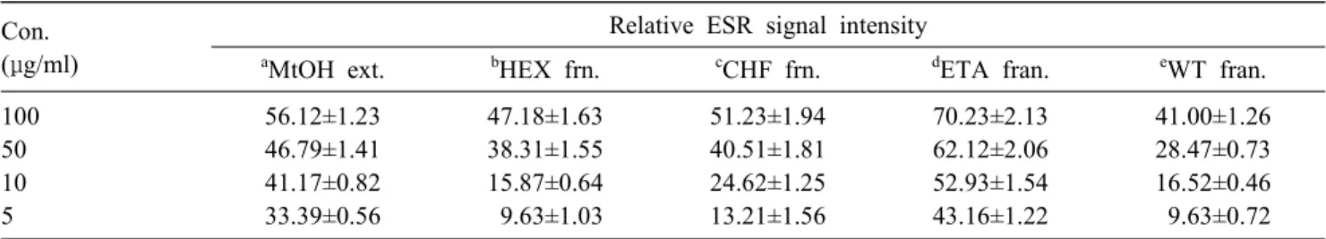
(6) Inhibition of Oxidative Damage by Phlorotannins from Ecklonia cava. 131. Table 2. Scavenging effect of phlorotannins from EC extract and solvent fractions on hydroxyl radical Relative ESR signal intensity. Con. (μg/ml). a. MtOH ext.. b. 100 50 10 5. 56.12±1.23 46.79±1.41 41.17±0.82 33.39±0.56. 47.18±1.63 38.31±1.55 15.87±0.64 9.63±1.03. a. c. HEX frn.. CHF frn.. 51.23±1.94 40.51±1.81 24.62±1.25 13.21±1.56. d. e. 70.23±2.13 62.12±2.06 52.93±1.54 43.16±1.22. 41.00±1.26 28.47±0.73 16.52±0.46 9.63±0.72. ETA fran.. WT fran.. Methanol extract, bHexane fraction, cChloroform fraction, dEthyl acetate faction, eWater fraction. Table 3. Scavenging effect of phlorotannins from EC extract and solvent fractions on alkyl radical Relative ESR signal intensity. Con. (μg/ml). a. MtOH ext.. b. 100 50 10 5. 81.70±2.30 72.44±1.83 8.23±0.24 6.96±0.86. 33.24±0.63 19.16±0.29 5.97±0.38 1.47±0.20. HEX frn.. c. CHF frn.. 18.27±0.76 14.72±1.03 6.83±0.46 2.91±0.14. d. e. 93.58±2.13 80.68±2.05 13.87±0.23 11.42±0.77. 68.12±1.21 34.00±0.94 2.65±0.76 0.15±0.05. ETA fran.. WT fran.. a. Methanol extract, bHexane fraction, cChloroform fraction, dEthyl acetate faction, eWater fraction. Inhibitory effect of phlorotannins from EC methanol extract and other organic solvent fractions on intracellular ROS level generated by hydrogen peroxide in human dermal fibroblasts The ability of phlorotannins from EC samples to scavenge ROS was assessed in time and concentration dependent manners in normal human dermal fibroblast. Following treatment of cells with hydrogen peroxide, intracellular ROS was measured using DCFH-DA that has widely used to monitor intracellular oxidant stress by which it converted into DCF emitting fluorescence. DCF fluorescence intensity was increased with incubation time following treatment of HDFs with 0.5 mM hydrogen peroxide compared with blank group in Fig. 3. The scavenging activity of intracellular ROS was increased in time dependent manner in all treatment groups. There was no significant difference in scavenging activity of intracellular ROS between methanol extract and ethyl acetate fraction at 100 μg/ml. However, ethyl acetate fraction exhibited higher scavenging activity than that of methanol extract with increasing the concentration of EC samples. While methanol extract of EC show no scavenging activity below 10 μg/ml effect, ethyl acetate fraction of EC showed a significant scavenging effect on intracellular ROS even at 5 μg/ml ( P < 0.01). These results indicate that intracellular ROS level induced by hydrogen peroxide in cells can be decreased by phlorotannins from EC ethyl acetate fraction.. Inhibitory effect of phlorotannins from EC methanol extract and solvent fractions on lipid peroxidation of live cells We next examined the inhibitory effect of phlorotannins from EC samples on lipid peroxidation of cells. AAPH was treated to cells to generate peroxy radical that initiate directly lipid peroxidation in a variety of membrane systems. To detect lipid peroxidation of cells, human dermal fibroblasts were labeled with DPPP that reacts with hydroperoxides of cell membrane and resulting DPPP oxide is fluorescent. As shown in Fig. 4, fluorescent intensity of live cells induced by AAPH was decreased by all treatment groups at 100 μg/ml. However, there was no significant inhibitory effect on lipid peroxidation of live cells in all groups at 5 μg/ml except for ethyl acetate fraction of EC that exhibited 30% inhibitory effect on lipid peroxidation. Furthermore, phlorotannins from ethyl acetate fraction of EC showed a dose dependent inhibitory effect on lipid peroxidation.. Inhibitory effect of phlorotannins from EC methanol extract and solvent fractions on glutathione level in human dermal fibroblasts. To investigate whether EC samples increase redox potential in cells, phlorotannins from EC samples were treated to human dermal fibroblasts in concentration and time dependent manners, and then intracellular.
(7)
(8)
(9) 134. KIM et al.. effect through increment of GSH level that protects cells by scavenging free radicals and by reducing hydrogen peroxide or organic peroxides, these reaction being catalyzed by glutathione s-transferases and by glutathione peroxidase, respectively. In conclusion, our results suggest that phlorotannins from EC extract could have a therapeutic potential in prevention and treatment of several diseases such as wrinkle formation and chronic inflammation related to oxidative stress.. 2.. 3.. 4. 요. 약. Phloroglucinol 단위체로 구성된 Oligomeric polyphenol인 Phlorotannins을 갈조류의 일종인 감태( Ecklonia cava )의 메탄올 추출물의 용매 분획으로부 터 분리하였다. 산화스트레스에 대한 감태추출물의 용매분획으로부터 Phlorotannins의 억제효능을 주름 형성과 연관성이 있는 사람피부섬유아세포(HDFs) 에서 조사되었다. ESR spectroscopy 분석에서, 감태 의 에틸아세테이트 분획으로 부터의 Phlorotannins에 서 DPPH radical, Hydroxyl radical 및 Alkyl radical 에 대하여 가장 높은 소거효능이 나타났다. 2’,7’-Dichlorofluorescin diacetate (DCFH-DA)와 Diphenyl1-pyrenylphosphine (DPPP)을 이용하여 세포내에서 활성산소종 및 지질과산화 수준을 측정하였다. 감태 의 다른 용매 분획과 비교하여 에틸아세테이트 용매 분획으로 부터의 Pphlorotannins의 존재에서 이들수 준이 유의성 있게 감소되었다 (P < 0.01). 더욱이, Phlorotannins은 세포내의 Glutathione (GSH) 함량도 시간에 따라 증가시켰다. 그러므로, 이러한 결과들은 감태의 Phlorotannins이 산화적 스트레스와 연관성이 있는 주름형성 같은 여러가지 질환의 예방 및 치료에 잠재적인 효능이 있다는 것을 암시하고 있다. Acknowledgement. The authors acknowledge Marine Bioprocess Research Center of Marine Bio 21 Project, funded by the Ministry of Maritime Affairs and Fisheries, Republic of Korea, for the support provided through the research grant, B-2005-02.. 5.. 6.. 7.. 8.. 9.. 10.. 11.. 12.. 13.. References. 1. Ahn, M. J., Yoon, K. D., Min, S. Y., Lee, J. S., Kim, J. H., Kim, T. G., Kim, S. H., Kim, N. G., Huh, H. and Kim, J. W. 2004. Inhibition of HIV-1 reverse transcriptase and protease and protease by phlorotannins from. 14.. the brown algae Ecklonia cava. Biol. Pharm. Bull. 27,544-547. Auddy, B., Ferreira, M., Blasina, F., Lafon, L., Arredondo, F., Dajas, F., Tripathi, P. C., Seal, T. and Mukherjee, B. 2002. Screening of antioxidant activity of three Indian medicinal plants, traditionally used for the management of neurodegenarative diseases. J. Ethnopharmacol. 84, 131-138. Aviram, M., Kaplan, M., Rosenblat, M. and Fuhrman, B. 2005. Dietary antioxidants and paraoxonases against LDL oxidation and atherosclerosis development. Handb. Exp. Pharmacol. 170, 263-300. Choi, C. W., Kim, S. C., Hwang, S. S., Choi, B. K., Ahn, H. J., Lee, M. Y., Park, S. H. and Kim, S. K. 2002. Antioxidant activity and free radical scavenging capacity between Korean medicinal plants and flavonoids by assay-guided comparison. Plant Sci. 163, 1161 1168. Dhalla, N. S., Temsah, R. M. and Netticadan, T. 2000. Role of oxidative stress in cardiovascular diseases. J. Hypertens 18, 655-73. Eaton, P. 2006. Protein thiol oxidation in health and disease: Techniques for measuring disulfides and related modifications in complex protein mixtures. Free Radic. Biol. Med. 40, 1889-1899. Eddeb, M. F. and Pickering, A. T., 2000. Comparison of relative antioxidant activities of British medicinal plant species in vitro. J. Ethnopharmacol. 72, 47-51. Formica, J. V. and Regelson, W. 1995. Review of the biology of quercetin and related bioflavonoids. Food Chem. Toxicol. 33, 1061-1080. Fukuyama, Y., Kodama, M., Miura, I., Kinzyo, Z., Mori, H., Nakayama, Y. and Takahashi, M. 1990. Anti-plasmin inhibitor. VI. Structure of phlorofucofuroeckol A, a novel phlorotannin with both dibenzo-1,4-dioxin and dibenzofuran elements, from Ecklonia kurome Okamura. Chem. Pharm. Bull. 38, 133-135. Hansen, M. B.., Nielsen, S. E. and Berg, K., 1989. Re-examination and further development of a precise and rapid dye method for measuring cell growth/cell kill. J. Immunological Methods 119, 203-210. Harper, M. E., Bevilacqua, L., Hagopian, K., Weindruch, R. and Ramsey, J. J. 2004. Ageing, oxidative stress, and mitochondrial uncoupling. Acta Physiologica Scandinavica 182, 321-331. Henrotin, Y. E., Bruckner, P. and Pujol, J. P. 2003. The role of reactive oxygen species in homeostasis and degradation of cartilage. Osteoarthritis Cartilage 11, 747-755. Heo, S. J., Park, P. J., Park, E. J., Kim, S. K. and Jeon, Y. J. 2005. Antioxidant activity of enzymatic extracts from a brown seaweed Ecklonia cava by electron spin resonance spectrometry and comet assay. Eur. Food Res. Technol. 221, 41-47. Hiramoto, K., Johkoh, H., Sako, K. I. and Kikugawa, K. 1993. DNA breaking activity of the carbon-centered radical generated from 2,2¢-azobis(2 amidinopropane) hydrochloride (AAPH). Free Rad. Res. Commun. 19, 323-332.. –.
(10) Inhibition of Oxidative Damage by Phlorotannins from Ecklonia cava. 15. Kang, K. A., Lee, K. H., Chae, S., Koh, Y. S. and Yoo, B. S., Kim, J. H., Ham, Y. M., Baik, J. S., Lee, N. H. and Hyun, J. W. 2005. Triphlorethol-A from Ecklonia cava protects V79-4 lung fibroblast against hydrogen peroxide induced cell damage. Free Radic. Res. 39, 883-892. 16. Kang, K. A., Lee, K. H., Chae, S., Zhang, R., Jung, M. S., Lee, Y., Kim, S. Y., Kim, H. S., Joo, H. G., Park, J. W., Ham, Y. M., Lee, N. H. and Hyun, J. W. 2005. Eckol isolated from Ecklonia cava attenuates oxidative stress induced cell damage in lung fibroblast cells. FEBS Lett. 579, 6295-6304. 17. Kang, H. S., Chung, H. Y., Kim, J. Y., Son, B. W., Jung, H. A. and Choi, J. S. 2004. Inhibitory phlorotannins from the edible brown alga Ecklonia stolonifera on total reactive oxygen species (ROS) generation. Arch. Pharmacal. Res. 27, 194-198. 18. Kang, K. A., Zhang, R., Lee, K. H., Chae, S., Kim, B. J., Kwak, Y. S., Park, J. W., Lee, N. H. and Hyun, J. W. 2006. Protective effect of triphlorethol-A from Ecklonia cava against ionizing radiation in vitro. J. Radiat. Res. 47, 61-68. 19. Koivikko, R., Loponen, J., Honkanen, T. and Jormalainen, V. 2005. Contents of soluble, cell-wall-bound and exuded phlorotannins in the brown alga Fucus vesiculosus, with implications on their ecological functions. J. Chem. Ecol. 31, 195-212. 20. Ma, W., Wlaschek, M., Tantcheva-Poor, I., Schneider, L. A., Naderi, L., Razi-Wolf, Z., Schuller, J. and Scharffetter- Kochanek, K. 2001. Chronological ageing and photoageing of the fibroblasts and the dermal connective tissue. Clin. Exp. Dermatol. 26, 592-599. 21. Mathy-Hartert, M., Martin, G., Devel, P., Deby-Dupont, G., Pujol, J. P., Reginster, J. Y. and Henrotin, Y. 2003. Reactive oxygen species downregulate the expression of pro-inflammatory genes by human chondrocytes. Inflamm. Res. 52, 111-118. 22. Nanjo, F., Goto, K., Seto, R., Susuki, M., Sakai, M. and Hara, Y. 1996. Scavenging effects of tea catechins and their derivatives on 1,1-diphenyl-2-picrylhydrazyl radical. Free Rad. Biol. Med. 21, 895-902. 23. Okimotoa, Y., Watanabea, A., Nikia, E., Yamashitab, T. and Noguchia, N. 2000. A novel fluorescent probe di-. 24.. 25.. 26.. 27.. 28.. 29.. 30.. 31.. 32.. 33.. 135. phenyl-1-pyrenylphosphine to follow lipid peroxidation in cell membranes. FEBS Lett. 474, 137-140. Park, D. C., Ji, C. I., Kim, S. H., Jung, K. J., Lee, T. G.., Kim, I. S., Park, Y. H. and Kim, S. B. 2003 Characteristics of tyrosinase inhibitory extract from Ecklonia stolonifera. J. Fish. Sci. Technol. 3, 195-199. Poot, M., Verkerk, A., Koster, J. F. and Jongkind, J. F. 1986. De novo synthesis of glutathione in human fibroblasts during in vitro ageing and in some metabolic diseases as measured by a flow cytometric method. Biochim. Biophys. Acta. 883, 580-584. Rahman., Biswas S. K. and Kode, A. 2006. Oxidant and antioxidant balance in the airways and airway diseases. Eur. J. Pharmacol. 533, 222-229. Rice-Evans, C., Miller, N. J. and Paganga, G.. 1997. Antioxidant properties of phenolic compounds, Trends in Plant Sci. 2,154-159. Rosen, G. M. and Rauckman, E. J. 1984. Spin trapping of superoxide and hydroxyl radicals. In: Packer L, editor. Methods in Enzymology. Orlando: Academic Press Inc., pp198-209. Shin, H. C., Hwang, H. J., Kang, K. J. and Lee, B. H. 2006. An antioxidative and antiinflammatory agent for potential treatment of osteoarthritis from Ecklonia cava. Arch. Pharm. Res. 29, 165-171. Takahashi, M., Shibata, M. and Niki, E. 2001. Stimuation of lipid peroxidation of live cells using a fluorescent probe, diphenyl-1-pyrenylphosphine. Free Rad. Biol. Med. 31, 164-174. Wettern, M. and Weber, A. 1979. Some remarks on algal carotenoids and their interconversion into animal carotenoids. In: Hoppe, HA, Levring T, Tanaka Y, editors. Marine Algae in Pharmaceutical Science. Walter de Gruyter: Berlin, New York, pp 551-558. Waterman, P. G. and Mole, S. 1994. Analysis of phenolic plant metabolites. Blackwell Scientific, Oxford, pp 66103. Yung, L. M., Leung, F. P., Yao, X., Chen, Z. Y. and Huang, Y. 2006. Reactive oxygen species in vascular wall. Cardiovasc. Hematol. Disord. Drug Targets. 6,1-19..
(11)
수치
관련 문서
