• 검색 결과가 없습니다.

Clinical and Angiographic Outcomes of the First Korean-made Sirolimus-Eluting Coronary Stent with Abluminal Bioresorbable Polymer

N/A
N/A
Protected

Academic year: 2022

Share "Clinical and Angiographic Outcomes of the First Korean-made Sirolimus-Eluting Coronary Stent with Abluminal Bioresorbable Polymer"

Copied!
9
0
0

로드 중.... (전체 텍스트 보기)

전체 글

(1)

ABSTRACT

Background and Objectives: This trial evaluated the safety and efficacy of the Genoss drug- eluting coronary stent.

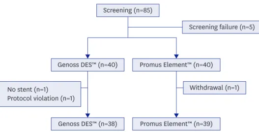
Methods: This study was a prospective, multicenter, randomized trial with a 1:1 ratio of Genoss drug-eluting stent (DES)™ and Promus Element™. Inclusion criteria were the presence of stable angina, unstable angina, or silent ischemia. Angiographic inclusion criteria were de novo coronary stenotic lesion with diameter stenosis >50%, reference vessel diameter of 2.5–4.0 mm, and lesion length ≤40 mm. The primary endpoint was in-stent late lumen loss at 9-month quantitative coronary angiography follow-up. Secondary endpoints were in-segment late lumen loss, binary restenosis rate, death, myocardial infarction (MI), target lesion revascularization (TLR), target vessel revascularization (TVR), and stent thrombosis during 9 months of follow-up.

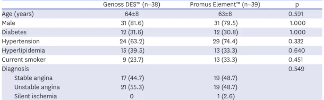
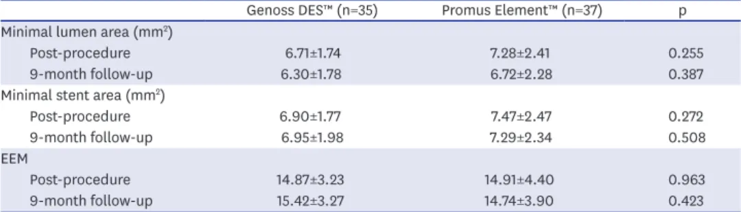
Results: We enrolled 38 patients for the Genoss DES™ group and 39 patients for the Promus Element™ group. In-stent late lumen loss at 9 months was not significantly different between the 2 groups (0.11±0.25 vs. 0.16±0.43 mm, p=0.567). There was no MI or stent thrombosis in either group. The rates of death (2.6% vs. 0%, p=0.494), TLR (2.6% vs. 2.6%, p=1.000), and TVR (7.9% vs. 2.6%, p=0.358) at 9 months were not significantly different.

Conclusion: This first-in-patient study of the Genoss DES™ stent showed excellent angiographic outcomes for in-stent late lumen loss and major adverse cardiac events over a 9-month follow-up.

Keywords: Drug-eluting stents; Coronary artery disease; Sirolimus

Original Article

Hyoung-Mo Yang, MD

1

, Kyoung-Woo Seo, MD

1

, Junghan Yoon, MD

2

, Hyo-Soo Kim, MD

3

, Kiyuk Chang, MD

4

, Hong-Seok Lim, MD

1

,

Byoung-Joo Choi, MD

1

, So-Yeon Choi, MD

1

, Myeong-Ho Yoon, MD

1

, Seung-Hwan Lee, MD

2

, Sung Gyun Ahn, MD

2

, Young Jin Youn, MD

2

, Jun-Won Lee, MD

2

, Bon-Kwon Koo, MD

3

, Kyung Woo Park, MD

3

, Han-Mo Yang, MD

3

, Jung-Kyu Han, MD

3

, Ki-Bae Seung, MD

4

, Wook-Sung Chung, MD

4

, Pum-Joon Kim, MD

4

, Yoon-Seok Koh, MD

4

, Hun-Jun Park, MD

4

, and Seung-Jea Tahk, MD, PhD,

1

1

Department of Cardiology, Ajou University School of Medicine, Suwon, Korea

2

Department of Cardiology, Yonsei University Wonju Christian Hospital, Wonju, Korea

3

Department of Internal Medicine, Seoul National University College of Medicine, Seoul, Korea

4

Department of Internal Medicine, Seoul St. Mary's Hospital, College of Medicine, The Catholic University of Korea, Seoul, Korea

Clinical and Angiographic Outcomes of the First Korean-made Sirolimus- Eluting Coronary Stent with Abluminal Bioresorbable Polymer

Received: May 6, 2017 Revised: Jul 24, 2017 Accepted: Jul 28, 2017 Correspondence to Seung-Jea Tahk, MD, PhD

Department of Cardiology, Ajou University School of Medicine, 164, World cup-ro, Yeongtong-gu, Suwon 16499, Korea.

E-mail: [email protected]

Copyright © 2017. The Korean Society of Cardiology

This is an Open Access article distributed under the terms of the Creative Commons Attribution Non-Commercial License (https://

creativecommons.org/licenses/by-nc/4.0) which permits unrestricted noncommercial use, distribution, and reproduction in any medium, provided the original work is properly cited.

Funding

This study was supported by Genoss Company Limited (Suwon, Korea).

Conflict of Interest

Tahk SJ received research grant from Genoss

Compnary Limitied, and other investigators

did not have any financial relationships or any

other biases or conflicts of interest related

with this study.

(2)

Author Contributions

Conceptualization: Tahk SJ, Yang HM; Data curation: Yoon J, Kim HS, Chang K, Lee SH, Ahn SG, Youn YJ, Lee JW, Koo BK, Park KW, Yang HM, Han JK, Seung KB, Chung WS, Kim PJ, Koh YS, Park HJ; Formal analysis: Tahk SJ, Yang HM; Funding acquisition: Yoon J, Kim HS, Chang K, Lee SH, Ahn SG, Youn YJ, Lee JW, Koo BK, Park KW, Yang HM, Han JK, Seung KB, Chung WS, Kim PJ, Koh YS, Park HJ; Validation:

Yoon J, Kim HS, Chang K, Lee SH, Ahn SG, Youn YJ, Lee JW, Koo BK, Park KW, Yang HM, Han JK, Seung KB, Chung WS, Kim PJ, Koh YS, Park HJ; Writing - original draft: Tahk SJ, Yang HM.

INTRODUCTION

Percutaneous coronary intervention (PCI) using a drug-eluting stent (DES) is a standard treatment for significant coronary artery occlusive disease. First-generation DESs have shown significantly lower restenosis rates compared to bare metal stents.

1-4)

Although DES reduced the rate of restenosis and target lesion revascularization (TLR), the risk of stent thrombosis increased.

5-7)

Stent thrombosis is a clinically serious complication because of its high mortality rate. It is related to early discontinuation of antiplatelet agents, resistance to antiplatelet agents, delayed endothelization, hypersensitivity reaction to polymers, and several clinical or procedural factors.

6-8)

As one possible cause of stent thrombosis is a hypersensitivity reaction to a durable polymer, the Nobori™ stent (Terumo Corporation, Tokyo, Japan) and BioMatrix™ (Biosensors International, Singapore) used a bioresorbable polymer that did not remain in the stent after a certain time period and showed good clinical outcomes compared to a durable polymer DES.

9)10)

The Genoss DES™ stent (Genoss Company Limited, Suwon, Korea) is the first sirolimus- eluting stent with biodegradable polymers made in Korea. The objectives of this first-in- patient prospective randomized study were to evaluate the efficacy and safety of the Genoss DES™ stent in comparison to the Promus Element™ (Boston Scientific, Natick, MA, USA).

METHODS

Patient population

This study was a prospective, multicenter, single blind (patient blind), randomized study with 1:1 ratio of patients using the Genoss DES™ stent and Promus Element™, conducted from March 2013 to April 2015. The study enrolled 80 patients at 4 Korean centers.

Inclusion criteria were the presence of stable angina, unstable angina, or silent ischemia in patients between 20 and 80 years of age. Angiographic inclusion criteria were de novo coronary stenotic lesion with diameter stenosis (DS) >50%, reference vessel diameter (RVD) of 2.5 to 4.0 mm, and a lesion length ≤40 mm. Exclusion criteria were the presence of acute myocardial infarction (MI), cardiogenic shock, low ejection fraction (<40%), contraindication to antiplatelet agents, and chronic total occlusion lesion, restenosis, left main disease, and stenosis of the graft vessel. The protocol was approved by the Institutional Review Board of the Ajou University Hospital, Yonsei University Wonju Christian Hospital, Seoul National University Hospital, and Seoul St. Mary's Hospital, and all patients provided written informed consent.

The Genoss DES™ sirolimus-eluting stent

The Genoss DES™ is a sirolimus-eluting cobalt-chromium coronary stent system for the

treatment of coronary artery diseases. The stent platform features open-cell 2 links with a

uniform architecture that permits a high level of flexibility with good radial force, shortening,

and conformability. Two kinds of strut thickness (70 and 78 µm) are adopted, depending on

the stent diameter (2.25–2.50 and 2.75–4.00 mm, respectively). To achieve favorable clinical

outcomes, the initial burst release of drug from DES is needed to suppress vascular injury

and inflammation induced by catheter manipulation and stent implantation. Accordingly,

the Genoss DES™ is designed to release approximately 70% of its initial drug payload (1.15

(3)

µg/mm

2

) within the first 30 days following implantation. To achieve the desired drug release profile and minimize the amount of polymer, a 4 µm ultra-thin coating is applied to only the abluminal side of the stent. This bioresorbable coating consists of a proprietary blend of poly-DL-lactide-co-glycolide (PLGA) and poly-DL-lactide (PLA), which are almost fully resorbed within 9 months.

Study procedure

PCI was performed using standard interventional techniques. Intravenous heparin was administered to maintain an activated clotting time of more than 250 seconds, and

antiplatelet therapy was used before the procedure with aspirin (100–325 mg) and clopidogrel (loading dose of 300–600 mg) or ticlopidine (500 mg/day). Dual antiplatelet therapy was maintained during the 9 months of follow-up. Glycoprotein IIb/IIIa receptor antagonists were used at the operator's discretion. After predilation, Genoss DES™, or Promus Element™

was implanted under rated burst pressure, and post-dilatation was performed if needed. This study permitted 2 stents to overlap in cases of long lesions.

Follow-up

Clinical follow-up was performed at the time of discharge, and following a period of 1, 5, and 9 months. All patients were planned to undergo a follow-up angiography and intravascular ultrasound (IVUS) at 9±1 months.

Quantitative coronary angiography and intravascular ultrasound Quantitative coronary angiography (QCA) analyses were performed by 2 independent observers blinded to treatment group using the Cardiovascular Angiography Analysis System II (CAAS II; Pie Medical, Maastricht, Netherlands). The percent DS, minimal luminal diameter, RVD, and lesion length were measured and calculated before the intervention, at the end of the procedure, and at the 9-month follow-up.

IVUS was performed at the end of the index procedure and at follow-up, and analyzed by 2 independent observers blinded to clinical and procedural information according to the American College of Cardiology clinical expert consensus document on standards for acquisition, measurement, and reporting of IVUS studies.

11)

All IVUS analyses were performed using computerized planimetry software (EchoPlaque 3.0; Indec Systems, Santa Clara, CA, USA).

Study endpoints

The primary endpoint was in-stent late lumen loss at 9-month angiography follow-up determined by QCA. Secondary endpoints were in-segment late lumen loss, binary restenosis rate, death, MI, TLR, target vessel revascularization (TVR), and stent thrombosis during 9-month follow-up.

Definition

MI was defined as chest pain and increased creatine kinase-myocardial band level

to more than twice the upper limit of the normal range with or without a Q-wave on

electrocardiography. Binary restenosis was defined as luminal narrowing of more than 50%,

and TLR and TVR were defined as repeated PCI or bypass surgery of a previously stented

site or vessel, respectively. Stent thrombosis was defined using the Academic Research

Consortium definition.

12)

(4)

Statistical analysis

This study was a non-inferiority trial of the Genoss DES™ stent for in-stent late lumen loss at 9-month angiography follow-up compared to the Promus Element™ stent. We assumed an in-stent late lumen loss of 0.20±0.28 mm in the Promus Element™ stent group based on prior study.

13)

We calculated 35 patients per group, using a one side α level 0.025 and non- inferiority margin of 0.19 mm with a statistical power of 80%. Assuming approximately 10%

of patients would drop out, we calculated 40 patients for each group.

Data are presented as mean±standard deviations for continuous variables, and frequency (percentage) for categorical variables. Comparison of continuous variables was performed using Student's t-test, and χ

2

or Fisher's exact tests were used for categorical variables. A p value <0.025 was considered statistically significant for the primary endpoint, and <0.050 for the secondary endpoints. Statistical analyses were performed using PASW software (version 18.0; SPSS Inc., Chicago, IL, USA).

RESULTS

Between March 2013 and April 2015, 85 patients were screened for enrollment in the study.

Five patients did not meet inclusion criteria and a total of 80 patients were randomly assigned to the Genoss DES™ or Promus Element™ group at a 1:1 ratio. In the Genoss DES™ group, 2 patients were excluded as one patient did not receive stent implantation and the other patient had a protocol violation. In the Promus Element™ group, 1 patient was excluded owing to the patient's withdrawal from the study (Figure 1).

Baseline clinical and angiographic characteristics are shown in Tables 1 and 2, respectively.

There were no significant differences in clinical characteristics including age and clinical diagnosis between the 2 groups (Table 1). Target vessels were not significantly different, but the left anterior descending artery was targeted more often in the Promus Element™ group.

Stent overlap was performed in 8 (21.1%) patients in the Genoss DES™ group and in 3 (7.7%) patients in the Promus Element™ group (Table 2).

Screening (n=85)

Screening failure (n=5)

Genoss DES™ (n=38) Promus Element™ (n=39) Genoss DES™ (n=40) Promus Element™ (n=40)

No stent (n=1)

Protocol violation (n=1)

Withdrawal (n=1)

Figure 1. Flow chart of patient enrollment.

DES = drug-eluting stent.

(5)

Angiographic follow-up at 9 months was performed for all patients, and IVUS follow-up was performed in 35 (92%) patients in the Genoss DES™ group and in 37 (95%) patients in the Promus Element™ group. QCA results are shown in Table 3. There were no significant differences in RVD. For the primary endpoint, in-stent late lumen loss at 9 months was not significantly different between the groups (0.11±0.25 mm for Genoss DES™ vs. 0.16±0.43 mm for Promus Element™, p=0.567) nor was in-segment late lumen loss (0.11±0.26 mm for Genoss DES™ vs. 0.15±0.43 mm for Promus Element™, p=0.558). Angiographic restenosis at 9 months was observed in 1 (2.6%) patient in the Genoss DES™ group and in 2 (5.1%) patients in the Promus Element™ group (p=1.000). IVUS minimal lumen area after stenting during the index procedure (6.95±1.98 vs. 7.47±2.47 mm

2

, p=0.272) and after 9 months (6.95±1.98 vs. 7.29±2.34 mm

2

, p=0.508) were not significantly different (Table 4). For clinical outcomes, there were no MI or stent thrombosis events during 9-month follow-up. One death occurred in the Genoss DES™ group due to aggravated renal function 6 months after the index procedure. However, the rate of death, MI, TLR, and TVR at 9 months were not significantly different (Table 5).

DISCUSSION

This prospective, multicenter, randomized first-in-patient study showed that in-stent late lumen loss of Genoss DES™ stents was not inferior to Promus Element™ stents as determined by QCA at 9-month angiography follow-up. Moreover, there were no differences in clinical outcomes and no stent thrombosis during 9 months of follow-up.

Table 1. Baseline clinical characteristics

Genoss DES™ (n=38) Promus Element™ (n=39) p

Age (years) 64±8 63±8 0.591

Male 31 (81.6) 31 (79.5) 1.000

Diabetes 12 (31.6) 12 (30.8) 1.000

Hypertension 24 (63.2) 29 (74.4) 0.332

Hyperlipidemia 15 (39.5) 13 (33.3) 0.640

Current smoker 9 (23.7) 13 (33.3) 0.451

Diagnosis 0.549

Stable angina 17 (44.7) 19 (48.7)

Unstable angina 21 (55.3) 19 (48.7)

Silent ischemia 0 1 (2.6)

Data are shown as mean±standard deviation or number (%).

DES = drug-eluting stent.

Table 2. Angiographic and procedural characteristics

Genoss DES™ (n=38) Promus Element™ (n=39) p

Target vessel 0.068

Left anterior descending 18 (47.3) 26 (66.7)

Left circumflex 4 (10.5) 6 (15.4)

Right coronary 16 (42.1) 7 (17.9)

AHA/ACC classification 0.089

A 4 (10.5) 7 (17.9)

B1 6 (15.8) 14 (35.9)

B2 10 (26.3) 8 (20.5)

C 18 (47.4) 10 (25.6)

Stent diameter (mm) 3.14±0.26 3.15±0.30 0.970

Stent length (mm) 25.5±8.6 24.1±5.0 0.395

Mean stent number 1.2 1.1 0.099

Stent overlap 8 (21.1) 3 (7.7) 0.114

Data are shown as mean±standard deviation or number (%).

ACC = American College of Cardiology; AHA = American Heart Association; DES = drug-eluting stent.

(6)

Table 3. QCA analysis

Genoss DES™ (n=38) Promus Element™ (n=39) p RVD (mm)

Pre-procedure 3.24±0.41 3.12±0.43 0.199

Post-procedure 3.30±0.30 3.33±0.30 0.652

9-month follow-up 3.17±0.35 3.16±0.39 0.932

Minimal lumen diameter (mm) In-stent

Pre-procedure NA NA NA

Post-procedure 2.95±0.35 3.03±0.30 0.269

9-month follow-up 2.84±0.42 2.87±0.56 0.743

In-segment

Pre-procedure 0.83±0.29 0.88±0.45 0.572

Post-procedure 2.90±0.27 3.00±0.36 0.159

9-month follow-up 2.79±0.40 2.85±0.56 0.615

DS (%) In-stent

Pre-procedure NA NA NA

Post-procedure 10.6±3.48 9.16±4.85 0.140

9-month follow-up 14.18±9.31 13.73±14.30 0.871

In-segment

Pre-procedure 74.41±8.62 72.65±12.14 0.465

Post-procedure 12.10±3.93 10.12±4.94 0.056

9-month follow-up 15.46±9.39 14.54±14.08 0.738

Acute gain (mm)

In-stent 1.79±0.40 1.82±0.36 0.742

In-segment 1.74±0.39 1.78±0.38 0.579

Late loss (mm)

In-stent 0.11±0.25 0.16±0.43 0.567

In-segment 0.11±0.26 0.15±0.43 0.558

Lesion length (mm) 23.8±8.1 22.8±4.9 0.531

Restenosis 1 (2.6) 2 (5.1) 1.000

Data are shown as mean±standard deviation or number (%).

DES = drug-eluting stent; DS = diameter stenosis; NA = not available; QCA = quantitative coronary angiography;

RVD = reference vessel diameter.

Table 4. IVUS analysis results

Genoss DES™ (n=35) Promus Element™ (n=37) p Minimal lumen area (mm

2

)

Post-procedure 6.71±1.74 7.28±2.41 0.255

9-month follow-up 6.30±1.78 6.72±2.28 0.387

Minimal stent area (mm

2

)

Post-procedure 6.90±1.77 7.47±2.47 0.272

9-month follow-up 6.95±1.98 7.29±2.34 0.508

EEM

Post-procedure 14.87±3.23 14.91±4.40 0.963

9-month follow-up 15.42±3.27 14.74±3.90 0.423

Data are shown as mean±standard deviation.

DES = drug-eluting stent; EEM = external elastic membrane; IVUS = intravascular ultrasound.

Table 5. Clinical outcomes

Genoss DES™ (n=38) Promus Element™ (n=39) p

Death 1 (2.6) 0 0.494

Cardiac 0 0 1.000

Non-cardiac 1 (2.6) 0 0.494

MI 0 0 1.000

TLR 1 (2.6) 1 (2.6) 1.000

TVR 3 (7.9) 1 (2.6) 0.358

Stent thrombosis 0 0 1.000

Data are shown as number (%).

DES = drug-eluting stent; MI = myocardial infarction; TLR = target lesion revascularization; TVR = target vessel

revascularization.

(7)

The Genoss DES™ stent is the first DES made in Korea. It consists of a cobalt-chromium alloy stent platform, an abluminal coating of bioresorbable polymers, and a drug-eluting coating with sirolimus. Sirolimus is an anti-proliferative and anti-inflammatory agent that inhibits progression from the G1 to the S phase of the cell cycle. Coronary DES using sirolimus has demonstrated good clinical outcomes.

4)14)

For the Genoss DES™ stent, animal studies using pigs were performed to determine the optimal dose of the drug, comparing high and low doses of sirolimus and paclitaxel (unpublished data). Consequently, sirolimus was loaded on the stent at a concentration of 1.15 µg/mm

2

. The stent was designed to release approximately 70% of the total drug amount within 30 days after implantation. To control the drug release profile, it possesses an ultra-thin (4 µm) bioresorbable layer applied to an abluminal stent surface. This bioresorbable layer consists of a proprietary blend of PLGA and PLA, which are completely resorbable within 6 months, to reduce the possibility of stent thrombosis due to polymer hypersensitivity. In addition, the cobalt-chromium alloy stent platform reduces strut thickness and maximizes flexibility.

In-stent late luminal loss at 9 months with the Genoss DES™ stent was 0.11±0.25 mm, comparable with previous DES trials (Table 6). Moreover, this study included a number of patients at high risk of unstable angina or complex lesions. The proportion of patients with unstable angina was around 50% in both groups and that of American Heart Association (AHA)/American College of Cardiology (ACC) type C lesions was 47.4% for the Genoss DES™

group and 25.6% for the Promus Element™ group. Angiographic in-stent restenosis was observed in only 2.6% of patients in the Genoss DES™ group, even though stent overlap was permitted such that mean lesion length was very high (25.5 mm for the Genoss DES™

group) compared with previous studies (Table 6). Considering patient and angiographic characteristics, in-stent late lumen loss of the Genoss DES™ stent at 9-month follow-up was acceptable in real clinical practice.

For clinical outcomes, there were no differences in death, MI, TLR, or TVR between the 2 groups. TLR occurred in only 2.6% of patients in both groups at 9-month angiography follow-up. In the Genoss DES™ group, 2 cases of TVR without TLR occurred, which were new lesions in the proximal portions of the right coronary artery independent of previously stented sites. Although not statistically significant, the stent overlap rate at the target lesion was higher in the Genoss DES™ group than in the Promus Element™ group (21.1% vs. 7.7%).

However, the clinical event rate of the Genoss DES™ group was very low. The Genoss DES™

stent did not develop stent thrombosis during the 9-month follow-up despite complex lesion characteristics.

This study has several limitations. For assumptions about sample size, the non-inferiority margin was relatively high and the number of patients was small. This made the statistical Table 6. Comparison of angiographic outcomes with previous studies

In-stent late loss (mm) Lesion length (mm)

Platinum QCA

13)

0.17±0.25 15.4±7.0

NOBORI 1

15)

0.11±0.30 10.56 

SPIRIT I

16)

0.10±0.21 10.1±2.6

RESOLUTE

17)

0.22±0.27 15.61±6.13

Genoss DES™ (current study) 0.11±0.25 25.5±8.6

Data are shown as mean±standard deviation.

DES = drug-eluting stent; QCA = quantitative coronary angiography.

(8)

power weak. However, the Genoss DES™ stent showed excellent in-stent late lumen loss compared to current second-generation DESs. In addition, as this is a first-in-patient trial, more studies with larger populations should be conducted in real clinical practice.

In conclusion, this first-in-patient study of the Genoss DES™ stent showed excellent angiographic outcomes for in-stent late lumen loss and major adverse cardiac events during the 9-month follow-up.

REFERENCES

1. Morice MC, Serruys PW, Sousa JE, et al. A randomized comparison of a sirolimus-eluting stent with a standard stent for coronary revascularization. N Engl J Med 2002;346:1773-80.

PUBMED | CROSSREF

2. Moses JW, Leon MB, Popma JJ, et al. Sirolimus-eluting stents versus standard stents in patients with stenosis in a native coronary artery. N Engl J Med 2003;349:1315-23.

PUBMED | CROSSREF

3. Stone GW, Ellis SG, Cox DA, et al. A polymer-based, paclitaxel-eluting stent in patients with coronary artery disease. N Engl J Med 2004;350:221-31.

PUBMED | CROSSREF

4. Stone GW, Moses JW, Ellis SG, et al. Safety and efficacy of sirolimus- and paclitaxel-eluting coronary stents. N Engl J Med 2007;356:998-1008.

PUBMED | CROSSREF

5. Joner M, Finn AV, Farb A, et al. Pathology of drug-eluting stents in humans: delayed healing and late thrombotic risk. J Am Coll Cardiol 2006;48:193-202.

PUBMED | CROSSREF

6. Virmani R, Guagliumi G, Farb A, et al. Localized hypersensitivity and late coronary thrombosis secondary to a sirolimus-eluting stent: should we be cautious? Circulation 2004;109:701-5.

PUBMED | CROSSREF

7. Lüscher TF, Steffel J, Eberli FR, et al. Drug-eluting stent and coronary thrombosis: biological mechanisms and clinical implications. Circulation 2007;115:1051-8.

PUBMED | CROSSREF

8. Nebeker JR, Virmani R, Bennett CL, et al. Hypersensitivity cases associated with drug-eluting coronary stents: a review of available cases from the Research on Adverse Drug Events and Reports (RADAR) project. J Am Coll Cardiol 2006;47:175-81.

PUBMED | CROSSREF

9. Zhang YJ, Ye F, Iqbal J, et al. NOBORI™ biodegradable-polymer biolimus-eluting stent versus durable- polymer drug-eluting stents: a meta-analysis. Int J Cardiol 2014;174:151-3.

PUBMED | CROSSREF

10. Stefanini GG, Byrne RA, Serruys PW, et al. Biodegradable polymer drug-eluting stents reduce the risk of stent thrombosis at 4 years in patients undergoing percutaneous coronary intervention: a pooled analysis of individual patient data from the ISAR-TEST 3, ISAR-TEST 4, and LEADERS randomized trials. Eur Heart J 2012;33:1214-22.

PUBMED | CROSSREF

11. Mintz GS, Nissen SE, Anderson WD, et al. American College of Cardiology Clinical Expert Consensus Document on Standards for Acquisition, Measurement and Reporting of Intravascular Ultrasound Studies (IVUS). A report of the American College of Cardiology Task Force on Clinical Expert Consensus Documents. J Am Coll Cardiol 2001;37:1478-92.

PUBMED | CROSSREF

12. Cutlip DE, Windecker S, Mehran R, et al. Clinical end points in coronary stent trials: a case for standardized definitions. Circulation 2007;115:2344-51.

PUBMED | CROSSREF

13. Meredith IT, Whitbourn R, Scott D, et al. PLATINUM QCA: a prospective, multicentre study assessing clinical, angiographic, and intravascular ultrasound outcomes with the novel platinum chromium thin-strut PROMUS Element everolimus-eluting stent in de novo coronary stenoses. EuroIntervention 2011;7:84-90.

PUBMED | CROSSREF

(9)

14. Windecker S, Serruys PW, Wandel S, et al. Biolimus-eluting stent with biodegradable polymer versus sirolimus-eluting stent with durable polymer for coronary revascularisation (LEADERS): a randomised non-inferiority trial. Lancet 2008;372:1163-73.

PUBMED | CROSSREF

15. Chevalier B, Silber S, Park SJ, et al. Randomized comparison of the Nobori Biolimus A9-eluting coronary stent with the Taxus Liberté paclitaxel-eluting coronary stent in patients with stenosis in native coronary arteries: the NOBORI 1 trial--Phase 2. Circ Cardiovasc Interv 2009;2:188-95.

PUBMED | CROSSREF

16. Serruys PW, Ong AT, Piek JJ, et al. A randomized comparison of a durable polymer Everolimus-eluting stent with a bare metal coronary stent: the SPIRIT first trial. EuroIntervention 2005;1:58-65.

PUBMED

17. Meredith IT, Worthley S, Whitbourn R, et al. Clinical and angiographic results with the next-generation resolute stent system: a prospective, multicenter, first-in-human trial. JACC Cardiovasc Interv 2009;2:977-85.

PUBMED | CROSSREF

수치

Figure 1. Flow chart of patient enrollment.
Table 1. Baseline clinical characteristics
Table 4. IVUS analysis results

참조

관련 문서

Purpose: This study was aimed to compare the functional outcomes of the continuous and discontinuous fractures, retrospectively, in multiple thoracolumbar

The struggle was successful, and in January 1926 the first of four papers on “Quantization as an Eigenvalue

The purpose of this study was to analyze the impaction pattern of the impacted mandibular third molar and the relationship with the inferior alveolar nerve

First, it was identified that students who participated in Saturday sport activity showed higher emotional development that students who did not, and showed difference in

The aim of this study was not only to compare the differences of ego-resilience between students with high experience and students with low experience in

In the POLARIS-2 and POLARIS-3 studies 14 , it was not determined that SOF/VEL/VOX for 8 weeks was non-inferior to SOF/VEL for 12 weeks for the treatment of non-cirrhotic

Higher open conversion rate and prolonged postoperative hospital stay in LATE group were not caused by the delayed operation in this study it seems to be caused by that

In this study, the application of 6-mL fibrin glue helped to obtain better clinical outcomes by decreasing the total drain volume and flow rate of exudate during the first