505
Open Access
Comparison of Transthoracic Echocardiography With
N-Terminal Pro-Brain Natriuretic Peptide as a Tool for Risk Stratification of Patients Undergoing Major Noncardiac Surgery
Sung-Ji Park, MD
1, Jin-Ho Choi, MD
1,2, Soo-Jin Cho, MD
1, Sung-A Chang, MD
1, Jin-Oh Choi, MD
1, Sang-Cheol Lee, MD
1, Seung Woo Park, MD
1, Jae K Oh, MD
1,3, Duk-Kyung Kim, MD
1, and Eun-Seok Jeon, MD
11Departments of Medicine and 2Emergency Medicine, Cardiovascular Imaging Center, Cardiac and Vascular Center, Samsung Medical Center, Sungkyunkwan University School of Medicine, Seoul, Korea
3Division of Cardiovascular Diseases, Mayo Clinic College of Medicine, Rochester, Minnesota, USA
ABSTRACT
Background and Objectives: The role of preoperative transthoracic echocardiography (TTE) for the risk stratification has not been well investigated yet. We compared the predictive power of TTE with N-terminal pro-brain natriuretic peptide (NT- proBNP), a representative biomarker that predicts perioperative cardiovascular risk, and investigated whether these tests have incremental value to the clinically determined risk. Subjects and Methods: We evaluated the Revised Cardiac Risk In- dex (RCRI), TTE, and NT-proBNP in 1,923 noncardiac surgery cases. The primary endpoint was a perioperative major cardio- vascular event (PMCE), which was defined by any single or combined event of secondary endpoints including myocardial in- farction, development of pulmonary edema, or primary cardiovascular death within 30 days after surgery. Results: All echo- cardiographic parameters including left ventricular ejection fraction, regional wall motion score index, and transmitral early diastolic velocity/tissue Doppler mitral annular early diastolic velocity (E/E’) were predictive of PMCE (c-statistics=0.579±
0.019 to 0.589±0.015), but none of these parameters were better than the clinically determined RCRI (c-statistics=0.594±0.019) and were inferior to NT-proBNP (c-statistics=0.748±0.019, p<0.001). The predictive power of RCRI {adjusted relative risk (RR)=
1.4} could be improved by addition of echocardiographic parameters (adjusted RR=1.8, p<0.001), but not to that extent as by addition of NT-proBNP to RCRI (adjusted RR=3.7, p<0.001). Conclusion: TTE was modestly predictive of perioperative cardiovascular events but was not superior to NT-proBNP. Moreover, it did not have incremental value to the clinically deter- mined risk. The results of our study did not support the use of routine echocardiography before noncardiac surgery. (Korean Circ J 2011;41:505-511)
KEY WORDS: Cardiovascular disease; Postoperative complications; Echocardiography; Natriuretic peptides.
Received: September 15, 2010 Revision Received: January 14, 2011 Accepted: January 24, 2011
Correspondence: Jin-Ho Choi, MD, Department of Medicine, Cardio- vascular Imaging Center, Cardiac and Vascular Center, Samsung Medi- cal Center, Sungkyunkwan University School of Medicine, 50 Irwon- dong, Gangnam-gu, Seoul 135-710, Korea
Tel: 82-2-3410-6547, Fax: 82-2-3410-3849 E-mail: [email protected]
• The authors have no financial conflicts of interest.
cc This is an Open Access article distributed under the terms of the Cre- ative Commons Attribution Non-Commercial License (http://creativecom- mons.org/licenses/by-nc/3.0) which permits unrestricted non-commer- cial use, distribution, and reproduction in any medium, provided the origi- nal work is properly cited.
Introduction
Most clinical cardiovascular risk indices are shown to have
modest predictive power in patients undergoing major non- cardiac surgery.1) Preoperative transthoracic echocardiogra- phy (TTE) is one of non-invasive cardiac evaluation tests that are frequently expected to increase the predictive power. Al- though poor left ventricular (LV) systolic or diastolic function is known to be predictive of postoperative heart failure or dea- th,2-4) the routine use of preoperative echocardiography in cli- nically stable patients is not usually recommended by the cur- rent guidelines.1)
Recent studies have shown that natriuretic peptides can pre- dict postoperative cardiovascular events.5-8) Currently, little data is available on the direct comparison of imaging- or bio- marker-based predictors, or the incremental value of these pre- dictors to the clinically determined risk. We compared direct- ly the predictive power of N-terminal pro-brain natriuretic
peptide (NT-proBNP) with TTE for the postoperative major cardiovascular events, and investigated whether additional ev- aluation of these risk predictors has incremental value to the clinical risk stratification.
Subjects and Methods
Study population
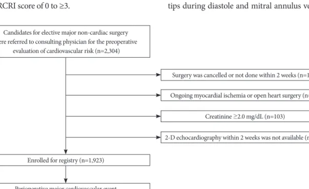
The study population was derived from our prospective, un- selected, consecutive cohort of preoperative cardiac consult- ation for elective noncardiac surgery.5) Of the 2,304 cohort patients, 1,923 (83.5%) patients had echocardiography with- in 2 weeks before surgery and constituted the study group (Fig.
1). Patients with moderate or severe valvular stenosis were not included in this study population. This study protocol was ap- proved by the institutional review board of Samsung Medical Center.
Data collection
Clinical perioperative cardiovascular risk was assessed ac- cording to the Revised Cardiac Risk Index (RCRI) modified by Lee, a well-validated and widely used point score-based risk prediction index.9)10) Briefly, RCRI calculates periopera- tive risk by sum of points. Each risk factor, including high-risk surgical procedures, history of ischemic heart disease, pulmo- nary edema, cerebrovascular disease, insulin-dependent dia- betes and serum creatinine >2.0 mg/dL, is assigned one point.
The risk of major cardiac event including myocardial infarc- tion, pulmonary edema, primary cardiac arrest and complete heart block predicted by RCRI was known to be 0.4% to 11%
according to an RCRI score of 0 to ≥3.
Basic laboratory tests including electrocardiography, chest X-ray, and NT-proBNP were evaluated within 2 weeks before surgery. Blood samples for NT-proBNP were collected into li- thium heparin tubes and stored at -70°C until further analy- sis. Plasma NT-proBNP levels were measured using an Elec- sys pro-BNP reagent kit (Roche Diagnostics, Indianapolis, In, USA) and an Elecsys 2010 analyzer (Roche Diagnostics, India- napolis, IN, USA).
Transthoracic echocardiography
Two-dimensional (2-D) TTE was performed within 2 weeks before surgery at the discretion of the physician or if the pati- ents had two or more of the following cardiovascular risk fac- tors: diabetes mellitus, hypertension, aged 65 years or greater, current smoking status, or hypercholesterolemia. TTE was performed with a commercially available echocardiographic instrument (Vivid 7, GE Medical Systems, Milwaukee, WI, USA or Sequoia 512, Acuson, Mountain View, CA, USA). A standard M-mode, 2-D echocardiogram and echocardiograph- ic Doppler study were performed. All TTE recordings were interpreted by staff cardiologists.
The routine standard echocardiographic examination in- cluded measurements of thickness of the ventricular septum and LV posterior wall, end-systolic and end-diastolic LV dia- meters from M-mode or 2-D imaging. Left atrial (LA) volume measurement and standard pulsed wave Doppler evaluation of diastolic function were carried out as previously describ- ed.11) Both LV mass and LA volume were indexed to body surface area. Mitral inflow velocities were obtained by pu- lsed wave Doppler sample volume between the mitral leaflet tips during diastole and mitral annulus velocities were ob-
Candidates for elective major non-cardiac surgery were referred to consulting physician for the preoperative
evaluation of cardiovascular risk (n=2,304)
Enrolled for registry (n=1,923)
Perioperative major cardiovascular event (PMCE) occurred in 14.6% (n=280)
Acute myocardial infarction in 5.2% (n=100)
Pulmonary edema in 12.5% (n=241)
Primary cardiovascular death in 0.7% (n=14)
Surgery was cancelled or not done within 2 weeks (n=118) Ongoing myocardial ischemia or open heart surgery (n=29)
Creatinine ≥2.0 mg/dL (n=103)
2-D echocardiography within 2 weeks was not available (n=131)
Fig. 1. Study flowchart.
tained from the septal portion of the mitral annulus by tis- sue Doppler imaging. All measurements were performed on 3 cardiac cycles and were then averaged.
Quantitative LV systolic ejection fraction (LVEF) and regio- nal wall motion index (RWMI) was obtained from the digital- ly stored records. Diastolic dysfunction could be evaluated in a subgroup of 1,132 patients (58.9%) who were examined us- ing tissue Doppler imaging with acceptable quality, and did not have non-sinus rhythm or left bundle branch block.
Clinical outcome
All patients were followed until discharge or up to 30 days of hospitalization after surgery. Primary endpoint was a pe- rioperative major cardiovascular event (PMCE), which was defined by any single or combined event of secondary end- points including myocardial infarction, development of pul- monary edema, or primary cardiovascular death. Individual patients may have had more than one event, and all events were counted as an incidence. Myocardial infarction was defined by a rise in postoperative troponin I above the 99th percen- tile of the upper reference limit (0.78 ng/mL, Roche Diag- nostics, Switzerland), which was evaluated at the end of the day of surgery and 24 hours later. Pulmonary edema was di- agnosed after a formal reading of the chest X-ray by a radio- logist consistent with the complication. Primary cardiovascu- lar death was defined by sudden death that could not be ex- plained by any other non-cardiovascular postoperative com- plications.
Statistical analysis
Perioperative risk predictors including RCRI, NT-proBNP, and echocardiographic parameters were treated as continu- ous variables or ordered categorical variables. Receiver-op- erating characteristic (ROC) analysis was performed to cal- culate sensitivity, specificity, area under the curve (AUC), and the optimal cut-off value. The predictive power of each pre- dictor was compared using Hanley and McNail method.12) Independent predictors of PMCE in univariate analysis were categorized by optimal cut-off levels, and were used in mul- tivariate logistic models.13) The adjusted relative risk (RR) of each predictor and the combination of these predictors in an additive manner was evaluated. A p<0.05 (2-sided) was con- sidered significant. SPSS version 13.0 was used mostly. ROC curves were compared using Medcalc version 9.6.
Results
Baseline characteristics
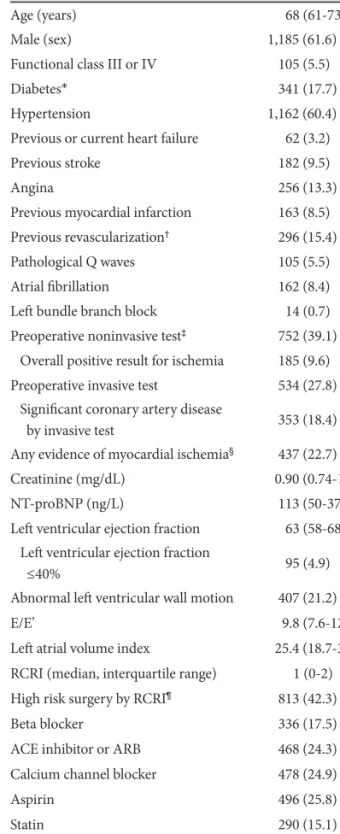
Preoperative clinical characteristics of the study popula- tion are shown in Table 1. 2-D TTE was performed within 2 weeks before surgery (10.8±8.2 days). NT-proBNP were ev- aluated within 2 weeks before surgery (7.4±7.2 days). Briefly,
Table 1. Clinical characteristics
Frequency (%) or median
with interquartile range
Age (years) 68 (61-73)
Male (sex) 1,185 (61.6)
Functional class III or IV 105 (5.5)
Diabetes* 341 (17.7)
Hypertension 1,162 (60.4)
Previous or current heart failure 62 (3.2)
Previous stroke 182 (9.5)
Angina 256 (13.3)
Previous myocardial infarction 163 (8.5) Previous revascularization† 296 (15.4)
Pathological Q waves 105 (5.5)
Atrial fibrillation 162 (8.4)
Left bundle branch block 14 (0.7)
Preoperative noninvasive test‡ 752 (39.1) Overall positive result for ischemia 185 (9.6) Preoperative invasive test 534 (27.8) Significant coronary artery disease
by invasive test 353 (18.4)
Any evidence of myocardial ischemia§ 437 (22.7)
Creatinine (mg/dL) 0.90 (0.74-1.06)
NT-proBNP (ng/L) 113 (50-377)
Left ventricular ejection fraction 63 (58-68) Left ventricular ejection fraction
≤40% 95 (4.9)
Abnormal left ventricular wall motion 407 (21.2)
E/E’ 9.8 (7.6-12.4)
Left atrial volume index 25.4 (18.7-33.0) RCRI (median, interquartile range) 1 (0-2) High risk surgery by RCRI¶ 813 (42.3)
Beta blocker 336 (17.5)
ACE inhibitor or ARB 468 (24.3)
Calcium channel blocker 478 (24.9)
Aspirin 496 (25.8)
Statin 290 (15.1)
Nitrate 80 (4.2)
IV inotropic agents 10 (0.5)
*Includes 68 (3.5%) patients with insulin-dependent diabetes, †In- cludes 215 (11.2%) cases of percutaneous coronary intervention and 81 (4.2%) cases of bypass surgery, ‡Includes 647 (33.6%) cases in which SPECT was performed, 138 (7.2%) cases in which tread- mill test was performed, and 45 (2.3%) cases in which stress echo- cardiography was performed, §Any positive result of non-invasive test or significant (>50%) stenosis of major coronary artery by in- vasive test, ¶Defined as intraperitoneal, intrathoracic, or suprain- guinal vascular surgery according to the Revised Cardiac Risk In- dex (RCRI modified by Lee). NT-proBNP: N-terminal pro-brain natriuretic peptide, ACE: angiotensin converting enzyme, ARB:
angiotensin receptor blocker
most patients had good functional status without overt heart failure (functional class I or II in 94.5% and no history of he- art failure in 96.8%). Evidence of myocardial ischemia which was determined by positive non-invasive test or significant coronary artery stenosis was found in 22.7%. Percutaneous coronary intervention (PCI) or coronary artery bypass sur- gery (CABG) before surgery had been performed in 15.4%.
Abnormal LV wall motion and LV systolic dysfunction de- fined by an ejection fraction of less than 40% was found in 21.2% of patients. Most patients received general anesthesia (97.5%). Patients who underwent urgent surgery within 24 hours after consultation because of altered clinical situation (4.4%) were not excluded from the analysis (Table 2).
Clinical outcomes
PMCE had developed in 280 patients (14.6%), including 100 (5.0%) acute myocardial infarction, 241 (12.5%) pulmo- nary edema, and 14 (0.7%) primary cardiovascular deaths
caused by 3 (0.2%) acute myocardial infarction, 2 (0.1%) st- ress induced cardiomyopathy, 4 (0.2%) aortic aneurysm rup- ture or dissection, 1 (0.1%) stroke, and 4 (0.2%) sudden death of unknown cause (Fig, 1). There were 5 deaths caused by po- stoperative disease progression or surgical complication (0.3%).
There were no differences in PMCE between the vascular surgery and non-vascular surgery groups in this study.
Receiver-operating characteristic analysis of perioperative risk predictors
ROC analysis using continuous variables showed that all
Table 2. Surgical procedure
Frequency (%)
Vascular surgery 523 (27.2)
Aorta 158 (8.2)0
Suprainguinal vascular 96 (5.0)0
Infrainguinal vascular 156 (8.1)0
Carotid endarterectomy 96 (5.0)0
Other vascular 17 (0.9)0
Non-vascular surgery 1,400 (72.8)
Thorax 85 (6.0)0
Abdomen 474 (33.9)
Head and neck 154 (11)0.
Orthopedic 415 (29.6)
Prostate 78 (5.6)0
Neurosurgery 36 (2.6)0
Other surgery 158 (11.3)
General anesthesia 1,874 (97.5)
Urgent surgery 085 (4.4)0
Fig. 2. Comparison of risk predictors. The predictive power of each risk predictors for the perioperative major cardiovascular event was investigated and compared to each other by area under curve (AUC) of ROC analysis. AUC with 95% confidence intervals (CIs) are shown. NT-proBNP: N-terminal pro-brain natriuretic peptide, RC- RI: Revised Cardiac Risk Index, LVEF: left ventricular ejection frac- tion, RWMI: regional wall motion index, LA volume index: left atrial volume index, E/E’: transmitral early diastolic velocity/tissue Dop- pler mitral annular early diastolic velocity. *p<0.05 by Hanley and McNeil method, ROC: receiver-operating characteristic.
100 80 60 40 20
0
100-specificity
NT-proBNP 0.748 (95% CI=0.727-0.768) RCRI 0.622 (95% CI=0.599-0.644) LVEF 0.614 (95% CI=0.591-0.637) RWMI 0.603 (95% CI=0.580-0.626) LA volume index 0.593 (95% CI=0.599-0.659) E/E’ 0.567 (95% CI=0.536-0.591)
Sensitivity
0 20 40 60 80 100
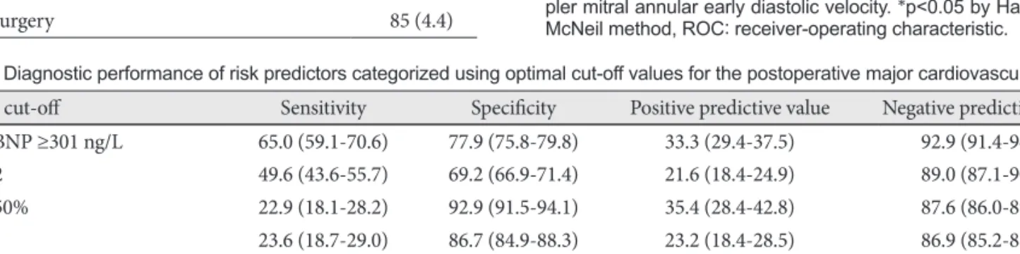
Table 3. Diagnostic performance of risk predictors categorized using optimal cut-off values for the postoperative major cardiovascular event Optimal cut-off Sensitivity Specificity Positive predictive value Negative predictive value NT-proBNP ≥301 ng/L 65.0 (59.1-70.6) 77.9 (75.8-79.8) 33.3 (29.4-37.5) 92.9 (91.4-94.2)
RCRI ≥2 49.6 (43.6-55.7) 69.2 (66.9-71.4) 21.6 (18.4-24.9) 89.0 (87.1-90.6)
LVEF <50% 22.9 (18.1-28.2) 92.9 (91.5-94.1) 35.4 (28.4-42.8) 87.6 (86.0-89.1)
E/E’ ≥13 23.6 (18.7-29.0) 86.7 (84.9-88.3) 23.2 (18.4-28.5) 86.9 (85.2-88.5)
LA volume index ≥33 27.1 (22.0-32.8) 83.2 (81.3-85.0) 21.6 (17.4-26.3) 87.0 (85.3-88.6)
Any RWMA exists 36.4 (30.8-42.4) 81.4 (79.5-83.3) 25.1 (20.9-29.6) 88.3 (86.5-89.8)
Any abnormal echocardiographic
parameters from the above 48.2 (42.2-54.2) 71.6 (69.3-73.7) 22.4 (19.2-26.0) 89.0 (87.2-90.7) The sensitivity, specificity, positive predictive value, and negative predictive values of each categorized risk predictors are shown. Because these values depend on the cut-off levels, values at the point of optimal cut-off levels calculated from ROC analysis were presented. NT-proB- NP: N-terminal pro-brain natriuretic peptide, RCRI: Revised Cardiac Risk Index, LVEF: left ventricular ejection fraction, LA: Left atrial, RWMA: regional wall notion abnormality
echocardiographic parameters including systolic and dia- stolic parameters were modestly predictive of PMCE; LVEF [AUC=0.614 {95% confidence interval (CI)=0.591-0.637}], RWM score index {AUC=0.603 (0.580-0.626)}, LA volume in- dex {AUC=0.593 (0.563-0.623)}, and E/E’ {AUC=0.567 (0.536- 0.597)} (p<0.05 for all). However, the predictive power of these parameters was not higher than RCRI {AUC=0.622 (0.599-0.644)} (p=0.020 between E/E’, p=not significant be- tween echocardiographic parameters) and was significantly lower than NT-proBNP {AUC=0.748 (0.727-0.768)} (p<
0.001 between echocardiographic parameters) (Fig. 2).
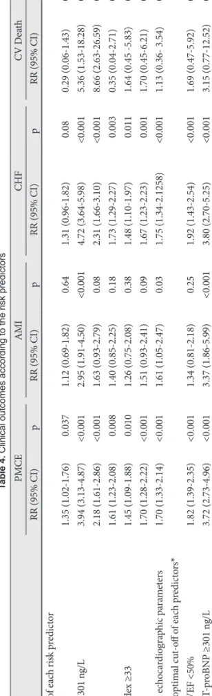
To evaluate the incremental value of other risk predictors to clinical risk index, RR adjusted to age, sex, and traditional clinical factors were calculated using the parameters catego- rized by optimal cut-off levels. The RR of RCRI cut-off (≥2) increased 2.8-fold after addition of NT-proBNP cut-off (≥301 ng/L) {RR=1.4 (95% CI=1.0-1.8) to 3.7 (2.7-5.0), p<0.001}.
Contrarily, the RR of RCRI cut-off (≥2) modestly increased after addition of LVEF cut-off (≤50%) {RR=1.4 (1.0-1.8) to 1.8 (1.4-2.4), p<0.001} and did not increase after addition of all the other echocardiographic parameters. Addition of both NT-proBNP and echocardiographic parameters did not result in a further increase in RR. The increase in RR by addi- tion of NT-proBNP to RCRI was also evident in secondary endpoints including AMI and pulmonary edema (Table 4).
Discussion
To our knowledge, this is the first study to compare the pre- dictive power of hemodynamic biomarker with that of imag- ing modality and investigated the incremental value of these modalities to the clinically determined risk. TTE, which is one of the widely used cardiovascular imaging modality, was inferior to NT-proBNP, a representative hemodynamic bio- marker, for the prediction of perioperative cardiovascular risk in non high-risk patients for major noncardiac surgery.
Moreover, TTE did not have incremental value to the clini- cally determined risk whereas NT-proBNP had incremental value to the clinically determined risk.
Most preoperative echocardiographic assessments have focused on the systolic function. Decreased LV ejection frac- tion has been repeatedly shown to be associated with periop- erative cardiovascular morbidity.2)3)14)15) In our study, LVEF of less than 50% was significantly but modestly predictive of PMCE. Although diastolic dysfunction is not uncommon in patients undergoing noncardiac surgery, routine preopera- tive evaluation does not include evaluation of diastolic dys- function for risk stratification. In our study, the presence of perioperative diastolic dysfunction was not related to the pe- rioperative cardiovascular risk. The result of our study could be partially explained by the difference between a general cardiovascular biomarker and an imaging modality. NT-
Table 4. Clinical outcomes according to the risk predictors PMCEAMICHFCV Death RR (95% CI)pRR (95% CI)pRR (95% CI)pRR (95% CI)p Optimal cut-off of each risk predictor RCRI ≥21.35 (1.02-1.76)0.0371.12 (0.69-1.82)0.641.31 (0.96-1.82)0.0800.29 (0.06-1.43)0.13 NT-proBNP ≥301 ng/L 3.94 (3.13-4.87)<0.0012.95 (1.91-4.50)<0.0014.72 (3.64-5.98)<0.0015.36 (1.53-18.28)0.009 LVEF <50%2.18 (1.61-2.86)<0.0011.63 (0.93-2.79)0.082.31 (1.66-3.10)<0.0018.66 (2.63-26.59)0.005 E/E’ ≥131.61 (1.23-2.08)0.0081.40 (0.85-2.25)0.181.73 (1.29-2.27)0.0030.35 (0.04-2.71)0.32 LA volume index ≥331.45 (1.09-1.88)0.0101.26 (0.75-2.08)0.381.48 (1.10-1.97)0.0111.64 (0.45 -5.83)0.45 RWMA ≥1.041.70 (1.28-2.22)<0.0011.51 (0.93-2.41)0.091.67 (1.23-2.23)0.0011.70 (0.45-6.21)0.43 Any abnormal echocardiographic parameters1.70 (1.33-2.14)<0.0011.61 (1.05-2.47)0.031.75 (1.34-2.1258)<0.0011.13 (0.36- 3.54)0.83 Combination of optimal cut-off of each predictors* RCRI ≥2 or LVEF <50%1.82 (1.39-2.35)<0.0011.34 (0.81-2.18)0.251.92 (1.43-2.54)<0.0011.69 (0.47-5.92)0.42 RCRI ≥2 or NT-proBNP ≥301 ng/L 3.72 (2.73-4.96)<0.0013.37 (1.86-5.99)<0.0013.80 (2.70-5.25)<0.0013.15 (0.77-12.52)0.10 RCRI ≥2 or NT-proBNP ≥301 ng/L or LVEF <50%3.96 (2.88-5.33)<0.0013.80 (2.02-6.99)<0.0014.16 (2.92-5.81)<0.0015.32 (1.07-25.92)0.041 The association of each risk predictor with clinical outcome is shown as adjusted relative risk (RR) with 95% confidence intervals (CIs). Significant univariate risk factors including significant uni- variate clinical factors-age, sex, functional status ≥3, diabetes, heart failure, stroke, evidence of ischemic heart disease or history of revascularization, emergency surgery, and vascular surgery; were included in multivariate logistic regression analysis. *Defined as at least one of three risk predictors is higher than cut-off values
proBNP is one of best independent predictors of cardiovas- cular impairment as well as a marker of myocardial ischemia and heart failure, which might better reflect the complex pathophysiology of perioperative cardiovascular stress rep- resented by a catecholamine surge with associated hemody- namic stress, systemic inflammation, and hypercoagulabili- ty.16-18) On the other hand, E/E’ is a specific marker for LV fill- ing pressure which could be affected significantly by perio- perative volume status.19)
Integration of both NT-proBNP and echocardiographic pa- rameters modestly improved the predictive power of the cli- nically determined risk, except for the risk of primary cardio- vascular death {RR=5.3 (95% CI=1.1-25.9), p=0.041}. There- fore, these two risk predictors might provide complementary prediction in the high-risk event or death.18) Investigation of the role of preoperative echocardiography in the high-risk group and comparison with biomarkers would be advisable in the near future.16)
Our study is not free from its several limitations as de- scribed below. First, in our patient cohort, only 4.9% of pa- tients had abnormal LV systolic function (LVEF <40%) and pa- tient mean age was 68 years. All patients in our study were low-risk non high-risk patients for major noncardiac surgery.
Therefore, these characteristics may limit the generalizability of our findings. Secondly, only patients who had undergone formal preoperative cardiovascular consultation were includ- ed. Thirdly, NT-proBNP and echocardiography were not ev- aluated on the same day. Fourthly, tissue Doppler study was done in only 59% of patients due to financial and clinical con- straints. However, given the strength of our results, it is un- likely that enrollment of more patients or tissue Doppler study in more number of patients would have changed the main re- sults of our study. Although it has been well validated and widely used in clinical practice, only the transmitral early dia- stolic velocity/tissue Doppler mitral annular early diastolic velocity (E/E’) has been used for the evaluation of diastolic dysfunction. The recently developed new methods for the ev- aluation of diastolic dysfunction such as strain, strain rate and LV torsion have not been evaluated and would be of interest in future studies.
In conclusion, preoperative echocardiography was modest- ly predictive of perioperative cardiovascular events but was inferior to NT-proBNP. Moreover, it did not have incremen- tal value to the clinically determined risk. Our results did not support the use of routine evaluation of echocardiography be- fore noncardiac surgery. However, preoperative echocardio- graphy before noncardiac surgery can provide independent information about the risk of postoperative cardiac complica- tions in selected patients.
Acknowledgments
This study was supported by grants from the Samsung Medical Center
Clinical Research Development Program, the In-Sung Foundation for Medical Research and a grant from the Korean Society of Cardiology (Industrial-Educational Cooperation, 2005).
REFERENCES
1) Fleisher LA, Beckman JA, Brown KA, et al. 2009 ACCF/AHA fo- cused update on perioperative beta blockade incorporated into the ACC/AHA 2007 guidelines on perioperative cardiovascular evalua- tion and care for noncardiac surgery. J Am Coll Cardiol 2009;54:
e13-118.
2) Halm EA, Browner WS, Tubau JF, Tateo IM, Mangano DT. Echo- cardiography for assessing cardiac risk in patients having noncardiac surgery. Study of Perioperative Ischemia Research Group. Ann Intern Med 1996;125:433-41.
3) Rohde LE, Polanczyk CA, Goldman L, Cook EF, Lee RT, Lee TH.
Usefulness of transthoracic echocardiography as a tool for risk strati- fication of patients undergoing major noncardiac surgery. Am J Car- diol 2001;87:505-9.
4) Kertai MD, Poldermans D, Bax JJ, Klein J, Van Urk H. Cardiac risk and perioperative management. J Cardiovasc Surg (Torino) 2003;
44:431-5.
5) Choi JH, Cho DK, Song YB, et al. Preoperative NT-proBNP and CRP predict perioperative major cardiovascular events in non-cardiac surgery. Heart 2010;96:56-62.
6) Feringa HH, Bax JJ, Elhendy A, et al. Association of plasma N-termi- nal pro-B-type natriuretic peptide with postoperative cardiac events in patients undergoing surgery for abdominal aortic aneurysm or leg by- pass. Am J Cardiol 2006;98:111-5.
7) Yun KH, Jeong MH, Oh SK, et al. Preoperative plasma N-terminal pro-brain natriuretic peptide concentration and perioperative cardio- vascular risk in elderly patients. Circ J 2008;72:195-9.
8) Karthikeyan G, Moncur RA, Levine O, et al. Is a pre-operative brain natriuretic peptide or N-terminal pro-B-type natriuretic peptide mea- surement an independent predictor of adverse cardiovascular out- comes within 30 days of noncardiac surgery? A systematic review and meta-analysis of observational studies. J Am Coll Cardiol 2009;54:
1599-606.
9) Lee TH, Marcantonio ER, Mangione CM, et al. Derivation and pro- spective validation of a simple index for prediction of cardiac risk of major noncardiac surgery. Circulation 1999;100:1043-9.
10) Devereaux PJ, Goldman L, Cook DJ, Gilbert K, Leslie K, Guyatt GH.
Perioperative cardiac events in patients undergoing noncardiac sur- gery: a review of the magnitude of the problem, the pathophysiology of the events and methods to estimate and communicate risk. CMAJ 2005;173:627-34.
11) Lang RM, Bierig M, Devereux RB, et al. Recommendations for chamber quantification: a report from the American Society of Echo- cardiography’s Guidelines and Standards Committee and the Cham- ber Quantification Writing Group, developed in conjunction with the European Association of Echocardiography, a branch of the Europe- an Society of Cardiology. J Am Soc Echocardiogr 2005;18:1440-63.
12) Stephan C, Wesseling S, Schink T, Jung K. Comparison of eight com- puter programs for receiver-operating characteristic analysis. Clin Chem 2003;49:433-9.
13) Zhang J, Yu KF. What’s the relative risk? A method of correcting the odds ratio in cohort studies of common outcomes. JAMA 1998;280:
1690-1.
14) Kontos MC, Brath LK, Akosah KO, Mohanty PK. Cardiac complic- ations in noncardiac surgery: relative value of resting two-dimensional echocardiography and dipyridamole thallium imaging. Am Heart J 1996;132:559-66.
15) Takase B, Younis LT, Byers SL, et al. Comparative prognostic value of clinical risk indexes, resting two-dimensional echocardiography, and dipyridamole stress thallium-201 myocardial imaging for periopera- tive cardiac events in major nonvascular surgery patients. Am Heart J 1993;126:1099-106.
16) Struthers A, Lang C. The potential to improve primary prevention in the future by using BNP/N-BNP as an indicator of silent ‘pancardiac’
target organ damage: BNP/N-BNP could become for the heart what mi- croalbuminuria is for the kidney. Eur Heart J 2007;28:1678-82.
17) Poldermans D, Hoeks SE, Feringa HH. Pre-operative risk assess- ment and risk reduction before surgery. J Am Coll Cardiol 2008;51:
1913-24.
18) Schouten O, Bax JJ, Poldermans D. Preoperative cardiac risk as- sessment in vascular surgery patients: seeing beyond the perioperative period. Eur Heart J 2008;29:283-4.
19) Dokainish H, Zoghbi WA, Lakkis NM, Quinones MA, Nagueh SF.
Comparative accuracy of B-type natriuretic peptide and tissue Dop- pler echocardiography in the diagnosis of congestive heart failure. Am J Cardiol 2004;93:1130-5.