© 2016 The Korean Ophthalmological Society
This is an Open Access article distributed under the terms of the Creative Commons Attribution Non-Commercial License (http://creativecommons.org/licenses /by-nc/3.0/) which permits unrestricted non-commercial use, distribution, and reproduction in any medium, provided the original work is properly cited.
Original Article
Oxidative stress is involved in the pathogenesis of many eye diseases, including dry eye syndrome, cataract, and age-related macular degeneration [1-3]. Oxidative stress impairs cellular function by damaging lipids, proteins, RNA, and DNA, in addition to causing inflammation and cell death [4,5]. 8-Hydroxydeoxyguanosine (8-OHdG), an oxidized DNA nucleoside, results from the oxidation of
guanine and is excreted in bodily fluids [6,7]. 8-OHdG is one of the most widely used biomarkers of cellular oxida- tive stress [6]. Malondialdehyde (MDA) is an endogenous genotoxic product of enzymatic and oxygen radical-in- duced lipid peroxidation [8].
Aqueous humor is continually produced by the ciliary body and is in direct contact with the anterior surface of the lens, iris, and corneal endothelial cells, before draining out of the eye via the trabecular meshwork [8,9]. Oxidative stress in aqueous humor reflects the balance among the ox- idative states of the lens, corneal endothelium, and retina.
Corneal endothelial cells are in direct contact with the aqueous humor [9] and can therefore secret their metabolic
Oxidative Stress Levels in Aqueous Humor from High Myopic Patients
Eun Bi Kim1, Ha Kyoung Kim1, Joon Young Hyon2,3, Won Ryang Wee2,Young Joo Shin1
1Department of Ophthalmology, Kangnam Sacred Heart Hospital, Hallym University College of Medicine, Seoul, Korea
2Department of Ophthalmology, Seoul National University College of Medicine, Seoul, Korea
3Department of Ophthalmology, Seoul National University Bundang Hospital, Seongnam, Korea
Purpose: To compare oxidative stress status in the aqueous humor of highly myopic eyes and control eyes.
Methods: Aqueous humor samples were collected from 15 highly myopic eyes (high myopia group) and 23 cat- aractous eyes (control group) during cataract surgery. Central corneal thickness, corneal endothelial cell den- sity, hexagonality of corneal endothelial cells, and cell area of corneal endothelial cells were measured using specular microscopy. Axial length was measured using ultrasound biometry. 8-Hydroxydeoxyguanosine (8- OHdG) and malondialdehyde levels were measured using enzyme-linked immunosorbent assay.
Results: 8-OHdG level was lower in the aqueous humor of myopic patients than in that of control group (p = 0.014) and was positively correlated with central corneal thickness and negatively correlated with axial length (r = 0.511, p = 0.02; r = -0.382, p < 0.001). There was no correlation between 8-OHdG level and corneal endothelial cell den sity, hexagonality, or cell area. Malondialdehyde level did not show any correlation with any parameters evaluated.
Conclusions: 8-OHdG might be a sensitive biomarker for evaluating oxidative stress status in the eye.
Oxidative stress level was lower in the aqueous humor of highly myopic eyes compared to that in control eyes, which indicates lower metabolic activity in these eyes.
Key Words: 8-Hydroxydeoxyguanosine, Axial eye length, Corneal pachymetry, High myopia, Malondialdehyde
Received: May 26, 2015 Accepted: August 31, 2015
Corresponding Author: Young Joo Shin, MD. Department of Ophthal- mology, Kangnam Sacred Heart Hospital, Hallym University College of Medicine, #1 Singil-ro, Yeongdeungpo-gu, Seoul 07441, Korea. Tel: 82-2- 829-5193, Fax: 82-2-848-4638, E-mail: [email protected]
products directly into the aqueous humor, whereas meta- bolic products from lens epithelial cells and the retina must diffuse through the anterior lens capsule and anterior hy- aloids membrane to reach the aqueous humor [10].
High myopia has been reported to be associated with various complications and is defined as a long axial length (AXL) [11,12]. Retinal degeneration is one of the major complications leading to severe vision loss [11,12]. Many causes of high myopic retinal degeneration, including oxidative stress and retinal ischemia, have been suggested [11,13]. An association between AXL and cytokine levels in aqueous humor has also been reported [14-16]. However, to the best of our knowledge, oxidative stress level in the aqueous humor of patients with high myopia has not yet been evaluated.
In this study, we investigated the oxidative stress status of aqueous humor in patients with high myopia and corre- sponding controls.
Materials and Methods
This comparative control study investigated aqueous hu- mor levels of 8-OHdG and MDA in 15 highly myopic eyes and 23 control eyes. Highly myopic eyes were defined as those with myopia of -6 diopters (D) or more or AXL 26 mm or longer. In the controls, aqueous humor samples were collected from senile cataract patients free from oth- er ocular or systemic diseases. The study protocol com- plied with the provisions of the Declaration of Helsinki and was reviewed and approved by the institutional review board/ethics committee of Hallym University Medical Center, Seoul, Korea. Patients were enrolled from the Oph- thalmic Centers in Hallym University Kangnam Sacred Heart Hospital between July and December 2010.
All patients underwent a complete ophthalmic examina- tion, including refraction, measurements of AXL and best-corrected visual acuity, indirect stereoscopic ophthal- moscopy, fluorescein angiography, and color fundus pho- tography. Central corneal thickness (CCT), corneal endo- thelial cell density, hexagonality of corneal endothelial cells, and cell area of corneal endothelial cells were mea- sured using specular microscopy. AXL was measured using ultrasound biometry (CineScan; Quantel Medical, Bozeman, MT, USA). The Lens Opacities Classification System II, which is based on photographic standards, was
used to assess cataracts [17,18]. Slit-lamp microscopic ex- aminations (BM-900; Haag-Streit, Koeniz, Switzerland) were performed by board-certified ophthalmologists or ophthalmologists in training.
Sample collection
In all patients, anterior chamber paracentesis was per- formed before incision during cataract surgery, and no ste- roids were administered before surgery. Undiluted aqueous humor samples were collected in sterile tubes and stored at -80°C until analysis.
Measurement of 8-OHdG and malondialdehyde using enzyme-linked immunosorbent assay
The aqueous humor levels of 8-OHdG and MDA were measured using the commercially available competitive 8-OHdG ELISA kit (Cayman Chemical, Ann Arbor, MI, USA) and MDA sandwich ELISA kit (Cell Biolabs Inc., San Diego, CA, USA), according to the respective manu- facturer’s instructions. Briefly, for the 8-OHdG assay, aqueous humor samples were incubated with 8-OHdG-ace- tylcholinesterase conjugate and an 8-OHdG monoclonal antibody in an enzyme immunoassay plate that had been pre-coated with goat anti-mouse antibodies. The enzyme immunoassay plate was incubated, washed, and developed with Ellman’s reagent, and absorbance was evaluated at 410 nm. For the MDA assay, aqueous humor samples were added to the microtiter plate precoated with an anti-MDA antibody. After incubation at 37°C for 1 hour and extensive washing, the plate was incubated for 1 hour with 100 μL of a biotinylated mouse anti-MDA antibody, followed by in- cubation for 1 hour with 100 μL of streptavidin-peroxidase conjugate. Then, TMB/E (3,3′,5,5′-tetramethylbenzidine) was added to each well and incubated for 5 to 10 minutes, after which the absorbance of plate was measured at 450 nm in a multi-plate reader (Spectramax Plus 384 plate reader; Molecular Devices, Sunnyvale, CA, USA). Serial dilutions of recombinant human 8-OHdG and MDA served as standards.
Statistics
Experimental data are expressed as mean ± standard deviation. The results were analyzed using the Mann-Whit-
ney U-test and Spearman’s correlation test. All statistical analyses were performed using SPSS ver. 14.0 (SPSS Inc., Chicago, IL, USA). A p-value less than 0.05 was consid- ered significant.
Results
Fifteen highly myopic eyes of 10 patients (high myopia group) and 23 cataract eyes of 23 patients (control group) were included in this study (Table 1). The mean age was 61.3 ± 10.3 years in the highly myopic group and 66.1 ± 12.1 years in the control group. The male to female ratio was 5 : 10 in the highly myopic group and 6 : 17 in the con- trol group. The mean refractive error (spherical equiva- lents) was -10.27 ± 3.78 D in the highly myopic group and +0.61 ± 1.66 D in the control group. Nucleosclerosis grade was not different between the control group (2.27 ± 0.70) and the highly myopic group (2.27 ± 0.68; p = 0.777, Mann-Whitney U-test). Intraocular pressure was also not different between the control group (13.78 ± 2.70 mmHg) and the highly myopic group (15.00 ± 2.98 mmHg; p = 0.314, Mann-Whitney U-test).
Oxidative stress marker levels in aqueous humor and axial length
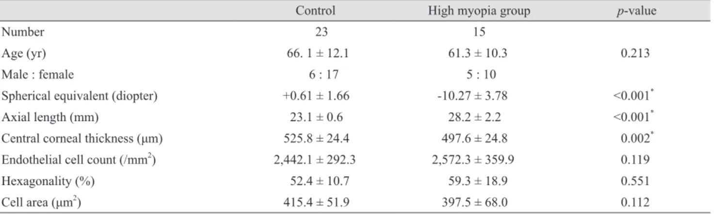
The levels of 8-OHdG and MDA in aqueous humor are shown in Fig. 1A-1D. 8-OHdG levels were lower in the high myopia group compared to the control group (p = 0.014, Mann-Whitney U-test) (Table 2). MDA level was not
significantly different between the control and high myo- pia groups. The level of 8-OHdG was dependent on AXL (r
= -0.382, p < 0.001, Spearman’s correlation coefficient).
MDA level did not correlate with AXL.
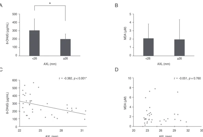
Correlations between 8-OHdG and ocular parameters The level of 8-OHdG correlated positively with CCT (r = 0.511, p = 0.02, Spearman’s correlation coefficient) (Fig.
2A). There was no correlation between 8-OHdG level and corneal endothelial cell den sity, hexagonality, or cell area (Fig. 2B-2D).
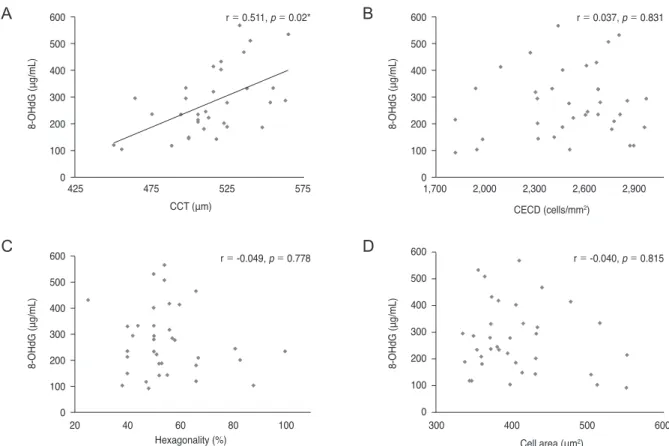
Correlations between malondialdehyde and ocular parameters
MDA level did not show any correlation with CCT, AXL, corneal endothelial cell den sity, hexagonality, or cell area (Fig. 3A-3D).
Discussion
Oxidative stress has been reported to be associated with various ocular diseases [11-16,19,20]. 8-OHdG has been used as a biomarker for oxidative DNA damage [20]. In this study, 8-OHdG level in aqueous humor was positively associated with CCT and negatively associated with AXL, whereas it was not associated with the measured parameters of corneal endothelial cells. These results suggest that 8-OHdG level in aqueous humor reflects the
Table 1. Demographic data
Control High myopia group p-value
Number 23 15
Age (yr) 66. 1 ± 12.1 61.3 ± 10.3 0.213
Male : female 6 : 17 5 : 10
Spherical equivalent (diopter) +0.61 ± 1.66 -10.27 ± 3.78 <0.001*
Axial length (mm) 23.1 ± 0.6 28.2 ± 2.2 <0.001*
Central corneal thickness (μm) 525.8 ± 24.4 497.6 ± 24.8 0.002*
Endothelial cell count (/mm2) 2,442.1 ± 292.3 2,572.3 ± 359.9 0.119
Hexagonality (%) 52.4 ± 10.7 59.3 ± 18.9 0.551
Cell area (μm2) 415.4 ± 51.9 397.5 ± 68.0 0.112
Values are presented as number or mean ± standard deviation.
*Statistically significant by Mann-Whitney U-test.
oxidative stress status of the whole eye, not only the corneal endothelium. Differences in CCT between myopia and emmetropia remain controversial [21].It has been re- ported that CCT does not correlate with degree of myopia [22]. Conversely, negative correlation between refraction degree and CCT has been reported [23]. In this study, CCT was thinner in patients with high myopia than it was in the controls.
High myopia is a degenerative disease [14]. A variety of
cytokines present in aqueous humor have been reported to be related to high myopia [14-16]. Elevated levels of matrix metalloproteinase-2 (MMP-2), tissue inhibitor of MMP-1 (TIMP-1), TIMP-2, and TIMP-3 have been found in eyes with elongated axes [14]. Concentration of transforming growth factor-β2 has been shown to positively correlate with AXL [16]. Conversely, concentration of vascular en- dothelial growth factor in aqueous humor has been nega- tively correlated with AXL [15]. High myopia results in retinal pigment epithelium degeneration [12]. It has been suggested that the metabolic activity in high myopia de- ceases due to retinal pigment epithelium degeneration [24- 26]. Ultraviolet light-blocking glasses worn to correct the myopia could reduce the oxidative stress level in the aque- ous humor.
Free radical production leading to oxidative stress is an initiating factor in the development of maturity-onset cata- ract [27,28]. Elevated level of MDA has been found in the cataract lenses and vitreous of myopic patients compared with non-myopic patients with cataract [29]. αA-crystallin,
8-OHdG (μg/mL)
500 400 300 200 100
0 <26 ≥26
22 25 28 31
AXL (mm)
AXL (mm)
r = -0.382, p < 0.001*
8-OHdG (μg/mL)
600 500 400 300 200 100 0
*
MDA (μM)
5 4 3 2 1
0 <26 ≥26
AXL (mm)
20 23 26 29 32 35
AXL (mm)
r = -0.051, p = 0.760
MDA (μM)
10 8 6 4 2 0
Fig. 1. Oxidative stress marker levels in aqueous humor and axial length (AXL). (A) 8-Hydroxydeoxyguanosine (8-OHdG) level in aque- ous humor was lower in the high myopia group compared to the control group (p = 0.014, Mann-Whitney U-test). (B) Malondialdehyde (MDA) levels were not different between control and high myopia groups. (C) 8-OHdG level decreased depending on AXL (r = -0.382, p
< 0.001, Spearman’s correlation coefficient). (D) MDA level did not correlate with AXL. *Statistically significant.
A
C
B
D
Table 2. Oxidative stress marker levels in aqueous humor of control and high myopia groups
Control
(n = 23) High myopia group (n = 15) p-value 8-OHdG level
(μg/mL) 311.6 ± 127.7 212.5 ± 103.2 0.014* MDA level (μM) 2.1 ± 1.7 1.9 ± 2.4 0.224 Values are presented as mean ± standard deviation.
8-OHdG = 8-hydroxydeoxyguanosine; MDA = malondialdehyde.
*Statistically significant by Mann-Whitney U-test.
the best-characterized structural protein of the human lens, acts as a chaperone under conditions of oxidative stress, thereby maintaining lens transparency [30]. High myopia has been reported as a risk factor for dark nuclear cataract, and CpG islands in the crystallin alpha A promot- er are hyper-methylated in lens epithelial cells of patients with high myopic dark nuclear cataract [31].However, the role of oxidative stress in high myopia is not well understood. In contrast to other studies [32], this study found that 8-OHdG level was lower in the highly myopic group compared to the control group and was negatively correlated with AXL. Reduced metabolism in degenerative high myopia [33] might lead to a decrease in oxidative stress level. Aqueous humor level of 8-OHdG was found to be higher in patients with exudative age-related macular degeneration, and the level correlated with macular lesion area [19]. One serious complication of high myopia causing vision loss is choroidal neovascularization [34]. Oxidative stress can be localized around choroidal neovasculariza- tion areas in high myopia patients. Furthermore, high my-
opia has been suggested as a risk factor for glaucoma [35].
Aqueous humor level of 8-OHdG increases and total anti- oxidant status decreases in the serum and aqueous humor of glaucoma patients [20].Glaucoma associated with high myopia might be different from that with normal AXL.
Age is an important factor for oxidative stress in eyes [2].
Cataract is a common cause of increased oxidative stress in aqueous humor [2]. However, in this study, cataract status was not different between the control and high myopia groups. One limitation of this study is that the comparison of 8-OHdG level in the aqueous humor between the high myopia group and non-cataract emmetropia group was not performed according to ethnics.
However, further study is necessary to compare 8-OHdG level in the aqueous humor between age-matched high my- opia and non-cataract emmetropia groups in order to support our conclusions.
In contrast, there were no significant differences in MDA level between the two groups. The production of MDA results from oxidative damage of lipids by free radi-
425 475 525 575
CCT (μm)
r = 0.511, p = 0.02*
8-OHdG (μg/mL)
600 500 400 300 200 100
0 1,700 2,000 2,300 2,600 2,900
CECD (cells/mm2)
r = 0.037, p = 0.831
8-OHdG (μg/mL)
600 500 400 300 200 100 0
20 40 60 80 100
Hexagonality (%)
r = -0.049, p = 0.778
8-OHdG (μg/mL)
600 500 400 300 200 100
0 300 400 500 600
Cell area (μm2)
r = -0.040, p = 0.815
8-OHdG (μg/mL)
600 500 400 300 200 100 0
Fig. 2. Correlation between 8-hydroxydeoxyguanosine (8-OHdG) and corneal endothelial cells. (A) 8-OHdG level was positively correlat- ed with central corneal thickness (CCT) (r = 0.511, p = 0.02, Spearman’s correlation coefficient). (B) 8-OHdG level did not correlate with corneal endothelial cell density (CECD). (C) 8-OHdG level did not correlate with cell hexagonality. (D) 8-OHdG level did not correlate with cell area. *Statistically significant by Spearman’s correlation coefficient test.
A
C
B
D
cals [36]; therefore, MDA level is frequently used as an in- dicator of oxidative damage to lipids resulting from free radicals [37]. There might be a potential reason for the dif- ferences between the 8-OHdG and MDA results. The mo- lecular weight of 8-OHdG (283 g/mol) is higher than that of MDA (72 g/mol); thus, it is possible that 8-OHdG is not able to be washed out into the trabecular meshwork but is retained in the anterior chamber. A previous study showed that 50% of 0.18-μm particles were caught in the trabecular meshwork and in the juxtacanalicular tissue [38]. In general, molecules having a molecular weight less than 500 g/mol are able to permeate the anterior chamber [39].
With increasing age, increased cross-linking might occur, leading to accumulation of extra-cellular material in the juxtacanalicular tissue, increased outflow resistance, and development of glaucoma [40]. Another reason for 8-OhdG accumulation is that is a very stable product of oxidative DNA damage following enzymatic cleavage after reactive oxygen species-induced 8-hydroxylation of guanine base in mitochondrial and nuclear DNA [41]. The half-life of
MDA is very short in the human body [42]. MDA has a room-temperature half-life in plasma of approximately 2 hours.
In this study, 8-OHdG level was evaluated only in aque- ous humor. However, 8-OHdG level in the vitreous might be more reflective of the oxidative stress level in the poste- rior segment, although a correlation between aqueous humor and vitreous levels has been reported [19]. Further study is necessary to compare 8-OHdG levels in vitreous of patients with high myopia.
In conclusion, 8-OHdG, a biomarker of oxidative stress, was found at lower levels in myopic patients compared to the control group. 8-OHdG level positively correlated with CCT and negatively correlated with AXL. There was no correlation between MDA level and CCT or AXL.
8-OHdG could be a sensitive marker for the evaluation of oxidative stress status of the eye. Oxidative stress level was lower in eyes with high myopia, which indicates lower metabolic activity in highly myopic eyes.
425 475 525 575
CCT (μm)
r = 0.272, p = 0.120
MDA (μM)
10 8 6 4 2 0
CECD (cells/mm2)
r = 0.151, p = 0.373
MDA (μM)
10 8 6 4 2 0
1,500 2,000 2,500 3,000 3,500
20 40 60 80 100
Hexagonality (%)
r = 0.078, p = 0.647
MDA (μM)
10 8 6 4 2 0
Cell area (μm2)
r = -0.150, p = 0.375
MDA (μM)
10 8 6 4 2 0
300 400 500 600
Fig. 3. Correlation between malondialdehyde (MDA) and corneal endothelial cells. (A) MDA level did not correlate with central corneal thickness (CCT). (B) MDA level did not correlate with corneal endothelial cell density (CECD). (C) MDA level did not correlate with cell hexagonality. (D) MDA level did not correlate with cell area.
A
C
B
D
Conflict of Interest
No potential conflict of interest relevant to this article was reported.
Acknowledgements
This study was supported by the National Research Foundation grant (2012R1A1A2040118) funded by the Ko- rea government (MEST) and Hallym University Research Fund 2014 (HURF-2014-52).
References
1. Wakamatsu TH, Dogru M, Matsumoto Y, et al. Evaluation of lipid oxidative stress status in Sjogren syndrome pa- tients. Invest Ophthalmol Vis Sci 2013;54:201-10.
2. Thiagarajan R, Manikandan R. Antioxidants and cataract.
Free Radic Res 2013;47:337-45.
3. Chen Y, Mehta G, Vasiliou V. Antioxidant defenses in the ocular surface. Ocul Surf 2009;7:176-85.
4. Ito K, Suda T. Metabolic requirements for the maintenance of self-renewing stem cells. Nat Rev Mol Cell Biol 2014;15:
243-56.
5. Markesbery WR, Lovell MA. Damage to lipids, proteins, DNA, and RNA in mild cognitive impairment. Arch Neu- rol 2007;64:954-6.
6. Wu LL, Chiou CC, Chang PY, Wu JT. Urinary 8-OHdG: a marker of oxidative stress to DNA and a risk factor for cancer, atherosclerosis and diabetics. Clin Chim Acta 2004;339:1-9.
7. Halliwell B, Whiteman M. Measuring reactive species and oxidative damage in vivo and in cell culture: how should you do it and what do the results mean? Br J Pharmacol 2004;142:231-55.
8. Janero DR. Malondialdehyde and thiobarbituric acid-reac- tivity as diagnostic indices of lipid peroxidation and perox- idative tissue injury. Free Radic Biol Med 1990;9:515-40.
9. Freddo TF. A contemporary concept of the blood-aqueous barrier. Prog Retin Eye Res 2013;32:181-95.
10. Danysh BP, Duncan MK. The lens capsule. Exp Eye Res 2009;88:151-64.
11. Silva R. Myopic maculopathy: a review. Ophthalmologica 2012;228:197-213.
12. Saw SM, Gazzard G, Shih-Yen EC, Chua WH. Myopia and associated pathological complications. Ophthalmic Physiol Opt 2005;25:381-91.
13. Bosch-Morell F, Sanz A, Diaz-Llopis M, Romero FJ. Lipid peroxidation products in human subretinal fluid. Free Rad- ic Biol Med 1996;20:899-903.
14. Jia Y, Hu DN, Zhu D, et al. MMP-2, MMP-3, TIMP-1, TIMP-2, and TIMP-3 protein levels in human aqueous hu- mor: relationship with axial length. Invest Ophthalmol Vis Sci 2014;55:3922-8.
15. Shin YJ, Nam WH, Park SE, et al. Aqueous humor concen- trations of vascular endothelial growth factor and pigment epithelium-derived factor in high myopic patients. Mol Vis 2012;18:2265-70.
16. Jia Y, Hu DN, Zhou J. Human aqueous humor levels of TGF- β2: relationship with axial length. Biomed Res Int 2014;2014:258591.
17. Chylack LT Jr, Wolfe JK, Singer DM, et al. The Lens Opacities Classification System III: the Longitudinal Study of Cataract Study Group. Arch Ophthalmol 1993;111:831-6.
18. Karbassi M, Khu PM, Singer DM, Chylack LT Jr. Evalua- tion of lens opacities classification system III applied at the slitlamp. Optom Vis Sci 1993;70:923-8.
19. Lau LI, Liu CJ, Wei YH. Increase of 8-hydroxy-2’-deox- yguanosine in aqueous humor of patients with exudative age-related macular degeneration. Invest Ophthalmol Vis Sci 2010;51:5486-90.
20. Sorkhabi R, Ghorbanihaghjo A, Javadzadeh A, et al. Oxi- dative DNA damage and total antioxidant status in glauco- ma patients. Mol Vis 2011;17:41-6.
21. Pedersen L, Hjortdal J, Ehlers N. Central corneal thickness in high myopia. Acta Ophthalmol Scand 2005;83:539-42.
22. Ortiz S, Mena L, Rio-San Cristobal A, Martin R. Relation- ships between central and peripheral corneal thickness in different degrees of myopia. J Optom 2014;7:44-50.
23. Wei W, Fan Z, Wang L, et al. Correlation analysis between central corneal thickness and intraocular pressure in juve- niles in Northern China: the Jinan city eye study. PLoS One 2014;9:e104842.
24. Bothman L. The relation of the basal metabolic rate to pro- gressive axial myopia. Am J Ophthalmol 1931;14:918-24.
25. Mao JF, Liu SZ, Dou XQ. Retinoic acid metabolic change in retina and choroid of the guinea pig with lens-induced myopia. Int J Ophthalmol 2012;5:670-4.
26. Ghazi NG, Green WR. Pathology and pathogenesis of reti- nal detachment. Eye (Lond) 2002;16:411-21.
27. Spector A. Oxidative stress-induced cataract: mechanism of action. FASEB J 1995;9:1173-82.
28. Lou MF. Redox regulation in the lens. Prog Retin Eye Res 2003;22:657-82.
29. Micelli-Ferrari T, Vendemiale G, Grattagliano I, et al. Role of lipid peroxidation in the pathogenesis of myopic and se- nile cataract. Br J Ophthalmol 1996;80:840-3.
30. Wang K, Spector A. Alpha-crystallin can act as a chaper- one under conditions of oxidative stress. Invest Ophthalmol Vis Sci 1995;36:311-21.
31. Zhu XJ, Zhou P, Zhang KK, et al. Epigenetic regulation of αA-crystallin in high myopia-induced dark nuclear cata- ract. PLoS One 2013;8:e81900.
32. Cherfan GM, Michels RG, de Bustros S, et al. Nuclear scle- rotic cataract after vitrectomy for idiopathic epiretinal membranes causing macular pucker. Am J Ophthalmol 1991;111:434-8.
33. Silverstone BZ, Syrkin N, Algur N, Berson D. A metabolic aspect of high myopia. Ann Ophthalmol 1985;17:546-51.
34. Leveziel N, Yu Y, Reynolds R, et al. Genetic factors for choroidal neovascularization associated with high myopia.
Invest Ophthalmol Vis Sci 2012;53:5004-9.
35. Xu L, Wang Y, Wang S, et al. High myopia and glaucoma susceptibility the Beijing Eye Study. Ophthalmology 2007;
114:216-20.
36. Kara A, Akman S, Ozkanlar S, et al. Immune modulatory and antioxidant effects of melatonin in experimental peri- odontitis in rats. Free Radic Biol Med 2013;55:21-6.
37. Khalili J, Biloklytska HF. Salivary malondialdehyde levels in clinically healthy and periodontal diseased individuals.
Oral Dis 2008;14:754-60.
38. Inomata H, Bill A, Smelser GK. Aqueous humor pathways through the trabecular meshwork and into Schlemm’s canal in the cynomolgus monkey (Macaca irus): an electron mi- croscopic study. Am J Ophthalmol 1972;73:760-89.
39. Schoenwald RD, Ward RL. Relationship between steroid permeability across excised rabbit cornea and octanol-wa- ter partition coefficients. J Pharm Sci 1978;67:786-8.
40. Bill A, Svedbergh B. Scanning electron microscopic stud- ies of the trabecular meshwork and the canal of Schlemm:
an attempt to localize the main resistance to outflow of aqueous humor in man. Acta Ophthalmol (Copenh) 1972;
50:295-320.
41. Cheng KC, Cahill DS, Kasai H, et al. 8-Hydroxyguanine, an abundant form of oxidative DNA damage, causes G→T and A→C substitutions. J Biol Chem 1992;267:166-72.
42. Gil HW, Seok SJ, Jeong DS, et al. Plasma level of malondi- aldehyde in the cases of acute paraquat intoxication. Clin Toxicol (Phila) 2010;48:149-52.