Veterinary Science
http://dx.doi.org/10.4142/jvs.2011.12.4.387
Received: 23 Mar. 2010, Revised: 18 Feb. 2011, Accepted: 20 May 2011
Original Article
*Corresponding author
Tel: +55-61-33847887; Fax: +55-61-33847887 E-mail: [email protected]
Cone beam computed tomography and intraoral radiography for diagnosis of dental abnormalities in dogs and cats
Marcello R. Roza
1,*, Luiz Antonio F. Silva
1, Mauricio Barriviera
2, Alessandro L. Januário
3, Ana Cristina B. Bezerra
4, Maria Clorinda S. Fioravanti
11
Department of Veterinary Medicine, Federal University of Goias, Goiânia 74001-970, Brazil
2
Department of Radiology, Faculty of Dentistry, Catholic University of Brasilia, Brasília 71966-700, Brazil
3
International Team for Implantology, São Paulo 04551-060, Brazil
4
Faculty of Health Sciences, University of Brasilia, Brasília 70910-700, Brazil
The development of veterinary dentistry has substantially improved the ability to diagnose canine and feline dental abnormalities. Consequently, examinations previously performed only on humans are now available for small animals, thus improving the diagnostic quality. This has increased the need for technical qualification of veterinary professionals and increased technological investments. This study evaluated the use of cone beam computed tomography and intraoral radiography as complementary exams for diagnosing dental abnormalities in dogs and cats. Cone beam computed tomography was provided faster image acquisition with high image quality, was associated with low ionizing radiation levels, enabled image editing, and reduced the exam duration. Our results showed that radiography was an effective method for dental radiographic examination with low cost and fast execution times, and can be performed during surgical procedures.
Keywords: cone beam computed tomography, dental, dogs, radiography, X-ray diagnosis
Introduction
The development of veterinary dentistry has substantially improved diagnostic quality and efficiency, resulting in more satisfactory indexes for treatments performed on the stomatognathic system of domestic animals. In addition to demands for technological investment on the part of equipment manufacturing companies, this veterinary specialty requires technical qualification of medical per- sonnel through training programs, continuing professional development, and the acquisition of state-of-the-art
equipment. As a result of these advancements, dental procedures that were previously performed without the aid of diagnostic resources (especially imaging studies) are currently used in the daily routine of veterinary clinics and hospitals. This has significantly contributed to the success of interventions with this diagnostic modality.
Despite the increased diagnostic quality resulting from scientific discoveries, the high cost of dental exams is a limiting factor that can prevent the implementation of new technologies in the veterinary environment. Therefore, intraoral radiography (IOR) remains the chosen method for imagenologic examination for assessing abnormalities of the teeth and other oral cavity structures. If possible, this procedure should be performed during the animals’ first dental appointment [22]. However, procedures such as tomography, which may seem economically impractical at first, might be routinely included in examination performed at veterinary clinics and hospitals if deemed necessary. Cone beam computed tomography (CBCT), routinely used in human medicine, has recently become a viable and cost-effective diagnostic alternative that can identify several different types of oral abnormalities in domestic animals [19].
Even though a previous report has shown that CBCT should only be recommended for diagnosing oral diseases in domestic animals [19], the first reports about the use of this technique in humans date back to the 1990s [5,10].
CBCT is a diagnostic modality in which the X-ray tube-detector system rotates 360
oaround the patient’s head and acquires images that will be analyzed by a specific computer program [5,20]. Volumetric data obtained by tomography consist of three-dimensional blocks of small cuboid structures known as voxels. In CBCT, this block is isometric in that the height, width, and depth are equal [3].
Although CBCT has been included in the diagnostic
routine for identifying dental diseases in human patients
since the end of the last century, scientific reports have
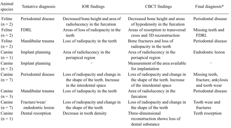
Table 1. Distribution of study subjects according to species, diagnoses, and odontological abnormalities for comparison between cone beam computed tomography (CBCT) and intraoral radiography (IOR) for canine and feline odontological assessment
Animal
species Tentative diagnosis IOR findings CBCT findings Final diagnosis*
Feline (n = 2)
Periodontal disease Decreased bone height and area of radiolucency in the furcation
Decreased bone height and areas of hypodensity in the furcation
Periodontal disease Feline
(n = 2)
FDRL Areas of loss of radiopacity in the teeth
Areas of resorption to transversal cross and 3D reconstruction
Missing teeth and FDRL
Feline (n = 2)
Mandibular trauma Loss of radiopacity in the teeth Bone fractures and loss of radiopacity in the teeth
Periodontal disease Canine
(n = 1)
Implant planning Area of radiolucency in the periapical region
Area of radiolucency in the periapical region
Endodontic lesion Canine
(n = 2)
Implant planning – Measurement of the area available
for implantation
– Canine
(n = 7)
Periodontal disease Loss of radiopacity and change in the shape of the teeth. Increase in the interdental space
Loss of radiopacity and change in the shape of the teeth. Increase of the interdental space
Missing teeth, fracture, ankylosis and tooth wear Canine
(n = 3)
Mandibular trauma Loss of radiopacity in the teeth Area of radiolucency in the furcation
Periodontal disease Canine
(n = 7)
Fracture/wear/
endodontic lesion
Loss of radiopacity and change in the shape of the teeth
Loss of radiopacity and change in the shape of the teeth
Tooth wear and fractures Canine
(n = 1)
Dental resorption Decrease in tooth density Three-dimensional
reconstruction shows loss of dental substance
Teeth resorption
*Number of animals of the same species with identical odontological abnormality during the exam (some patients were diagnosed with more than one abnormality). FDRL: feline dental resorptive lesion.