353 Original Article
Korean Circulation J 2005;35:353-356
ISSN 1738-5520
ⓒ 2005, The Korean Society of Circulation ORIGINAL ARTICLE
Risk Factors for Acute Cardioembolic Brain Stroke in Acute Myocardial Infarction
Si-Hoon Park, M.D. and Ji Min Jung, M.D.
Department of Cardiology and Ewha Medical Research Institute, College of Medicine, Ewha Womans University, Seoul, Korea ABSTRACT
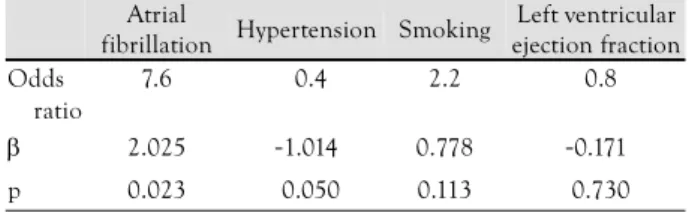
Background and Objectives:An ischemic brain stroke following acute myocardial infarction (AMI) has a poor clinical prognosis, which primarily results from a thromboembolism. We determined the risk factors of acute car- dioembolic brain stroke events that developed concurrently with, or soon after, the onset of AMI. Subjects and Methods:We evaluated 38 AMI patients, who developed subsequent acute cardioembolic brain stroke during their index admission, by comparing their clinical and angiographic characteristics with those of 1,443 consecutive patients that had not experienced a brain stroke. Strokes that occurred between the onset of the AMI and patient discharge were analyzed. The incidences of cardiovascular risk factors, and the clinical and angiographic characteristics, of patients admitted to Ewha Womans University Mokdong Hospital, with a diagnosis of AMI over a 10-year period, were compared. Results:In the univariate analysis, the frequencies of atrial fibrillation (21% vs. 4%, p=0.011) and hypertension (71% vs. 48%, p=0.030), and a left ventricular ejection fraction <40% (52% vs. 33%, p=0.039) were significantly higher in patients that had had an acute cardioembolic brain stroke. In a logistical regression analysis, atrial fibrillation was found to be a significant contributor to the subsequent development of an acute cardioembolic brain stroke in the AMI patients (p=0.023, β=2.025, odds ratio=7.6). Mean follow-up period, which was mainly determined as the time to death after the AMI, was shorter in the acute cardioembolic brain stroke patients (8.5 month vs. 24.3 month, p=0.002). The death rate during the mean follow-up period was much higher in these patients (50% vs. 29%). Conclusion:We found that the presence of atrial fibrillation at the time of admission for an AMI was associated with an increased risk of a subsequent acute cardioembolic brain stroke.
(Korean Circulation J 2005;35:353-356)
KEY WORDS:Myocardial infarction;Stroke, acute;Atrial fibrillation;Thromboembolism.
Introduction
Epidemiological studies have shown that an acute st- roke(AS) and acute myocardial infarction(AMI) have common risk factors, like age, gender and hypertension.1-5) Moreover, stroke after an AMI occurs due to patholo- gical conditions of the heart. The total incidence of strokes after an AMI, including patients without a throm- bolysis, ranges from 1.0 to 1.3%, and about 0.8% are ischemic strokes due to cardioembolism or an unknown etiology.6-10) No data concerning a cardioembolic brain stroke after the onset of AMI are available for the Korean population. Therefore, we evaluated the AMI patients
that subsequently experienced a cardioembolic brain stroke to identify the risk factors contributing to a car- dioembolic brain stroke.
Subjects and Methods
SubjectsWe evaluated 38 AMI patients, who developed a subse- quent cardioembolic stroke during their index admission, by comparing their clinical and angiographic characte- ristics with those of 1,443 consecutive patients that had not experienced a brain stroke. Patients that had suffe- red strokes between the onset of the AMI and their dis- charge were analyzed. The cardiovascular risk factors, and clinical or angiographic characteristics, of the pa- tients admitted to Ewha Womans University Hospital, with a diagnosis of acute myocardial infarction over a 10-year period, were assessed and compared between these two patient groups.
The AMI was diagnosed using the WHO definition;
Received:March 7, 2005 Revision Received:April 20, 2005 Accepted:May 11, 2005
Correspondence:Si-Hoon Park, M.D.,Department of Cardiology and Ewha Medical Research Institute, College of Medicine, Ewha Womans Univer- sity, 911-1 Mok-dong, Yangcheon-gu, Seoul 158-710, Korea Tel: 82-2-2650-5308, Fax: 82-2-2650-5424
E-mail: [email protected]
354·Korean Circulation J 2005;35:353-356
i.e., when patients had at least two of the following three criteria: a chest pain typical of myocardial ischemia, ini- tial and serial conventional electrocardiographic changes in the standard or precordial leads, and enzymatic evi- dence of myocardial necrosis.11) An acute stroke was de- fined using the WHO definition as, “rapidly developing clinical signs of focal(or global) disturbance of cerebral function lasting more than 24h(unless interrupted by surgery or death), with no apparent cause other than one of vascular origin”. Patients with intracerebral he- morrhage were excluded. The definition used for a car- dioembolic stroke was similar to that of the National Institute of Neurological Disorders and Stroke Classi- fication.12) Patients with a recorded history of hyperten- sion, or with three or more successive blood pressure measurement exceeding 140/90 mmHg during hospita- lization, were considered hypertensive. Diabetes mellitus was determined to be present if documented in medical records or if a patient required dietary sugar restriction, insulin, or oral hypoglycemic drugs during hospitaliza- tion, in addition to having positive criteria for diabetes.13) Statistical analysis
Data were expressed as means±SD for continuous variables, and as frequencies for variables on a nominal scale. For group comparisons Student’s t-, ANOVA or
chi-squared tests were used. Logistical regression analysis was used to determine the risk factors for cardioembo- lic stroke after an AMI. P<0.05 were considered signi- ficant.
Results
The mean time from admission due to the AMI to the taking of brain CT or MRI for the detection of brain stroke was 4.2±2.1 days. The frequencies of atrial fib- rillation(21% vs. 4%, p=0.011) and hypertension(71%
vs. 48%, p=0.030), and a left ventricular ejection frac- tion <40%(52% vs. 33%, p=0.039) were significantly higher in patients that had experienced a cardioembo- lic brain stroke(Table 1). Age, gender, frequency of an- terior myocardial infarction, ST segment myocardial infarction, previous myocardial infarction, a previous ischemic stroke, diabetes mellitus or hypercholesterole- mia were no different between the two groups. The drug histories, including aspirin, ticlopidine or clopidogrel, were similar in the two groups. The peak CK-MB level, frequency of thrombolytic treatment, left anterior des- cending artery(as an infarct-related artery), TIMI 3 flow of an infarct-related artery and extension of coronary disease, including normal or 1-vessel disease, were compa- rable between the two groups. In the logistical regression
Table 1. Characteristics of the patients with an acute myocardial infarction, according to the development of a cardioembolic brain stroke With cardioembolic brain stroke
(n=38)
Without cardioembolic brain stroke (n=1,443) p
Age (yrs) 70±11. 69±12 0.568
Male 53% 50% 0.806
Anterior myocardial infarction 44% 64% 0.108
STEMI 50% 60% 0.367
Atrial fibrillation on admission 21% 04% 0.011
Previous myocardial infarction 08% 12% 0.571
Previous ischemic stroke 11% 08% 0.643
DM 45% 25% 0.051
Hypertension 71% 48% 0.030
Hypercholesterolemia 0.174±51.00 178±410 0.740
Smoking 45% 69% 0.020
LVEF<40% 52% 33% 0.039
Peak CK-MB(IU/L) 100.1±143.3 250.7±488.1 0.069
Use of aspirin before admission 18% 14% 0.524
Use of ticlopiding/clopidogrel before admission 08% 14% 0.409
Thrombolysis 16% 17% 0.849
Coronary angiography 29% 69% 0.000
Coronary angioplasty 24% 58% 0.012
Normal or 1-vessel coronary artery 30% 31% 0.777
IRA in LAD 61% 62% 0.949
TIMI 3 flow in IRA 30% 27% 0.733
Mean follow-up time (months) 008.5±010.7 024.3±17.40 0.002
Follow-up patient 50% 46% 0.170
Mean±SD. STEMI: ST segment elevation myocardial infarction, DM: diabetes mellitus, LVEF: left ventricular ejection fraction, LAD: left anterior descending artery, IRA: infarct related artery
Si-Hoon Park, et al: Risk Factor for Embolic Brain Stroke in AMI·355
analysis, atrial fibrillation was identified as a significant contributory risk factor for the subsequent development of a cardioembolic stroke in patients with an AMI(p=
0.023, β=2.025, odds ratio=7.6)(Table 2). Follow-up was achieved in nearly half the patients, and the mean follow-up period, mainly determined by the time to death after the AMI, was shorter in the cardioembolic stroke group(8.5 month vs. 24.3 month, p=0.002) (Table 1). The death rate during the mean follow-up period was much higher in these patients(50% vs. 29%).
Discussion
This study demonstrated the presence of atrial fibril- lation at the time of admission for an AMI was associated with an increased risk of a subsequent acute cardioem- bolic brain stroke.
The determination of the risk factors and clinical features of a cardioembolic brain stroke are highly re- levant issues, given the high mortality and risk associa- ted with recurrent embolism, such that anticoagulation might be used as an effective prophylaxis. A cardiac source of an embolism has been reported in around one third of patients with cerebral infarction.14-22) The various sources of a cardiogenic embolism represent distinct types of heart disease, each with unique demographic features and risks of primary or secondary emboli.22) Re- cent data has shown that hypertrophic hypertensive car- diac disease, complicated by atrial fibrillation, is the most frequent cardiac source of emboli in cardioembolic stroke patients. Other important cardiac sources identified in- clude; isolated atrial fibrillation, rheumatic mitral valve disease, and systolic left ventricular dysfunction of ische- mic and non-ischemic causes. However, the incidences of traditional emboligenous-prone cardiac disorders, such as mitral valve prolapse and mitral annular calci- fication, have been reported to be low.17)
The total incidence of strokes after an AMI has been reported to be 1.0%.6-8) In the present study, 2.5% of the AMI cases experienced a cardioembolic brain stroke, which is somewhat higher than that reported previously.
A recent report showed that for patients <75 years old with an AMI, almost 60% of those that developed a stroke presented with an anterior wall myocardial infarc- tion, compared with only 38% of those not developing a stroke.23) These data support the hypothesis that, “em-
boli from the left ventricle following an AMI are an important cause of a peri-AMI associated stroke”. In the setting of an acute transmural infarction, patients with an anterior infarction and severe apical-wall-mo- tion abnormality(akinesis or dyskinesis) are at high risk of developing a left ventricular thrombus; these thrombi were diagnosed an average of five days after the infarc- tion.24) Left ventricular thrombi, which project into the lumen, are more likely to embolize, and a trend of an association between greater mobility and embolization has been reported.25) A cardioembolism tends to occur more in the acute phase rather than the chronic phase after an AMI.26) Although, in the present study, we could not check for either the existence of a left ventricular thrombus or the exact extent of the left ventricular re- gional wall motion abnormality, due to the retrospective nature of our study. A left ventricular ejection fraction
<40% was more prevalent in those that had experien- ced a cardioembolic brain stroke. Our study suggests that left ventricular wall motion abnormality might be as- sociated with cardioembolization, although this was not found to be significant in the logistical regression analy- sis.
Atrial fibrillation is widely accepted as one of most significant risk factors for a cardioembolic brain stroke.17) Recent data from cardioembolic brain stroke patients has shown that atrial fibrillation, without organic heart disease, was present in 22.1%, but with organic heart disease this figure was 57.7%.17) However, in those with ischemic heart disease only, severe left ventricular sys- tolic dysfunction was identified as a high risk cardiac source of embolism. The presence of intraventricular thrombosis, and the predominance of cases with a sinus rhythm, confirms the primary role of severe left ventri- cular systolic dysfunction, rather than atrial fibrillation, as a cardiologic substrate in patients with a cardioem- bolic stroke.17) This observation differed somewhat from the findings of the present study, which indicate that atrial fibrillation is the most significant risk factor for a cardioembolic brain stroke, even in those with an acute myocardial infarction, one of most important ischemic heart diseases. The difference in the follow-up dura- tion might be one of the possible reasons for these dis- crepancies. We followed the acute phase after AMI onset;
whereas, the previous report followed the patient over several years. The OPTIMAAL trial aimed to determine the frequency of atrial fibrillation, its impact on the cli- nical outcomes of patients with AMI and the left ven- tricular dysfunction.27) This trial included 5,477 patients with both an AMI and signs of left ventricular dysfunc- tion. Patients with atrial fibrillation at the baseline were found to have a higher risk of mortality than those without. New-onset atrial fibrillation was associated with an increased subsequent mortality over the first 30 days.
A recent study showed a small subset of patients that
Table 2. Logistical regression analysis for the occurrence of an acute cardioembolic stroke accompanied by an acute myocardial infarction
Atrial
fibrillation Hypertension Smoking Left ventricular ejection fraction Odds
ratio
7.6 -0.4 2.2 -0.8
β 2.025 -1.014 0.778 -0.171 p 0.023 -0.050 0.113 -0.730
356·Korean Circulation J 2005;35:353-356
presented with a stroke, but without any signs or symp- toms of AMI. 1.6% of the total AMI, 22% of the “stroke complicating MI” cases, presented with a stroke at the time of admission. The nature of these presentations highlights the importance of urgent EKG in all cases of an acute stroke.28)
The provision of long-term anticoagulation is currently a class Ia recommendation in patients with a documen- ted left ventricular thrombus, but a class IIa recommen- dation for patients with significant apical wall motion abnormalities.29) In view of these recommendation, and the findings of the present study, anticoagulation can not be overemphasized, in patients with atrial fibrillation following a large anterior AMI, with regard to the pre- vention of a cardioembolism.
The present study had several limitations; firstly, due to the small numbers of echocardiographic studies avai- lable, we could not evaluate the coexistence of a left ventricular thrombus, or regional wall motion abnor- malities, with a cardioembolic stroke. Secondly, the onset times of atrial fibrillation or brain stroke were not determined due to incomplete data. in the near future, we hope to conduct well-organized, prospective studies.
REFERENCES
1) Adams RJ, Chimowitz MI, Alpert JS, et al. Coronary risk eva- luation in patients with transient ischemic attack and ischemic stroke: a scientific statement for healthcare professionals from the Stroke Council and the Council on Clinical Cardiology of the American Heart Association/American Stroke Association. Cir- culation 2003;108:1278-90.
2) de Backer G, Ambrosioni E, Borch-Johnsen K, et al. European guidelines on cardiovascular disease prevention in clinical practice.
Atherosclerosis 2003;171:145-55.
3) Feigin VL, Lawes CM, Bennett DA, Anderson CS. Stroke epide- miology: a review of population-based studies of incidence, pre- valence, and case-fatality in the late 20th century. Lancet Neurol 2003;2:43-53.
4) Truelsen T, Mahonen M, Tolonen H, Asplund K, Bonita R, Vanuzzo D. Trends in stroke and coronary heart disease in the WHO MONICA Project. Stroke 2003;34:1346-52.
5) Yusuf S, Reddy S, Ounpuu S, Anand S. Global burden of car- diovascular diseases: part I. general considerations, the epide- miologic transition, risk factors, and impact of urbanization.
Circulation 2001;104:2746-53.
6) GUSTO Angiographic Investigators. The effects of tissue plasmi- nogen activator, streptokinase, or both on coronary-artery patency, ventricular function, and survival after acute myocardial infarction.
N Engl J Med 1993;329:1615-22.
7) Fibrinolytic Therapy Trialists’ (FTT) Collaborative Group. Indi- cations for fibrinolytic therapy in suspected acute myocardial infarction: collaborative overview of early mortality and major morbidity results from all randomized trials of more than 1000 patients. Lancet 1994;343:311-22.
8) Becker RC, Burns M, Gore JM, et al. Early assessment and in- hospital management of patients with acute myocardial infarction at increased risk for adverse outcomes: a nationwide perspective of current clinical practice. Am Heart J 1998;135:786-96.
9) Simoons ML, Maggioni AP, Knatterud G, et al. Individual risk assessment for intracranial haemorrhage during thrombolytic therapy. Lancet 1993;342:1523-8.
10) Mahaffey KW, Granger CB, Sloan AM, et al. Risk factors for in- hospital nonhemorrhagic stroke in patients with acute myocardial infarction treated with thrombolysis. Circulation 1998;97:757-64.
11) Gillum RF, Fortmann SP, Prineas RJ, Kottke TE. International diagnostic criteria for acute myocardial infarction and acute stroke. Am Heart J 1984;108:150-8.
12) National Institute of Neurological Disorders and Stroke. Special report from the National Institute of Neurological Disorders and Stroke: classification of cerebrovascular diseases III. Stroke 1990;
21:637-76.
13) Harris MI, Hadden WC, Knowler WC, Bennett PH. International criteria for the diagnosis of diabetes and impaired glucose tole- rance. Diabetes Care 1985;8:562-7.
14) Boon A, Cheriex E, Lodder J, Kessels F. Cardiac valve calcifica- tion: characteristics of patients with calcification of the mitral annulus or aortic valve. Heart 1997;78:472-4.
15) Gagliardi R, Benvenuti L, Frosini A, Ammannati F, Barletta GA, Fantini F. Frequency of echocardiographic abnormalities in pati- ents with ischemia of the carotid territory: a preliminary report.
Stroke 1985;16:118-20.
16) Fogelholm R, Melin J. Echocardiography in ischaemic cerebro- vascular disease. Br Med J 1987;295:305-6.
17) Pujadas Capmany R, Arboix A, Casanas-Munoz R, Anguera-Fer- rando N. Specific cardiac disorders in 402 consecutive patients with ischaemic cardioembolic stroke. Int J Cardiol 2004;95:129-34.
18) Bogousslavsky J, van Melle G, Regli F. The Lausanne Stroke Registry: analysis of 1,000 consecutive patients with first stroke.
Stroke 1988;19:1083-92.
19) Timsit SG, Sacco RL, Mohr JP, et al. Brain infarction severity differs according to cardiac or arterial embolic source. Neurology 1993;43:728-33.
20) al-Rajeh S, Larbi E, Bademosi O, et al. Stroke in a tertiary hos- pital in Saudi Arabia: a study of 372 cases. Eur Neurol 1991;31:
251-6.
21) Rothrock JF, Lyden PD, Brody ML, et al. An analysis of ische- mic stroke in an urban southern California population. Arch Intern Med 1993;153:619-24.
22) Norrving B, Lowenhielm P. Epidemiology of stroke in Lund-Orup, Sweden, 1983-85: incidence of first stroke and age-related chan- ges in subtypes. Acta Neurol Scand 1988;78:408-13.
23) Spencer FA, Gore JM, Yarzebski J, Lessard D, Jackson EA, Goldberg RJ. Trends (1986 to 1999) in the incidence and outco- mes of in-hospital stroke complicating acute myocardial infarction (The Worcester Heart Attack Study). Am J Cardiol 2003;92:383-8.
24) Asinger RW, Mikell FL, Elsperger J, Hodges M. Incidence of left- ventricular thrombosis after acute transmural myocardial infarc- tion: serial evaluation by two-dimensional echocardiography. N Engl J Med 1981;305:297-302.
25) Meltzer RS, Visser CA, Kan G, Roelandt J. Two-dimensional echocardiographic appearance of left ventricular thrombi with systemic emboli after myocardial infarction. Am J Cardiol 1984;
53:1511-3.
26) Stratton JR, Resnick AD. Increased embolic risk in patients with left ventricular thrombi. Circulation 1987;75:1004-11.
27) Lehto M, Snapinn S, Dickstein K, Swedberg K, Nieminen MS.
Prognostic risk of atrial fibrillation in acute myocardial infarction complicated by left ventricular dysfunction: the OPTIMAAL ex- perience. Eur Heart J 2005;26:350-6.
28) Balachandran V. Myocardial infarction presenting as stroke. J Asso Physicians India 1998;46:613-5.
29) Ryan TJ, Anderson JL, Antman EM, et al. ACC/AHA guidelines for the management of patients with acute myocardial infarction:
a report of the American College of Cardiology/American Heart Association Task Force on Practice Guidelines (Committee on Management of Acute Myocardial Infarction). J Am Coll Cardiol 1996;28:1328-428.