https://doi.org/10.12750/JET.2017.32.3.139
돼지 난자의 체외성숙에서 합성배양액에 첨가된 과당이 난자의 성숙 및 단위발생 배아의 체외발육에 미치는 영향
신혜지1, 김민지3, 이주형2, 이승태3, 박춘근3, 현상환4, 이은송1,2†
1강원대학교 수의과대학, 2강원대학교 동물의학종합연구소, 3강원대학교 동물생명과학대학, 4충북대학교 수의과대학
Effects of Fructose in a Chemically Defined Maturation Medium on Oocyte Maturation and Parthenogenetic Embryo Development in Pigs
Hyeji Shin
1, Minji Kim
3, Joohyeong Lee
2, Seung Tae Lee
3, Choon-Keun Park
3, Sang-Hwan Hyun
4and Eunsong Lee
1,2†1
College of Veterinary Medicine, Kangwon National University, Chuncheon 24341, Korea
2
Institute of Veterinary Science, Kangwon National University, Chuncheon 24341, Korea
3
Division of Applied Animal Science, College of Animal Life Science, Kangwon National University, Chuncheon 24341, Korea
4
Laboratory of Veterinary Embryology and Biotechnology (VETEMBIO), College of Veterinary Medicine, Chungbuk National University, Cheongju 28644, Korea
ABSTRACT
The objective of this study was to determine the effect of fructose that was supplemented to a chemically defined in vitro maturation (IVM) medium on oocyte maturation and embryonic development after parthenogenesis in pigs.
The base medium for in vitro maturation (IVM) was porcine zygote medium (PZM) that was supplemented with 0.05%
(w/v) polyvinyl alcohol (PVA) or 10% (v/v) porcine follicular fluid (pFF). In the first experiment, when immature pig oocytes were matured in a chemically defined medium that was supplemented with 5.5 mM glucose or with 1.5, 3.0 and 5.5 mM fructose, 3.0 mM fructose resulted in a higher nuclear maturation (91.5%) than 1.5 and 5.5 mM fructose (81.9 and 81.9%, respectively) but showed a similar result with 5.5 mM glucose (94.2%). However, there was no significant differences among groups in the embryo cleavage (89.4-92.4%), blastocyst formation (37.5-41.1%), and mean cell number of blastocyst (30.8-34.2 cells). Fructose at the concentration of 3.0 mM (1.08 pixels/oocyte) resulted in a higher intra-oocyte glutathione (GSH) content than 1.5 and 5.5 mM fructose (1.00 and 0.87 pixels/oocytes, respectively) while the cumulus cell expansion was not influenced. In the second experiment, effect of individual and combined supplementation of a chemically defined maturation medium with 5.5 mM glucose or 3.0 mM fructose was examined. No significant effect was found in the nuclear maturation (86.3-92.6%). Embryo cleavage was significantly increased by the combined supplementation with glucose and fructose (95.2%) compared to that with 3.0 mM fructose only (85.7%) while blastocyst formation (37.3-42.8%) and embryonic cell number (33.3-34.1 cells) were not altered. Effect of supplementation of pFF-containing medium with glucose and fructose + glucose was examined in the third experiment. No significant effect by the supplementation with glucose and fructose or glucose alone was observed in the nuclear maturation of oocytes (90.7-94.1%) and blastocyst formation (51.0-56.5%). Our results demonstrate that 3.0 mM fructose was comparable to 5.5 mM glucose in supporting in vitro oocyte maturation and embryonic development after parthenogenesis and could be used as an alternative energy source to glucose for in vitro maturation of pig oocytes.
(Key words: fructose, oocyte maturation, parthenogenesis, defined medium, pig)
†Correspondence: Eunsong Lee
Phone: 82-33-250-8670; Fax: 82-33-259-5625.
E-mail: [email protected].
서 론
최근 동물번식과 관련된 생명공학 기술이 발달함에 따라 가 축의 난자를 이용한 체외 수정란의 생산과 핵 치환 등 인공번식 기술을 통한 특수동물의 생산에 관한 연구가 활발히 수행되고 있다. 돼지에서 체외수정이나 체세포 핵이식 기술을 이용하여 동물을 생산하기 위해서는 고품질의 난자를 대량으로 생산할 수 있는 체외성숙 체계의 확립이 필요하다. 이를 위해서 미성숙 난자의 체외성숙에 있어 다양한 인자가 난자의 체외성숙 및 배 발육에 미치는 영향을 검토하는 연구가 광범위하게 진행되 고 있다. 체외성숙을 통해 생산된 난자의 품질이 체외수정이나 체세포 핵이식을 통해 만들어진 배아의 생존성, 체내 이식 후의 발육능 및 태아 생존성에 유의한 영향을 미친다는 것은 이미 잘 알려져 있다(Kim 등, 2010). 난자의 성숙 및 배 발육은 미성 숙 난자의 체외 성숙기간 동안 배양되는 환경 및 배지에 포함되 어 있는 무기염류, 아미노산 및 단백질의 종류에 따라 영향을 받는다. 배양액의 성분 중 에너지원은 그 종류에 따라 배아 발 육과 품질에 큰 영향을 미치는 것으로 알려져 있다(Gardner 1998). 동물의 난자는 성숙기간 동안 해당 분해작용과 5탄당 인산경로(pentose phosphate pathway)를 통하여 필수 에너지원 인 포도당을 대사한다(Downs와 Utecht, 1999). 포도당은 돼지 배아의 초기발육단계에서 해로운 영향을 끼치는 것으로 알려 져 있으나 분할단계 이후 치밀상실배와 배반포 형성에 있어서 매우 중요한 에너지원으로 작용한다(Flood와 Wiebold, 1988;
Kikuchi 등, 2002). 포도당과 같은 6탄당에는 과당과 갈락토오 스가 있다. 과당은 다양한 종의 포유동물 난관이나 자궁에 존재 하는 것으로 알려져 있으며(Haynes와 Lamming, 1967; Aitken, 1976), 소, 마우스 및 햄스터 배아의 발육에 필요한 에너지원으 로 효율적으로 사용될 수 있다고 보고되었다(Sakkas 등, 1993;
Guyader-Joly 등, 1996; Ludwig 등, 2001; Kwun 등, 2003). 돼지 의 체외성숙 배양액에 많이 사용되고 있는 돼지 난포액은 혈청 과 유사한 물질과 성분을 명확히 알 수 없는 다양한 미지의 물질을 포함하고 있으며, 그 구성성분은 난포의 크기, 발정주기 및 계절에 따라 달라진다(Wiesak 등, 1990). 실제로 난포액에는 다양한 종류의 아미노산과 약 1 mM 정도의 포도당이 포함되어 있는 것이 확인되었다(Brad 등, 2003; Hong과 Lee, 2007). 따라 서 배양액 내에 존재하는 특정 성분이 난자의 성숙과 배아 발육 에 미치는 영향이나 효과를 명확하게 조사하기 위해서는 혈청 이나 난포액보다는 성분이 명확한 polyvinyl alcohol (PVA)과 같은 합성물질로 대체하여 사용하는 것이 더욱 효과적이다 (Abeydeera 등, 1998). PVA는 혈청이나 혈청 알부민과 같은 macromolecule 물질을 대체할 수 있는 합성물질로 난자 또는 배아가 배양 접시에 달라 붙는 것을 방지하여 난자와 배아의 조작을 용이하게 하기 위하여 사용되고 있다(Duque 등, 2003;
Lee 등, 2004).
돼지나 소 미성숙난자의 체외성숙에서 포도당의 역할에 대 해서는 다양한 연구를 통하여 이로운 영향이 있는 것으로 보 고되었다(Downs와 Mastropolo, 1994). 포도당 이외에 배양액 에 첨가되는 과당이 소나 돼지 배아의 초기 발육 및 배반포 세포수에 미치는 영향에 대해서는 일부 연구가 진행되었으나 과당이 돼지 난자의 체외성숙 및 그 이후의 배 발육에 미치는 영향에 관한 연구결과는 많지 않다. 따라서 본 연구에서는 화 학적 조성이 명확한 단순합성 배양액을 이용하여, 체외성숙 배양액에 첨가된 과당이 난자의 성숙과 난자 세포질 내 글루 타치온(glutathione, GSH) 함량 및 단위발생 배아의 발육에 미 치는 영향을 조사하였다.
재료 및 방법
1. 난자의 채취 및 체외 성숙
도축장에서 성 성숙 이전의 미경산 돼지 난소를 채취 한 후 멸균 생리식염수로 세정하였다. 난소에서 난자를 회수하기 위 해 18G 주사침이 부착된 주사기를 이용하여 난소표면에 존재 하는 직경 3-8 mm의 난포로부터 난포 내용물을 흡인하였다.
난포 내용물을 원심관에 모은 후 5분 이상 정치하여 난자가 포함된 침전물을 회수한 후 0.05% (w/v) polyvinyl alcohol이 포함된 HEPES-buffered Tyrode’s medium (TLH-PVA)이 들어 있는 배양접시로 옮겨 실체현미경 하에서 난자를 선별, 회수 하였다. 체외성숙을 위한 배양액으로는 다양한 농도(1.5, 3.0, 5.5 mM)의 fructose 또는 5.5 mM glucose가 포함된 porcine zygote medium(PZM)-4에 15 ng/ml EGF, 5 IU/ml eCG, 5 IU/ml hCG, 0.57 mM cysteine, 0.91 mM sodium pyruvate 및 0.075 mg/ml kanamycin을 첨가하여 사용하였다. 성숙 배양액 0.5 ml가 들어있는 4-well dish의 각 well에 50-60개의 미성숙 난자를 넣어 39℃, 5% 탄산가스 인큐베이터 내에서 22시간 배양하였다. 그 후 호르몬이 첨가되지 않은 새로운 배양액으 로 난자를 옮겨 추가로 22시간 배양하였다.
2. 난구세포 팽창 정도의 측정
체외성숙 후 이전에 보고된 방법에 준하여 난구세포의 팽 창 정도를 측정하였다(Vanderhyden 등, 1990). 간단히 설명하 면, 난구세포 팽창이 전혀 없을 경우 0, 가장 바깥 쪽 세포층 이 둥글고 반짝이게 보이는 최소한의 변화가 관찰되는 상태 를 1, 난구세포의 바깥 층만이 팽창된 경우를 2, 방선관을 제 외한 모든 난구세포 층이 팽창된 경우를 3, 방선관을 포함한 모든 난구세포층이 팽창된 경우를 4로 판정하였다.
3. 난자의 활성화 처리
체외 성숙 후 난구세포를 제거 하기 위하여 난자를 0.1%
(w/v) hyaluronidase가 첨가된 TLH-BSA로 옮겨 부드럽게 피 펫팅함으로써 난구세포를 완전히 제거하였다. 실체현미경 하 에서 제1극체가 배출된 난자만을 선별한 후 0.1 mM CaCl2와 0.05 mM MgCl2가 포함된 280 mM mannitol액에 3분간 정치 하였다. 그 후 mannitol액이 도포된 1 mm 간격의 전극 사이에 난자를 위치시킨 120 V/mm DC의 조건으로 60 μsec 동안 2회 통전하여 난자의 활성화를 유도하였다. 전기 자극이 끝난 후 7.5 μg/ml의 cytochalasin B가 첨가된 체외배양액으로 옮겨 탄 산가스 인큐베이터 내에서 4시간 동안 배양함으로써 후활성 화 처치를 실시하였다.
4. 체외성숙 난자의 세포질 내 glutathione (GSH) 함량 측정 난자 세포질 내 GSH 함량은 이전에 보고된 방법에 준하여 측정하 였다(King 등, 2004; Sakatani 등, 2007). 간략히 설명하면, 제1극 체를 배출한 체외성숙난자를 선별하여 10 μM CellTracker Blue CMF2HC (4-chloromethyl-6.8-difluoro-7-hydroxycoumarin; Invitrogen) 가 들어 있는 TLH-PVA 배양액에서 30분간 배양하였다. 배양 후 난자를 0.1% (w/v) PVA가 포함된 phosphate-buffered saline (PBS) 미소적으로 옮긴 후 자외선 필터(370 nm)가 장착된 형광현 미경(TE-300; Nikon, Tokyo, Japan) 하에서 관찰하였다. 디지털 카메라를 이용하여 난자의 형광 이미지를 TIFF 형식의 그래픽 파일로 저장한 후 난자의 형광강도를 ImageJ 소프트웨어(version 1.45r; National Institutes of Health, Bethesda, MD, USA)를 이용 하여 분석하였다.
5. 단위발생 배아의 체외배양 및 배반포 세포수 산정 단위발생 배아의 체외배양을 위한 배양액으로는 modified PZM-3 배양액을 이용하였다(Lee 등, 2017). 체외배양액으로 30 µl의 미소적을 만든 후 mineral oil을 도포하였다. 각 미소 적에 30-40개의 단위발생 배아를 위치시킨 후 39℃, 5% 탄산 가스, 5% 산소가스, 90% 질소가스가 들어있는 인큐베이터 내 에서 7일간 배양하였다. 체외배양 2일 및 7일 후에 각각 배아 의 분할과 배반포 발육여부를 관찰하였다. 배반포의 평균 세 포수를 조사하기 위하여 체외배양 7일째에 발육한 배반포를 bisbenzimide로 염색한 후 형광현미경 하에서 염색된 핵의 수 를 세어 세포수를 산정하였다.
6. 실험설계
실험 1에서는 체외성숙 배양액으로 단순합성 배양액에 육 탄당인 5.5 mM 포도당과 1.5, 3.0 및 5.5 mM의 과당을 첨가 하여 난자의 성숙을 유도한 후 난자의 핵 성숙, 난구세포 팽 창 정도, 난자 세포질 내 GSH 함량과 단위발생 배아의 체외
발육에 미치는 영향을 검토하였다. 실험 2에서는 5.5 mM 포 도당과 3.0 mM 과당의 단독 및 병용 처리 효과를 검토하였 다. 실험 3에서는 10% (v/v) 돼지 난포액이 첨가된 배양액에 5.5 mM 포도당과 3.0 mM 과당을 단독 또는 병용 첨가하여 난자의 체외성숙에 이용하였다. 이 실험에서 난포액이 포함된 배양액에는 난포액에 1.0 mM의 포도당이 포함된 것으로 가 정하여 포도당의 최종 농도가 5.5 mM이 되도록 조정하였다.
체외성숙 후 난자의 핵 성숙 및 단위발생 배아의 체외발육능, 배반포 세포수를 비교, 평가하였다.
7. 통계처리
실험결과의 유의성을 검정하기 위한 통계처리는 Statistical Analysis System(version9.3; SAS Institute, Cary, NC, USA)을 이용하여 수행하였다. 각 데이터는 일반선형모델(general linear model)을 이용하여 분석하였고, least significant difference (LSD) test를 이용하여 p<0.05 수준에서 처치군간의 통계학적 유의성을 검정하였다. 실험결과는 평균±표준오차(standard error of the mean; SEM)로 표시하였다.
결 과
1. 단순합성 체외성숙 배양액에 첨가된 과당이 난자의 성숙 및 단위발생 배아의 체외발육에 미치는 영향
체외성숙용 합성 배양액에 다양한 농도의 과당을 첨가하여 난자의 체외성숙을 유도하였다(Table 1). 난자의 핵 성숙은 1.5 및 5.5 mM 처리군(각각 81.9%와 81.9%)에 비해 3.0 mM 과당 처리군(91.5%)에서 유의적으로(P<0.05) 높은 결과를 나 타내었다. 그러나 5.5 mM 포도당 처리군(94.2%)과는 유의적 차이가 없었다. 단위발생 배아의 분할(89.4-92.4%)과 배반포 형성률(37.5-41.1%)은 포도당 및 과당 처리에 의해 유의적인 차이를 보이지 않았다. 한편, 포도당, 1.5, 3.0 및 5.5 mM 과당 이 포함된 배양액에서 체외성숙된 난자의 세포질 내 GSH 함 량은 각각 1.03 ± 0.06, 1.00 ± 0.06, 1.08 ± 0.06 및 0.87 ± 0.07 pixels/oocyte로 3.0 mM 과당 첨가군이 5.5 mM 첨가군 에 비해 유의적으로 높은 함량을 보였으나 포도당 첨가군과 는 차이가 없었으며, 난구세포의 팽창정도는 모든 처치군간에 유의적인 차이가 나타나지 않았다(Fig. 1 and Table 2).
2. 합성배양액 내 포도당과 3.0 mM 과당의 단독 및 병용 처 리가 돼지 난자의 핵 성숙 및 배 발육에 미치는 영향 합성배양액에 포도당과 3.0 mM 과당을 단독으로 또는 병 용 첨가한 후 난자의 체외성숙을 유도한 결과 난자의 핵 성숙 률은 포도당과 과당의 단독처리군이 각각 92.6%와 86.3%, 병
Table 1. Effects of fructose in a chemically defined maturation medium on oocyte maturation and embryonic development after parthenogenesis (PA)
Treatment
No. of oocytes matured
% of oocytes that reached MII
No. of PA oocytes cultured
% of embryos developed to
No. of cells in blastocyst
≥ 2-cell Blastocyst
5.5 mM glucose 157 94.2 ± 0.8a 145 90.1 ± 4.0 39.4 ± 4.7 31.8 ± 1.2 1.5 mM Fructose 153 81.9 ± 3.1b 124 89.4 ± 3.3 39.3 ± 4.1 30.8 ± 1.4 3.0 mM Fructose 153 91.5 ± 3.9a 136 90.6 ± 3.6 41.1 ± 3.3 34.2 ± 1.6 5.5 mM Fructose 155 81.9 ± 1.2a 128 92.4 ± 3.6 37.5 ± 5.2 32.8 ± 1.4
Four replicates.
Porcine zygote medium-4 was used as a base medium for in vitro maturation.
ab
Values in the same column with different superscript letters are different (P<0.05).
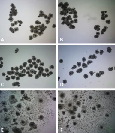
Fig. 1. Morphology of cumulus-oocyte complexes (COCs) matured for 44 h in a chemically defined maturation medium (A-D) that was supplemented with 1.5 mM fructose (A), 3.0 mM fructose (B), 5.5 mM fructose (C), and 5.5 mM glucose (D) or in a maturation medium containing porcine follicular fluid that was further supplemented with 3.0 mM fructose (E) and 5.5 mM glucose (F).
용처리군은 91.8%로 유의적 차이는 없었다. 단위발생 배아의 분할은 과당의 단독처리(85.7%)보다 병용 처리했을 경우 유 의적으로(P<0.05) 높은 수치(95.2%)를 보였다. 배반포 형성률 (37.3-42.8%)과 배반포 세포수(33.3-34.1 cells)에는 유의적인 영향이 없었다(Table 3).
3. 난포액이 포함된 배양액 내 포도당 및 과당의 단독 또는 병용 첨가가 난자의 핵 성숙 및 단위발생 배아의 체외발 육에 미치는 효과
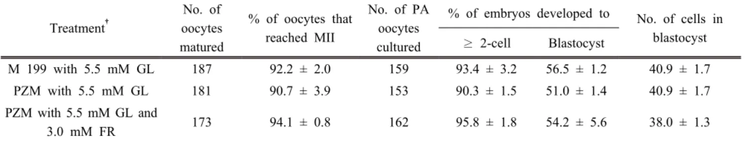
난포액이 포함된 배양액에 첨가된 포도당 및 과당은 난자 의 핵 성숙(90.7-94.1%), 단위발생 배아의 분할(90.3-95.8%), 배반포 형성률(51.0-56.5%) 및 배반포 세포수(38.0-40.9 cells) 에 유의적 영향을 미치지 않았다(Table 4).
Table 2. Intra-oocyte glutathione (GSH) content and cumulus expansion score of oocytes matured in a chemically defined maturation medium supplemented with glucose or various concentrations fructose
Treatment
No. of metaphase II oocytes examined for GSH
content
Relative level (pixels/oocyte) of
GSH content Cumulus cell expansion
5.5 mM glucose 46 1.03 ± 0.06ab 2.75 ± 0.13
1.5 mM fructose 46 1.00 ± 0.06ab 2.42 ± 0.11
3.0 mM fructose 46 1.08 ± 0.06a 2.51 ± 0.12
5.5 mM fructose 46 0.87 ± 0.07b 2.60 ± 0.12
Four replicates.
Porcine zygote medium-4 was used as a base medium for in vitro maturation.
†
Cumulus cell expansion was scored as 0 (no response), 1 (minimum observable response with the cells in the outermost layer of the cumulus become round and glistening), 2 (the expansion of outer cumulus cell layers), 3 (the expansion of all cumulus cell layers except corona radiata), and 4 (the expansion of all cumulus cell layers).
abValues in the same column with different superscript letters are different (P<0.05).
Table 3. Effects of glucose and/or fructose supplemented to a chemically defined maturation medium on oocyte maturation and embryonic development after parthenogenesis (PA)
Treatment
No. of oocytes matured
% of oocytes that reached MII
No. of PA oocytes cultured
% of embryos developed to
No. of cells in blastocyst
≥ 2-cell Blastocyst
5.5 mM glucose 190 92.6 ± 2.1 183 91.8 ± 1.1ab 39.2 ± 5.4 33.3 ± 1.3 3.0 mM fructose 174 86.3 ± 3.3 163 85.7 ± 2.4b 37.3 ± 2.5 33.7 ± 1.4 Glucose + fructose 191 91.8 ± 1.3 185 95.2 ± 1.3a 42.8 ± 2.4 34.1 ± 1.2
Three replicates.
Porcine zygote medium-4 was used as a base medium for in vitro maturation.
ab
Values in the same column with different superscript letters are different (P<0.05).
Table 4. Effects of glucose (GL) with or without fructose (FR) in a maturation medium containing porcine follicular fluid (pFF) on oocyte maturation and embryonic development after parthenogenesis (PA)
Treatment†
No. of oocytes matured
% of oocytes that reached MII
No. of PA oocytes cultured
% of embryos developed to No. of cells in blastocyst
≥ 2-cell Blastocyst
M 199 with 5.5 mM GL 187 92.2 ± 2.0 159 93.4 ± 3.2 56.5 ± 1.2 40.9 ± 1.7 PZM with 5.5 mM GL 181 90.7 ± 3.9 153 90.3 ± 1.5 51.0 ± 1.4 40.9 ± 1.7 PZM with 5.5 mM GL and
3.0 mM FR 173 94.1 ± 0.8 162 95.8 ± 1.8 54.2 ± 5.6 38.0 ± 1.3
Three replicates.
M199; medium 199 (Invitrogen), PZM, porcine zygote medium.
†
In vitro maturation medium was supplemented with 10% (v/v) pFF.
고 찰
많은 동물 종에서 체외 배양액 구성 성분 중 포도당은 난자 의 성숙과 배아 발달을 위한 필수 에너지 원으로서 광범위하 게 이용 되어 왔다. 소의 수정란 발육에서 포도당은 배반포
형성에 기여하는 중요한 에너지원임이 증명되었다(Rieger와 Loskutoff, 1994). 본 연구에서 난포액이나 혈청이 포함되지 않은 합성 체외성숙 배양액을 이용하여 과당이 돼지 난자의 성숙 및 단위발생 이후의 배 발육에 미치는 영향을 검토한 결 과 3.0 mM 과당은 포도당에 필적하는 효과를 보이는 것으로
나타났다.
포도당은 몇 가지 당 분해과정을 거쳐 세포, 난자 및 수정 란의 에너지원으로 사용된다. 해당과정의 경로는 두 개의 분 리 된 과정으로 구성되는데, 첫 번째 단계는 포도당을 과당 -1,6-인산(F1,6BP)으로 전환 시키는데 에너지를 요구한다. 두 번째 단계에서는 F1,6BP가 젖산으로 분해되면서 에너지와 NADH가 생산된다. 과당이 F1,6BP으로 전환되는 데에는 포 도당이 해당과정으로 들어가 대사되는 것이 비해 더 적은 에 너지를 필요로 하는데(Styler, 1995), 이러한 사실은 과당이 포 도당에 비해 더 용이하게 사용될 수 있다는 것을 시사한다.
본 연구에서 성분이 명확한 합성 체외 배양액에 3.0 mM 과당을 첨가하여 난자의 체외성숙을 유도한 결과 5.5 mM 포 도당과 유사한 수준의 핵 성숙률을 보이는 것을 확인하였다.
난자의 세포질 성숙도를 나타내는 하나의 지표로 사용되는 난자 내 GSH 함량을 비교해 보았을 때도 유사한 결과가 나타 났다. GSH는 저분자 thiol 화합물로서 활성산소종의 유해한 작용을 경감시킴으로써 세포 또는 난자를 보호한다. 난자 세 포질 내 GSH 함량은 종종 소 및 돼지 난자의 세포질 성숙의 마커로 사용 되어왔으며, 세포질 내 GSH 함량은 난자의 성숙 과 배아 발생에 영향을 미친다는 보고가 있다(Abeydeera 등, 1998; De Matos 등, 2000). 본 연구에서 3.0 mM 과당이 1.5 및 5.5 mM에 비해 난자 성숙에 이로운 효과를 보인 정확한 이유는 알 수 없다. 이에 관해서는 난포액 내 과당의 존재여 부 또는 돼지 난자에서 탄수화물 대사와 관련한 추가적인 연 구가 필요할 것으로 생각된다. 난구세포는 유해한 환경으로부 터 난자를 보호하고 난자와 배양액의 영양분과 신호를 전달 하는 중요한 역할을 하며, 또한 수정 과정에 관여한다(Zhang 등, 1995; Yamauchi와 Nagai, 1999; Yong과 Lee, 2007). 난구 세포의 팽창은 체외성숙 난자의 중요한 형태학적 기준 중 하 나이며 난자 성숙의 간접적인 지표로 사용되어 왔다(Eppig 등 1982; Marei 등, 2012). 본 연구에서 PVA가 포함된 체외성숙 배양액에 첨가된 포도당과 과당은 난구세포 팽창에 유의한 영향을 나타내지 않았다. 이 결과는 난구세포 팽창이 배양액 내 에너지원보다는 혈청이나 난포액 등 거대분자 물질이나 다른 미지의 물질에 더 영향을 받기 때문인 것으로 추측된다.
한편, Ludwig 등 (2001)은 햄스터 배아의 체외발육에 미치는 과당과 포도당의 영향을 검토한 연구에서 포도당보다는 과당 이 더 적절한 에너지원인 것 같다는 결과를 보고하고 있다.
본 연구에서는 포도당과 과당의 병용처리에 의해 분할이 유 의적으로 증가했으나 배반포 형성률에는 차이가 없었다. 과당 과 포도당의 병용처리가 어떻게 분할을 촉진시켰는지는 정확 히는 알 수 없으나 포도당과 과당이 난자의 에너지 대사과정 세포질 성숙에 이로운 영향을 미쳤고 이로 인해 배아의 초기 분할에 영향을 미친 것으로 판단된다.
돼지 난포액이 첨가된 배양액을 이용하여 포도당과 과당의 효과를 검토한 결과 난자의 성숙이나 단위발생 후 배 발육은 에너지원의 종류에 따라 영향을 받지 않았다. 돼지의 혈청이 나 난포액에는 일정 농도의 포도당이 존재하며, 이외에도 성 분을 명확히 알 수 없는 다양한 물질들이 포함되어 있다(Brad 등, 2003; Hong과 Lee, 2007). 따라서 난포액에 포함되어 있는 포도당, 아미노산 및 미지의 물질들의 작용으로 인해 추가로 첨가된 포도당이나 과당의 효과가 나타나지 않은 것으로 사 료된다.
난자가 성숙하는 동안 과도한 포도당의 첨가는 난자의 활 성산소종의 함량을 증가시키고 GSH 함량을 감소시키며, 배 아 발달능력에 손상을 유발 할 수 있다(Hashimoto 등, 2000).
또한, 높은 포도당 농도에서 배아를 배양하면 대사 이상이 발 생하여 ATP 저장량이 감소하고 유전자 발현에 악영향을 미 침으로써 배아의 사멸 또는 태아의 잠재적인 기형을 초래할 수 있다고 보고되었다(Moley, 2001). 착상 전 배아의 발육을 위한 배양액에서 포도당을 과당으로 대체하는 것이 이롭다는 결과가 햄스터(Ludwig 등, 2001)와 소(Kwun 등, 2003)에서 보고되었다. 본 연구에서 포도당을 과당으로 대체하는 것은 배반포 형성을 유의하게 촉진시키지는 않았지만 합성배양액 에 첨가되었을 경우 난자의 성숙과 난자 내 GSH 함량에 있어 포도당에 필적하는 효과를 보였다. 결론적으로, 돼지 난자의 체외성숙에 있어서 과당은 포도당을 대체하여 돼지 난자의 성숙 및 이후의 배 발육을 지원할 수 있는 실질적인 에너지원 으로 사용될 수 있을 것으로 사료된다.
ACKNOWLEDGMENTS
This research was supported by Agricultural Biotechnology Development Program (IPET 31060-05), Ministry of Agriculture, Food and Rural Affairs, Republic of Korea and 2014 Research Grant from Kangwon National University (No.120141468).
REFERENCES
Abeydeera LR, Wang WH, Prather RS and Day BN. 1998.
Maturation in vitro of pig oocytes in protein-free culture media: fertilization and subsequent embryo development in vitro. Biol. Reprod. 58:1316-1320.
Aitken RJ. 1976. Uterine secretion of fructose in the red deer.
J. Reprod. Fertil. 46:439-440.
Brad AM, Herrick JR, Lane M, Gardner DK and Krisher RL.
2003. Glucose and lactate concentrations affect the metabolism of in vitro matured porcine oocytes. Purdue University Swine Res. Rep. 136.
De Matos DG and Furnus CC. 2000. The importance of having high glutathione (GSH) level after bovine in vitro maturation on embryo development: effect of bmercaptoethanol, cysteine and cystine. Theriogenology 53:761-771.
Downs SM and Utecht AM. 1999. Metabolism of radiolabeled glucose by mouse oocytes and oocyte-cumulus cell complexes. Biol. Reprod. 60:1446-1452.
Downs SM and Mastropolo AM. 1994. The participation of energy substrates in the control of meiotic maturation in murine oocytes. Dev. Biol. 162:154-168.
Duque P, Hidalgo CO, Gómez E, Pintado B, Facala E and Díez C. Macromolecular source as dependent on osmotic pressure and water source: effects on bovine in vitro embryo development and quality. Reprod. Nutri, Dev.
43:487-496.
Eppig JJ. 1982. The relationship between cumulus cell-oocyte coupling, oocyte meiotic maturation, and cumulus expansion.
Dev. Biol. 89:268-272.
Flood MR and Wiebold JL. 1988. Glucose metabolism by preimplantation pig embryos. J. Reprod. Fertil. 84:7-12.
Gardner DK. 1998. Changes in requirements and utilization of nutrients during mammalian preimplantation embryo development and their significance in embryo culture.
Theriogenology 49:83-102.
Guyader-Joly C, Khatchadourian C and Menezo Y. 1996.
Comparative glucose and fructose incorporation and conversion by in vitro produced bovine embryos. Zygote 4:85-91.
Hashimoto S, Minami N, Yamada M and Imai H. 2000. Excessive concentration of glucose during in vitro maturation impairs the developmental competence of bovine oocytes after in vitro fertilization relevance to intracellular reactive oxygen species and glutathione contents. Mol. Reprod. Dev. 56:520-526.
Haynes NB and Lamming GE. 1967. The carbohydrate content of sow uterine flushings. J. Reprod. Fertil. 14:335-337.
Hong J and Lee E. 2007. Intrafollicular amino acid concentration and the effect of amino acids in a defined maturation medium on porcine oocyte maturation, fertilization, and preimplantation development. Theriogenology 68:728-735.
Kikuchi K, Onishi A, Kashiwazaki N, Iwamoto M, Noguchi J
and Kaneko H. 2002. Successful piglet production after transfer of blastocysts produced by a modified in vitro system. Biol. Reprod. 66:1033-1041.
Kim J, You J, Hyun SH, Lee G, Lim J and Lee E. 2010.
Developmental competence of morphologically poor oocytes in relation to follicular size and oocyte diameter in the pig.
Mol. Reprod. Dev. 77:330-339.
King, N., Korolchuk, S., McGivan, J., and Suleiman, M. 2004.
A new method of quantifying glutathione levels in freshly isolated singlesuperfused rat cardiomyocytes. J. Pharmacol.
Toxicol. Methods 50:215-222.
Kwun J, Chang K, Lim J, Lee E, Lee B and Kang S. 2003.
Effects of exogenous hexoses on bovine in vitro fertilized and cloned embryo development: improved blastocyst formation after glucose replacement with fructose in a serum-free culture medium. Mol. Reprod. Dev. 65:167-174.
Lee ES, Fukui Y, Lee BC, Lim JM, and Hwang WS. 2004.
Promoting effect of amino acids added to a chemically defined medium on blastocyst formation and blastomere proliferation of bovine embryos cultured in vitro. Anim.
Reprod. Sci. 84:257-267.
Ludwig TE, Lane M and Bavister BD. 2001. Differential effect of hexoses on hamster embryo development in culture. Biol. Reprod. 64:1366-1374.
Marei WF, Ghafari F and Fouladi-Nashta AA. 2012. Role of hyaluronic acid in maturation and further early embryo development of bovine oocytes. Theriogenology 78:670-677.
Moley KH. 2001. Hyperglycemia and apoptosis: mechanisms for congenital malformations and pregnancy loss in diabetic women. Trends Endocrinol. Metab. 12:78-82.
Rieger D, and Loskutoff NM. 1994. Changes in the metabolism of glucose, pyruvate, glutamine and glycine during maturation of cattle oocytes in vitro. J. Reprod.
Fertil. 100:257-262.
Sakatani M, Suda I, Oki T, Kobayashi S, Kobayashi S and Takahashi M. 2007. Effects of purple sweet potato anthocyanins on development and intracellular redox status of bovine preimplantation embryos exposed to heat shock.
J. Reprod. Dev. 53:605-614.
Sakkas D, Urner F, Menezo Y and Leppens G. 1993. Effects of glucose and fructose on fertilization, cleavage, and viability of mouse embryos in vitro. Biol. Reprod. 49:1288-1292.
Styler L. 1995. Biochemistry. New York: WH Freeman and Co.
Vanderhyden BC, Caron PJ, Buccione R and Eppig JJ. 1990.
Developmental pattern of the secretion of cumulus expansion-enabling factor by mouse oocytes and the role of oocytes in promoting granulosa cell differentiation. Dev.
Biol. 140:307-317.
Wiesak T, Hunter MG and Foxcroft GR. 1990. Differences in follicular morphology, steroidogenesis and oocyte maturation in naturally cyclic and PMSG/hCG-treated prepubertal gilts.
J. Reprod. Fertil. 89:633-641.
Yamauchi N and Nagai T. 1999. Male pronuclear formation in denuded porcine oocytes after in vitro maturation in the presence of cysteamine. Biol. Reprod. 61:828-833.
Yong HY and Lee E. 2007. Presence of intact cumulus cells during in vitro fertilization inhibits sperm penetration but improves blastocyst formation in vitro. J. Emb. Trans.
22:1-7 (in Korean).
Zhang L, Jiang S, Wozniak PJ, Yang X and Godke RA. 1995.
Cumulus cell function during bovine oocyte maturation, fertilization, and embryo development in vitro. Mol. Reprod.
Dev. 40:338-344.
Received September 20 2017, Revised September 22, 2017, Accepted September 26, 2017