Korean J Vet Res
(2005) 45(4) : 501~505
501
면역기능 증강 신물질에 대한 마우스의 면역학적 및 혈액학적 변화
정 지 윤*
서울대학교 수의과대학 (게재승인: 2005년 11월 28일)
Changes of immunostimulatory effects by Immu-Forte on mice
Ji-Youn Jung*
College of Veterinary Medicine, Seoul National University, Seoul 151-742, Korea
(Accepted
:November 28, 2005)
Abstract : Immu-Forte composed of chitosan,
β-glucan, manno-oligosaccharide and pangamic acid was evaluated for its effectiveness as a nonspecific immunostimulator in mice. The effects of Immu-Forte were determined by analysis of cytokines using ELISA and phenotype of leukocyte subpopulations using monoclonal antibodies specific to mouse leukocyte differentiation antigens and flow cytometry. All T cells, all B cells, CD4 T cells, CD8 T cells, macrophages, IL-2, IL-4, IL-12 and IFN-r in Immu-Forte A-treated group increased in 1 months posttreatment and were significantly higher (
p< 0.05) than that of control at 1 months posttreatment. All T cells, all B cells, CD4 T cells, CD8 T cells, macrophages and IL-2 in Immu- Forte EX-treated low and middle dose groups increased in 1 months posttreatment and were significantly higher (
p< 0.05) than that of control at 1 months posttreatment. In the Immu-Forte soybean-treated group, NK cells and IL-4 were significantly higher in middle dose-treated group, and IL-2, IL-4 and IFN-r were significantly higher in low dose-treated group. In the Immu-Forte F-treated group, all T cells, all B cells, CD4 T cells, CD8 T cells, macrophages, NK cells, IL-2, IL-4, IL-12 and IFN-r in high dose-treated group and all T cells, all B cells, CD4 T cells, CD8 T cells, macrophages, IL-2, IL-4, IL-12 and IFN-r in middle dose-treated group and NK cells, IL-2, IL-4, IL-12 and IFN-r in low dose-treated group were significantly higher (
p< 0.05) than that of control at 1 months posttreatment. In conclusion, this study has demonstrated that Immu-Forte had an immunostimulatory effect on mice through proliferation and activation of mouse immune cells.
Key words : cytokines, Immu-Forte, leukocyte, nonspecific immunostimulator
서 론
최근축산업이집단사육형태로발전되어감에따라 효율성이증대된반면질병에대한노출및질병전파의 위험성은더욱높아지게되었으며이러한질병을예방
,
치료하기위한약제의사용이증가하고있다
[1].
이러한질병감염의 위험성을제거하기위하여백신의개발 및백신치료제에대한프로그램의개발
,
항균제의사료첨가에의한사전감염차단등의다양한방법이이용
되고있다
[7].
그러나이러한질병예방과치료를위한방법중에서백신의이용은포괄적인질병방어보다는 제한적인특정질병원인체에대해서만효과를거둘수 있을뿐만아니라병원성이높은미생물의감염시에는그
나마도기대에미치지못하는결과를가져오게된다
[3].
또한
,
최근에는사료첨가용이나치료약제로이용되고있는항균제의무절제한사용과휴약기간의미준수로인
*Corresponding author: Ji-Youn Jung
College of Veterinary Medicine, Seoul National University, Seoul 151-742, Korea [Tel: +82-2-880-1298, Fax: +82-2-876-7610, E-mail: [email protected]]
한항균제다제내성균의출현을초래하게되었고이에 따른항균제의축산물내잔류로인한인체에의영향이
크게우려되고있는실정이다
[1, 3].
따라서
,
이러한질병에대한예방제및치료제로서의대체물질개발에대한연구가전세계적으로활발히이 루어지고있으며
,
이러한연구분야중에동물생체에잔 류하지않고인체에영향을주지않는생체면역증강제에대한연구가활발히이루어지고있다
[2, 6].
본연구는면역기능을증강시키는생물제제인
Immu-
Forte
의면역체계활성화에따른생체내에서의변화를알아보고자본연구를수행하였다
.
재료 및 방법
시험 동물
면역기능증강성물질투여에의한마우스에서의면 역학적세포및사이토카인의변화및혈액학적변화를
알아보기위하여
5
주령의암컷Balb/c
마우스70
마리를오리엔트㈜에서구입하였다
.
본시험에사용될마우스는면역기능시험에있어서지금까지많이사용되어왔 기때문에비교할많은기초자료가있어서선택하였다
.
실험실에순화시키는기간을약
1
주일간두었으며,
그기간중에일반증상을관찰하여건강한동물만을시험 에제공하였다
.
본시험은온도22
±3
oC,
상대습도50
±10%,
환기 회수10-12
회/hr,
조명시간12
시간(07 : 00- 19 : 00),
조도150-300Lux
로설정된서울대학교수의과 대학동물사육장동물실험실5
호실에서실시하였다.
시험 면역증강제
본실험에제공된시험물질은
Immu-Forte EX, Immu- Forte
된장고추장, Immu-Forte A
및Immu-Forte F
로서Immu-Forte EX
는 전통 누룩유래의 삭카로마이세스( Saccharomyces )
베타-1, 3-
및베타-1, 6-
글루칸,
비타민B,
비타민C,
글루타치온,
판가민산으로구성된물질이며
, Immu-Forte
된장고추장은키토산,
베타-
글루간,
키토 올리고당,
이소플라본(Isoflavon)
으로 구성 되었고,
Immu-Forte A
는전통메주유래의라이조프스키토산( Rhizopus chitosna),
삭카로마이세스( Saccharomyces )
베 타-1, 3-
및베타-1, 6-
글루칸,
라이조프스키토올리고당( Rhizopus chitooligosaccharides)
으로 구성되었으며, Immu-Forte F
는삭카로마이세스( Saccharomyces )
베타-1, 3-
및베타-1, 6-
글루간,
만노오리고당(manno oligosac- charide),
비타민B,
비타민C,
글라타치온,
판가민산으로 구성되었다.
각각의물질은사람에게적용하는용량을 중간그룹의용량으로설정하고그용량의2
배수를고용량으로
,
절반을저용량으로두고실험을실시하였다. EX
는성인
65 kg
을기준으로하였을때1
일섭취용량이75 ml
이었으며, Immu-Forte
된장고추장은5 g, Immu- Forte A
는0.5g,
사료첨가제로개발되어진Immu-Forte F
는톤당2 kg
을1
일섭취량으로기준을삼았다.
마우스 비장에서의 면역세포 분석검사
시험물질들을
1
개월간매일투여한후마우스에서비장을적출하여면역세포의증가유무및사이토카인의
변화를
Flow cytometry
를이용하여분석하였다.
분석한세포및사이토카인은
T cell, B cell, CD4 T cell, CD8 T cell, IL-2, IL-4, IL-12, IFN-r, IgG, IgM, macrophage, NK cell
이다.
이러한세포및사이토카인의변화를측정하기 위하여세포는1
개또는2
개의형광색소(FITC
및phy-
coerythrin)
를이용하여표식이되어있도록실험을설계하였으며
,
염색이완료된재료는flow cytometry
를이용하여총
2000
개이상의세포들을검사하여세포수를측정하였으며 측정과 자료분석은
FACSCalibur
및CellQuest program
을이용하여실시하였다.
또한, IL-2,
IL-4, IL-12, IFN-r
의 정량적인 수치를 얻기 위하여sandwitch-ELISA
를이용하여혈중사이토카인을측정하였다
.
혈액학적 변화 분석
시험물질에의한생체내에서의혈액학적변화를측정 하기위하여
1
개월투여후마우스의안와에서혈액을채취하였으며
,
혈청을분리후혈액학적분석을실시하였다.
통계학적인 처리
대조군을비롯한
Immu-Forte EX, Immu-Forte
된장고 추장, Immu-Forte A
및Immu-Forte F
간의평균치유의성을검정하기위하여분석치에대하여
Microcal Origin
6.0(Microcal Software, USA)
을사용하여ANOVA test
를 실시하였으며,
동일시험기간에서의대조군과시험물질처리군들의비교를위하여
Student's t-test
를실시하여유의성을검증하였다
.
결 과
면역증강
생체내에서의자기방어기전은대식세포등의항원전 달세포와
T
림파구를중심으로하는일련의면역반응에 의해이루어진다[4, 5].
이러한체내면역증강성을관찰하기위하여체내임파구의활성및증가여부를
1
개 월간의뢰한물질인Immu-Forte EX, Immu-Forte
된장 고추장, Immu-Forte A, Immu-Forte F
를투여한후, flowc-
ytometry
를이용하여측정하였다.
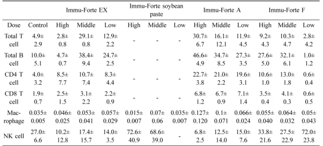
전체
T
림파구의수에있어서는Immu-Forte EX
에서대 조군과비교할때중간용량과저용량에서각각5.8
배,
2.6
배의증가를보였으며, Immu-Forte A
물질에서는농도의존적으로고용량
(6.2
배),
중간용량(3.2
배),
저용량(2.4
배)
의증가를보였고, Immu-Forte F
에서는고용량과중간용량에서각각
1.8
배, 2.0
배의증가를나타내었다.
이러한결과를미루어볼때생체내에서면역능력에중요
한역할을하는
T
림파구가Immu-Forte
된장고추장을제외한
Immu-Forte EX, Immu-Forte A, Immu-Forte F
에서증가하는것을알수있었고
,
특히Immu-Forte A
물 질에서가장효과적으로증가하는것을알수있었다.
따라서
,
세포성면역에있어서핵심적인역할을하는T
림파구의숫적 증가에
Immu-Forte A, Immu-Forte EX,
Immu-Forte F
가유의적으로효과를나타내는것으로추측된다
.
체액성면역에있어서핵심적인역할을수행하는전
체
B
림파구에있어서도Immu-Forte A
에서는농도의존적으로유의적인증가를나타내었고
,
그수치에있어서도다른시험물질과비교하였을때가장높은수치를나 타내었다
.
따라서,
체액성면역에 있어서도Immu-Forte A, Immu-Forte EX, Immu-Forte F
순으로유의적인면역 증강성을나타내는것을알수있었고, T
림파구에서분류되는
CD4 T
림파구및CD8 T
림파구에서도숫적으로
Immu-Forte A, Immu-Forte EX, Immu-Forte F
순으로유의적인면역증강성을나타내고있다
.
대식세포의숫적증가에 있어서도유의적인증가를 보이는물질은
Table 1. The changes of immun-cells in the spleen of mice at 1 months posttreatment of Immu-Forte Immu-Forte EX Immu-Forte soybean
paste Immu-Forte A Immu-Forte F
Dose Control High Middle Low High Middle Low High Middle Low High Middle Low Total T
cell 4.9±
2.9 2.8±
0.8 29.1±
0.8 12.9±
2.2 - - - 30.7±
6.7 16.1±
12.1 11.9±
4.5 9.2±
4.3 10.3±
4.7 2.8±
4.2 Total B
cell 10.0±
5.1 4.7±
0.7 38.4±
9.4 24.7±
2.5 - - - 46.6±
4.9 34.7±
8.5 27.3±
3.5 27.6±
5.0 32.1±
6.1 1.0±
1.2 CD4 T
cell 4.0±
3.2 8.5±
7.7 10.7±
7.4 8.3±
4.4 - - - 22.7±
3.8 21.0±
2.2 19.6±
3.1 10.6±
1.0 13.0±
1.8 0.6±
0.4 CD8 T
cell 1.9±
0.7 2.5±
1.5 3.1±
2.2 2.2±
0.9 - - - 6.8±
1.2 6.7±
0.9 7.1±
1.4 3.5±
0.4 4.1±
0.3 0.6±
0.5 rophage Mac- 0.035±
0.005 0.046±
0.025 0.053±
0.041 0.057±
0.029 0.015±
0.007 0.07±
0.06 0.035±
0.007 0.127±
0.120 0.1±
0.071 0.066±
0.024 0.055±
0.040 0.064±
0.032 0.05±
0.043 NK cell 27.0± 6.6 10.2±
12.8 17.4±
15.7 14.0±
3.5 72.6±
40.9 68.6±
39.0 - 6.8±
2.5 12.5±
14.0 15.0±
7.6 33.8±
21.6 27.5±
22.9 72.0±
23.8
−
: P < 0.05, No significantly difference from the control
Table 2. The changes of cytokines in the spleen of mice at 1 months posttreatment of Immu-Forte ng/ml Immu-Forte EX Immu-Forte soybean
paste Immu-Forte A Immu-Forte F
Dose Control High Middle Low High Middle Low High Middle Low High Middle Low IL-2 0.301± 0.051 0.097±
0.156 0.042±
0.048 0.195±
0.386 0.146±
0.107 0.172±
0.233 0.806±
2.260 0.688±
0.633 0.456±
0.428 1.578±
1.513 1.622±
2.750 4.578±
17.188 0.732±
2.310 IL-4 0.151± 0.0351 0.195±
0.101 0.155±
0.040 0.415±
0.184 0.148±
0.078 0.652±
0.723 0.580±
0.265 0.776±
0.228 0.766±
0.458 1.786±
0.565 8.596±
10.990 4.996±
6.637 0.688±
0.496 IL-12 0.217± 0.106 0.090±
0.104 0.154±
0.352 0.252±
0.025 0.136±
0.128 0.064±
0.134 0.132±
0.328 0.486±
0.322 0.140±
0.163 1.794±
1.635 2.716±
7.361 2.236±
6.111 0.427±
0.420 IFN-r 0.224± 0.053 0.427±
0.066 0.087±
0.011 0.230±
0.357 0.012±
0.005 0.234±
0.894 0.86±
0.248 0.228±
0.017 0.796±
0.118 1.818±
0.186 2.438±
0.935 7.452±
3.098 1.214±
0.448
Immu-Forte EX, Immu-Forte A, Immu-Forte F
였으며,
자 연살해세포에서는Immu-Forte
된장고추장의중간그룹과 고용량그룹에서유의적인숫적증가를나타냈다.
이러한점을미루어보아시험물질
4
가지모두기본적으로 생체내의면역능을향상시키는것으로판단되어진다.
이러한판단을확인하기위하여
sandwitch-ELISA
를이용한사이토카인의변화를 측정해본결과
(Table2),
자연 살해세포의growth factor
이면서면역세포들의활성을 촉진하는IL-2
의경우Immu-Forte A
와Immu-Forte F
에서유의적인증가를보였으며
, IgE
의생산및Th2
세포의성숙에있어서중요한역할을하는
IL-4
에서는전군에서유의적인변화가관찰되었고
,
세포매개면역에있어서초기반응에서중요한작용을하는
IL-12
는Immu-
Forte
된장고추장과Immu-Forte F
에서대조군과비교하여유의적인증가를나타내었다
.
이러한결과를종합한결과
, Table 1
과Table 2
의전체 적인수치를검토해보았을때면역증강성은Immu-Forte A
에서가장강력하게증가하는것을판단되어지며, Immu- Forte EX, Immu-Forte F, Immu-Forte
된장고추장순으로체내에서의면역증강성을높여주는것으로여겨진다
.
혈액학적 분석
Table 3
의혈액학적인검사에서alkaline phosphatase, T protein, albumin, creatine, ALT, AST, total bilirubin,
potasium
을검사해본결과대조군과비교했을때투여군전군에서정상적인범위내수치를나타내는것으로 나타났다
.
이러한정상적인수치로서의결과는앞에서 면역학적인세포의숫적변화및사이토카인의변화가 질병이나감염에의한변화가아닌정상적인실험동물 에서나타난결과라는것을나타내는것으로서이러한 혈액학적변화의결과가정상적이라는것이필수불가 결한요소라고할수있다.
결론적으로면역세포및사 이토카인의변화가물질의면역증강효과에의한것으로 판단되어진다.
콜레스테롤
1
개월간Immu-Forte EX
를투여후혈중콜레스테롤수치를검사해본결과
Table 3
에서대조군(153
±15.29 mg/
Table 3. The changes of blood-chemistry in 1 months posttreatment of Immu-Forte Alkaline
phosphatase (U/l)
Total Protein
(g/dl) Albumin
(g/dl) Creatinine (mg/dl)
Alanine transferase
(U/l)
Aspartate transferase
(U/l)
Total Bilirubin
(mg/dl) K
+(mmol/l)
Total Cho- lesterol (mg/dl) Control 143.6±9.2 5.6±0.2 2.9±0.1 0.3±0.1 131.6±43.0 160.2±16.7 0.8±0.2 5.8±0.7 153.2±15.3 Immu-Forte EX
(High) 168.3±16.6 5.6±0.6 2.8±0.3 0.5±0.3 114.3±43.8 196.5±19.5 0.9±0.4 5.9±0.2 133.0±11.6 Immu-Forte EX
(Middle) 143.5±41.1 5.2±0.6 2.5±0.2 0.6±0.2 158.3±32.5 200.0±30.4 1.0±0.6 5.5±0.4 140.5°æ16.7 Immu-Forte EX
(Low) 147.7±24.7 5.3±0.3 2.7±0.1 0.7±0.1 129.7±59.1 169.0±18.3 1.0±0.2 5.8±0.5 132.0±9.6 Immu-Forte
soybean paste
(High) 129.6±17.4 5.8±0.1 2.8±0.1 0.3±0.1 125.8±44.0 120.6±27.9 06±0.2 5.4±0.5 142.6±6.4 Immu-Forte
soybean paste
(Middle) 155.0±11.7 5.5±0.3 2.9±0.0 0.4±0.2 127.6±38.9 156.8±20.8 0.4±0.1 6.1±1.1 133.2±10.6 Immu-Forte
soybean paste
(Low) 145.2±20.4 5.6±0.2 3.0±0.2 0.3±0.1 105.8±58.5 181.8±16.9 0.7±0.5 6.3±1.2 144.4±2.4 Immu-Forte A
(High) 134.0±11.1 5.6±0.2 2.9±0.2 0.4±0.1 122.8±56.9 136.4±58.7 0.8±0.4 5.8±1.1 140.0±7.1 Immu-Forte A
(Middle) 152.4±8.6 5.7±0.4 2.9±0.3 0.8±0.7 116.4±89.0 171.0±73.5 0.9±0.5 5.6±1.5 144.2±5.5 Immu-Forte A
(Low) 153.0±7.5 5.7±0.1 2.9±0.0 0.3±0.2 87.0±41.1 122.0±41.5 0.9±0.3 5.0±0.9 149.4±10.2
dl)
과비교하였을때,
고용량(133
±11.57 mg/dl),
중간용량(140.5
±16.66 mg/dl),
저용량(132
±9.64 mg/dl)
으로서혈중콜레스테롤의감소가보였으나통계학적인유의적감 소로는보이지않는다
.
이러한결과로부터의뢰한Immu-
Forte EX
의혈중콜레스테롤저하효과는없는것으로사료된다
.
고 찰
체내면역증강성을관찰하기위하여체내임파구의 활성 및증가 여부를
1
개월간Immu-Forte EX, Immu- Forte
된장고추장, Immu-Forte A, Immu-Forte F
를투여 한후, flowcytometry
및sandwitch ELISA
를이용하여측정하였다
.
전체적인수치를검토해보았을때분석결과전체적 인면역증강성은
Immu-Forte A
에서가장강력하게증가하는것을나타냈으며
, Immu-Forte EX, Immu-Forte F,
Immu-Forte
된장고추장순으로체내에서의면역증강성을높여주는것으로나타났다
.
혈액학적인검사에서
alkaline phosphatase, T protein, albumin, creatine, ALT, AST, total bilirubin, potasium
을 검사해본결과대조군과비교했을때투여군전군에서 정상적인범위내수치를나타내는것으로나타났다.
1
개월간Immu-Forte EX
를투여후혈중콜레스테롤수치를검사해본결과대조군
(153
±15.29 mg/dl)
과비교하였을때
,
고용량(133
±11.57 mg/dl),
중간용량(140.5
±16.66 mg/dl),
저용량(132
±9.64 mg/dl)
으로서혈중콜레스테롤의감소가보였으나통계학적인유의적감소로는 보이지않는다
.
결 론
Immu-Forte A, Immu-Forte EX, Immu-Forte F, Immu-
Forte
된장고추장순으로체내에서의면역증강성을높여주는것으로나타났는데
,
이러한체내면역세포와사이 토카인의변화가혈액학적인검사에서정상범위즉,
질병이나감염이없는동물생체내에서일어난변화이므
로면역세포와사이토카인의변화가면역증강에의한 변화라고판단할수있었다
.
또한
,
혈중콜레스테롤수치는Immu-Forte EX
와대조 군과 비교하여유의적인 변화가없는 것으로미루어Immu-Forte EX
는혈중콜레스테롤수치를저하시키는효과는없는것으로사료된다
.
감사의 글
본연구는