학 술 논 문
62
약물지연방출을 위한 스테레오컴플렉스 PLA 원반형 마이크로입자
박채원· 박상효 · 김우철 · 기재홍*
연세대학교 보건과학대학 의공학부
Stereocomplex Poly(lactic acid) Discoidal Microparticles for Sustained Drug Release
Chaewon Park, Sanghyo Park, Woo Cheol Kim and Jaehong Key*
Department of Biomedical Engineering, Yonsei University Gangwon-do, Korea
(Manuscript received 9 September 2019 ; revised 21 February 2020 ; accepted 27 February 2020)
Abstract: Controlled drug release is important for effective treatment of cancer. Poly(DL-lactide-co-glycolide) acid (PLGA) is a Food and Drug Administration (FDA) approved polymer and have been extensively studied as drug deliv- ery carriers with biodegradable and biocompatible properties. However, PLGA drug delivery carriers are limited due to the initial burst release of drug. Certain drugs require an early rapid release, but in many cases the initial rapid release can be inefficient, reducing therapeutic effects and also increasing side effects. Therefore, sustained release is important for effective treatment. Poly Lactic Acid stereo complex (PLA SC) is resistant to hydrolysis and has high stability in aqueous solutions. Therefore, in this work, PLGA based discoidal polymeric particles are modified by Poly Lactic Acid stereocomplex (PLAsc DPPs). PLAsc DPPs are 3 μm in diameter, also showing a relatively sustained release profile. Fluorescein 5(6)-isothiocyanate (FITC) released from PLAsc DPPs was continuously observed until 38 days, which showed the initial release of FITC from PLAsc DPPs was about 3.9-fold reduced as compared to PLGA based DPPs at 1 hour.
Keywords: Drug delivery, Carriers, PLA stereo complex, Sustained release
I. 서 론
암 치료는 주로 화학요법에 의해 시행된다[1,2]. 그러나 항암제는 암세포 뿐만 아니라 비 특이적 약물 반응에 의해 정상세포에 영향을 줄 수 있다[3]. 특히 항암제의 종양 조 직에 대한 낮은 전달률은 환자에 대한 더 높은 복용량을 요 구하게 된다. 항암제의 표준 복용량은 정상 세포에 독성과 다중 약물 내성의 발생률을 증가시키고 다른 장기에 손상을 줄 수 있다[2,4,5]. 따라서 이러한 부작용을 최소화하기 위해 다양한 약물 전달에 관한 연구가 이루어지고 있다[6]. 예를
들면 마이셀(Micelle), 실리카 나노 입자 등을 이용한 우수 한 약물전달 치료효과가 보고되었다[2,6-9]. 입자를 이용한 약물전달 시스템은 입자의 화학적, 물리적 특성을 변화시켜 생체 이용률과 약물 적재량, 신체 내 약물의 안정성, 약물의 제 어 방출, 표적 전달 등을 향상시키는 방향으로 발전되어왔 다[6]. 이 중 약물의 제어 방출(controlled release)은 약물의 효능 개선, 독성 감소, 환자의 편의성을 포함하여 많은 이점을 제 공한다[10,11]. 예를 들어 상처 치료 초기에 약물의 급속 방 출(burst release)은 즉각적인 완화를 제공한다[11,12]. 한 편 지연 방출(sustained release)은 급속 방출 동안의 약물 낭비를 피하고 독성을 감소시킨다[11]. 따라서 목적에 따른 적절한 방출 프로파일의 조절은 효과적인 약물치료를 나타 낼 수 있다.
Poly(DL-lactide-co-glycolide) acid (PLGA) 와 Poly Lactic Acid (PLA) 는 Food and Drug Administration (FDA) 승인을
Corresponding Author : Jaehong Key1, Yeonsedae-gil, Heungeop-myeon, Wonju-si, Gangwon-do, Republic of Korea
Tel: +82-033-760-2857 E-mail: [email protected]
63 받은 생분해성 고분자이며, 약물전달시스템에서 널리 응용되고
있다[13]. PLA의 거울상 이성질체인 Poly(L-lactic acid) (PLLA) 와 Poly(D-lactic acid) (PDLA)를 혼합하여 Poly(lactic acid) stereo complex(PLA SC) 를 형성하는 것이 가능하 다[14]. 이러한 PLA SC는 PLLA, PDLA 단일 결정과 비 교하여 높은 용융 온도, 용매에 대한 우수한 안정성과 낮은 용해도, 높은 결정화율, 우수한 기계적 특성, 가수분해에 대 한 저항성을 나타낸다[15,16]. 이러한 PLA SC의 특성을 입 자에 적용하면 약물 방출을 제어할 수도 있다. 예를 들어, Laura Pastorino et al. 은 적재된 약물의 90%를 15분만에 방 출하던 키토산 기반의 66 μm 구형 입자 표면에 PLA SC를 코 팅하여 360시간 동안 34%를 방출하는 특성을 보고하였다[17].
한편 폐로 약물을 전달하는 방법에는 주로 흡입 방식이나 정맥 주입 방식이 보고되고 있다[18-20]. 정맥 주입 시, 나노 입 자와 같은 초소형 약물전달체는 폐에 대한 약물전달률이 매우 낮은 것으로 보고되어 있다[18-20]. 반면 마이크로 입자는 주로 폐에 축적되는 것으로 알려져 있다[21]. 또한 0.7 μm, 1.0 μm, 2.5 μm, 3.0 μm 크기의 입자 중 3.0 μm 크기의 입자가 폐에 가 장 많이 축적되며, 원반형, 구형, 반구형, 원통형의 입자 중 원 반형의 입자가 폐에 가장 많이 축적된다고 보고되었다[22].
한편 폐에 축적된 마이크로 입자는 장기간 분해되지 않아 폐 색전증(pulmonary thromboembolism)과 같은 부작용을 일으 킬 수 있다[23,24]. 따라서 입자의 크기, 모양에 따라 생체분 포(bio-distribution)가 변화되고, 또한 입자의 생분해성과 생체적합성에 따라 인체에 미치는 독성이 변화하게 된다.
본 연구에서는 폐에 약물을 지속적으로 전달하기 위한 약 물전달체를 개발하고자 하였다. 본 연구팀은 기존 PLGA기 반의 3 μm 원반형 약물전달체가 폐에 집중적으로 전달된다는 결과를 보고하였다[25]. 따라서 본 연구에서는 약물전달체를 활용하여 폐에 약물을 지연방출하는 목적으로 PLA SC를 3 μm 원반형 PLGA 입자에 적용하여 생분해성 특성을 유
지하면서 약물 방출을 지연시킬 수 있는 약물전달체를 보고 하였다. 또한 이러한 입자의 약물 지연방출효과를 증명하기 위하여 Fluorescein 5(6)-isothiocyanate (FITC)를 입자에 적재하였다. 그리고 Dynamic light Scattering (DLS)과 형광현미경으로 입자의 크기와 형태를 확인하였다. 약물 방 출 프로파일을 비교하기 위하여 PLA SC를 적용한 입자와 PLGA 입자로부터 방출되는 FITC의 형광 양을 측정하였 다[26,27].
II. 연구 방법
1. 재료
입자를 구성하는 Poly(D,L-lactide-co-glycolide) (PLGA) (acid terminated, lactide:glycolide 50:50, Mw 24,000- 38,000), Poly(L-lactide) (PLLA) (average Mn 10,000, PDI ≤ 1.1), Poly(D,L-lactide) (PDLA) (average Mn 10,000, PDI ≤ 1.2), Fluorescein 5(6)-isothiocyanate (FITC)를 SIGMA-ALDRICH Co 에서 구매하여 합성하였다.
2. PLAsc-DPPs 생산공정 (1) PDMS 형판 생산
Electron beam lithography 방식으로 제작 된, 직경 3 μm 디스크 모양의 홈이 패인 실리콘 주형에 PDMS와 elastomer 를 무게 비 10:1로 혼합하여 첨가하여 동일한 크기와 모양의 기 둥이 새겨진 PDMS 형판을 생산하였다.
(2) PVA 형판 생산
증류수 800 ml과 PVA가루 48 g을 교반하였다. PVA가 루가 다 녹으면 용액을 충분히 식힌 후 PDMS 형판에 100 ml 첨가하여 실리콘 주형과 동일한 패턴의 PVA 형판을 생산 하였다.
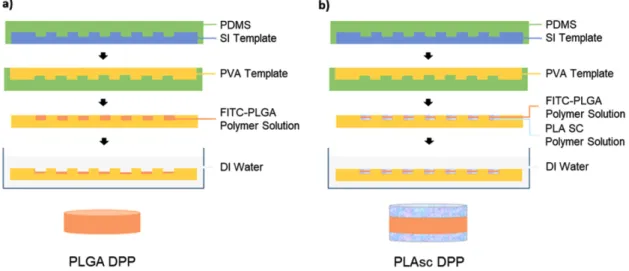
그림 1. DPPs의 생산공정 모식도. a) PLGA DPP, b) PLAsc DPP
Fig. 1. Schematic diagram of the fabrication of discoidal polymeric particles (DPPs). a) PLGA DPP, b) PLAsc DPP
64
(3) 입자 생산
입자 생산의 개략적인 과정은 도표에 설명되어 있다(그림 1). DCM 1 ml 에 녹인 PLLA 10 mg과 DCM 1 ml에 녹인 PDLA 10 mg 을 혼합하여 PLA SC를 합성시켜 PLA SC 용액을 생산하였다. 적재물의 방출 프로파일을 보기 위해 DMSO 50 µl 에 녹인 FITC 2 mg와 Dichloromethane 100 µl 과 Chloroform 100 µl에 녹인 PLGA 70 mg를 혼 합하여 FITC-PLGA 용액을 준비하였다. PVA 형판에 PLA SC 용액을 첨가한 후, 유기용매를 건조시켰다. 차례로 FITC- PLGA 용액과 PLA SC 용액을 반복적으로 첨가하여 PLAsc- DPPs(PLA SC coated discoidal polymeric particles) 를 생산하였다. 단독 PLGA DPPs의 경우 FITC-PLGA 용액을 1 번 첨가한 후, 건조시켰다. 각 PVA 형판을 증류수에 넣고 교반하여 녹인 후, 나일론 필터(pore size: 100 μm)로 scum layers 를 제거하고 멤브레인 필터(pore size: 1.2 μm)로 불 순물을 제거하였다. 원심 분리하여 상층액을 제거한 후, 최종 동결건조 하였다.
3. DPPs의 특성 분석
각각의 DPPs를 1 mg/ml의 농도로 DI water에 재분산 후, Zetasizer (Nano zs90, Malvern Instruments, UK) 로 입 자의 크기를 측정하였다. PLAsc DPPs의 PLA SC층과 PLGA
층이 결합되었는지 확인하기 위해 위의 PLA SC 용액에 Cyanine 5.5 (Cy5.5) 를 첨가한 뒤 같은 방식으로 합성한 PLAsc DPPs 를 형광현미경으로 관찰하여 입자의 형광 발 현 및 모양과 크기를 확인하였다.
4. FITC 방출 실험
PLAsc DPPs 와 PLGA DPPs를 각각 1 mg/ml의 농도로 PBS 에 녹여 Dialysis bag에 넣고 약물 방출 실험을 진행 하였다. 정해진 시간에 방출된 FITC의 fluorescence를 측 정하고, 이를 바탕으로 시간에 따른 방출량을 계산하였다.
III. 연구 결과
1. DPPs의 물리적 특성
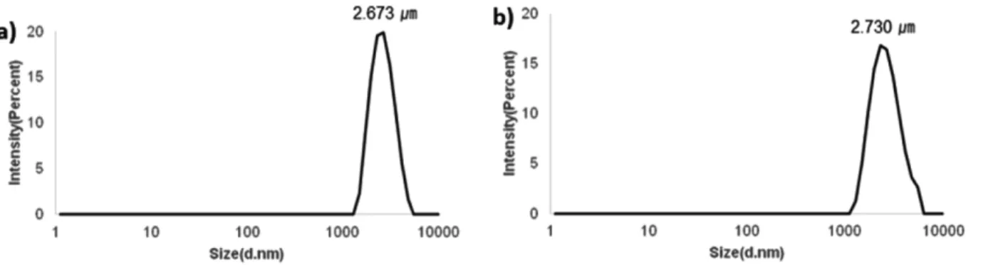
Zetasizer 측정 결과, PLGA DPPs의 평균 크기는 2.673 μm 이며 PLAsc DPPs의 평균 크기는 PLA SC의 코팅으로 인 하여 약 60 nm가 증가한 2.730 μm이었다(그림 2). 입자가 구형이 아닌 디스크 모양이기 때문에 모든 방면에서 크기가 일정하지 않아 DLS 측정에서 입자의 최대 길이 구간인 3 μm 보다 작게 측정되었다. 그림 3을 보면 입자가 직경 3 μm의 디스크 형태로 만들어진 것을 알 수 있다. 또한 FITC로 표지 된 PLGA와 Cy5.5로 표지 된 PLA SC의 사진을 겹쳤을 때, 입
그림 2. DPPs의 크기 분포. a) PLGA DDPs, b) PLAsc DDPs
Fig. 2. Size distribution of discoidal polymeric particles. a) PLGA DDPs, b) PLAsc DDPs
그림 3. DPPs의 형광 현미경 사진
Fig. 3. Fluorescence microscope image of PLAsc DPPs
65 자의 위치와 모양이 일치하는 것을 관찰하였다. 이는 PLA
SC 와 PLGA가 중첩된 구조라는 것을 의미한다.
2. FITC release profile
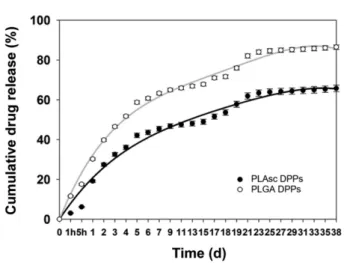
PLA SC 구조가 약물전달체로서 약물이 지효성을 갖는데 도움이 되는지 알아보고, 입자에 약물을 적재하고 투여했을 때 방출 프로파일을 예상하기 위해 PLGA DPPs와 PLAsc DPPs 에 같은 양의 FITC를 적재하였다. Plate reader기를 이용하여 확산 차이에 의해 입자에서 바깥의 PBS로 방출된 FITC 의 발현 (Excitation: 485/Emission: 528)을 시간에 따라 측정 하고 방출량을 계산하였다(그림 4). 실험은 38일간 지속되었으며 PLGA DPPs는 초기 1시간 이내에 적재 된 FITC 의 11.6%가 한 번에 방출되는 급속-방출 현상을 보이는 반면 PLAsc DPPs는 초기 1시간 이내에 적재 된 FITC의 3% 만이 방출 되었다. 이는 PLGA 입자 로부터 방출된 양과 비교했을 때, 74.14% 감소된 수치이다. 또한 실험을 중단 한 38일까지 PLGA DPPs는 85.5%, PLAsc DPPs는 65.7%
로 방출이 지속되었다. PLAsc DPPs가 안정성이 우수한 PLA SC 으로 구성됨으로 인해 입자의 분해를 막아 천천히 방출 되도록 유도한 것으로 보인다.
IV. 고찰 및 결론
본 연구에서는 안정성이 높은 PLA SC를 이용하여 약물을 지연 방출하는 약물전달체 PLAsc DPPs를 개발하였다. 합 성한 PLGA DPPs와 PLAsc DPPs의 평균 크기는 각각 2.673 μm, 2.730 μm이었다. 형광현미경 관찰 결과, 직경 3 μm 의 디스크 입자를 확인하였고, PLAsc DPPs에서 FITC로 표지 된 PLGA와 Cy5.5으로 표지 된 PLAsc의 위치와 모양이 일치하였다는 점을 통해 PLAsc DPPs가 PLGA와 PLA
SC 가 혼합된 구조라는 것을 확인하였다. 두 입자의 FITC 방출 실험 결과, 초반 1시간 PLAsc DPPs의 FITC 방출이 PLGA DPPs 와 비교하여 74.14% 감소하였다. PLAsc DPPs 에서 FITC의 초기 방출을 감소시키는 것으로 보아 PLA SC가 입자의 분해를 지연시키는 것으로 보여진다. 또한 PLGA DPPs 가 4일만에 적재된 FITC의 50%를 방출한 반면 PLAsc DPPs 는 50%를 방출하는데 16일이 소요되었고, 38일 이후 에도 65.7%로 방출이 계속 되는 것으로 보아 장기적으로도 완만히 방출이 지속되는 것을 알 수 있다. 따라서 본 입자를 폐 에 약물을 지속적으로 전달하기 위한 효과적인 약물전달체로 응용하는 가능성이 기대된다.
결론적으로 본 연구에서 보고한 약물전달체 PLAsc DPPs는 적재물의 급속 방출을 감소시키고 장기적으로 방출이 지속 되도록 한다는 것을 확인할 수 있었다. 이러한 연구를 바탕 으로 향후 다양한 약물에 대해서 본 약물전달체를 적용하여 폐 질환 치료용 약물전달체로 발전시킬 계획이다.
References
[1] TGJr CBR. Timeline: chemotherapy and the war on cancer.
Nat Rev Cancer. 2005;5:6572.
[2] Singh RP, Sharma G, Singh S, Bharti S, Pandey BL, Koch B, Muthu MS. Chitosan-folate decorated carbon nanotubes for site specific lung cancer delivery. Materials Science and Engi- neering:C. 2017;77:446-58.
[3] Mignani S, Bryszewska M, Klajnert-Maculewicz B, Zablocka M, Majoral J-P. Advances in combination therapies based on nanoparticles for efficacious cancer treatment: an analytical report. Biomacromolecules. 2014;16:1-27.
[4] Pérez-Herrero E, Fernández-Medarde A. Advanced targeted therapies in cancer: drug nanocarriers, the future of chemother- apy. European journal of pharmaceutics and biopharmaceu- tics. 2015;93:52-79.
[5] Senapati S, Mahanta A.K, Kumar S, Maiti P. Controlled drug delivery vehicles for cancer treatment and their performance.
Signal transduction and targeted therapy. 2018;3:7.
[6] Witharana C, Wanigasekara J. Drug Delivery Systems: A New Frontier in Nano-technology. International Journal of Medical Research & Health Sciences. 2017;6:11-4.
[7] Langer R. New methods of drug delivery. Science. 1990;249:
1527-33.
[8] Sonali, Agrawal P, Singh RP, Rajesh CV, Singh S, Vijayakumar MR, Pandey BL, Muthu MS. Transferrin receptor-targeted vitamin E TPGS micelles for brain cancer therapy: preparation, characterization and brain distribution in rats. Drug delivery.
2016;23:1788-98.
[9] Bhirde AA, Patel V, Gavard J, Zhang G, Sousa AA, Mase- dunskas A, Leapman RD, Weigert R, Gutkind JS, Rusling JF.
Targeted killing of cancer cells in vivo and in vitro with EGF- directed carbon nanotube-based drug delivery. ACS nano, 2009;3:307-16.
[10] Uhrich KE, Cannizzaro SM, Langer RS, Shakesheff KM. Poly- meric systems for controlled drug release. Chemical reviews.
1999;99:3181-98.
그림 4. DDPs의 FITC 방출 그래프
Fig. 4. FITC release profile of PLAsc DPPs and PLGA DPPs in PBS
66
[11] Huang X, Brazel CS. On the importance and mechanisms of burst release in matrix-controlled drug delivery systems. Journal of controlled release. 2001;73:121-36.
[12] Setterstrom JA, Tice TR, Myers WE. Development of encap- sulated antibiotics for topical administration to wounds. In Recent advances in drug delivery systems. 1984;185-98.
[13] Ulery BD, Nair LS, Laurencin CT. Biomedical applications of biodegradable polymers. Journal of polymer science Part B:
polymer physics. 2011;49:832-64.
[14] Ikada Y, Jamshidi K, Tsuji H, Hyon SH. Stereocomplex forma- tion between enantiomeric poly (lactides). Macromolecules.
1987;20:904-6.
[15] Monticelli O, Putti M, Gardella L, Cavallo D, Basso A, Prato M, Nitti S. New stereocomplex PLA-based fibers: effect of POSS on polymer functionalization and properties. Macromolecules.
2014;47:4718-27.
[16] Regnell Andersson S, Hakkarainen M, Inkinen S, Södergård A, Albertsson A-C. Customizing the hydrolytic degradation rate of stereocomplex PLA through different PDLA architec- tures. Biomacromolecules. 2012;13:1212-22.
[17] Pastorino L, Dellacasa E, Petrini P, Monticelli O. Stereocom- plex poly (lactic acid) nanocoated chitosan microparticles for the sustained release of hydrophilic drugs. Materials Science and Engineering:C. 2017;76:1129-35.
[18] Xie G, Sun J, Zhong G, Shi L, Zhang D. Biodistribution and toxicity of intravenously administered silica nanoparticles in mice. Archives of toxicology. 2010;84:183-90.
[19] Sarparanta M, Bimbo LM, Rytkönen J, Mäkilä E, Laaksonen TJ, Laaksonen Pi, Nyman M, Salonen J, Linder MB, Hirvonen J.
Intravenous delivery of hydrophobin-functionalized porous
silicon nanoparticles: stability, plasma protein adsorption and biodistribution. Molecular pharmaceutics. 2012;9:654-63.
[20] Almeida JPM, Chen AL, Foster A, Drezek R. In vivo biodis- tribution of nanoparticles. Nanomedicine. 2011;6:815-35.
[21] Jindal AB. The effect of particle shape on cellular interaction and drug delivery applications of micro-and nanoparticles. Inter- national journal of pharmaceutics. 2017;532:450-65.
[22] Decuzzi P, Godin B, Tanaka T, Lee SY, Chiappini C, Liu X, Ferrari M. Size and shape effects in the biodistribution of intra- vascularly injected particles. Journal of Controlled Release.
2010;141:320-7.
[23] Looney MR, Bhattacharya J. Live imaging of the lung. Annual review of physiology. 2014;76:431-45.
[24] Yu T, Greish K, McGill LD, Ray A, Ghandehari H. Influence of geometry, porosity and surface characteristics of silica nanopar- ticles on acute toxicity: their vasculature effect and tolerance threshold. ACS nano. 2012;6:2289-301.
[25] Park JY, Park S, Lee TS, Hwang YH, Kim JY, Kang WJ, Key J. Biodegradable micro-sized discoidal polymeric particles for lung-targeted delivery system. Biomaterials. 2019;218:119331, doi:10.1016/j.biomaterials.2019.119331.
[26] Mak WC, Richter H, Patzelt A, Sterry W, Lai KK, Renneberg R, Lademann J. Drug delivery into the skin by degradable par- ticles. European journal of pharmaceutics and biopharmaceu- tics. 2011;79:23-7.
[27] Liu Y, Miyoshi H, Nakamura M. Novel drug delivery system of hollow mesoporous silica nanocapsules with thin shells:
preparation and fluorescein isothiocyanate (FITC) release kinet- ics. Colloids and Surfaces B: Biointerfaces. 2007;58:180-7.