Anatomy and Biomechanics of the Elbow Joint
계명의대 정형외과학교실
조 철 현
- A sound understanding of elbow anatomy and biomechanics is necessary to treat common traumatic conditions of the elbow.
Elbow stability
1,2)Primary static constraints Ulnohumeral articulation
Anterior bundle of medial collateral ligament
Lateral collateral ligament complex (esp, lateral ulnar collateral ligament) Sencondary constraints
Radiocapitellar articulation
Common flexor & extensor tendon Capsule
Fig. 1. The ‘‘fortress’’ of static and dynamic
constraints to elbow instability. The three primary constraints are the ulnohumeral articulation, the anterior bundle of the medial collateral ligament (AMCL), and the lateral collateral ligament, especially the ulnar part known as the lateral ulnar collateral ligament (LUCL). The secondary constraints are the radiohumeral articulation, the common flexor-pronator (F-P) tendon, the common extensor tendon, and the capsule.The muscles that cross the elbow are the dynamic constraints.Elbow Anatomy
I. Osteoarticular anatomy
Trochleogingylomoid joint2,3)Ulnohumeral joint : hinged(gynglymoid) motion - flexion & extension Radioulnar joint ; trochoid motion - supination & pronation
1. Distal humerus (trochlea, capitellum)4)
: anterior angulation about 30o in lateral plane
: approximately 5o internal rotation with respect to the epicondylar line : approximately 6o of valgus in AP plane
Fig. 2. A, Lateral view shows 30° anterior rotation of the distal humeral
condyles. B, Axial view shows 5° to 7° internal rotation of the distal humerus articular surface. C, Anterior view shows 6° to 8° degrees of valgus tilt at the distal humerus.2. Proximal ulna3,4)
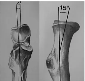
: 4o valgus angulation with the shaft of the ulna
: rotated posteriorly approximately 30o with respect to the long axis in lat. view.
3. Proximal radius5)
: radial neck makes an angle of 15o away from the radial tuberosity.
Fig. 3. A, Angular orientation of proximal ulna
in anteroposterior projection. B, Angular orien- tation of the radial head and neck with respect to the shaft of the radius.II. Capsuloligamentous anatomy
- anterior, posterior capsule, MCL, LCL complex
1. Capsule6)
intraarticular pressure - lowest at 70o to 80o of flexion capacity - 25 to 30 mL at 80o of flexion
2. Medial collateral ligament - valgus stability7-9) ant. bundle : primary static constraints
: taut after about 20o to 30o of flexion post. bundle : lax until about 60o of flexion transverse ligament
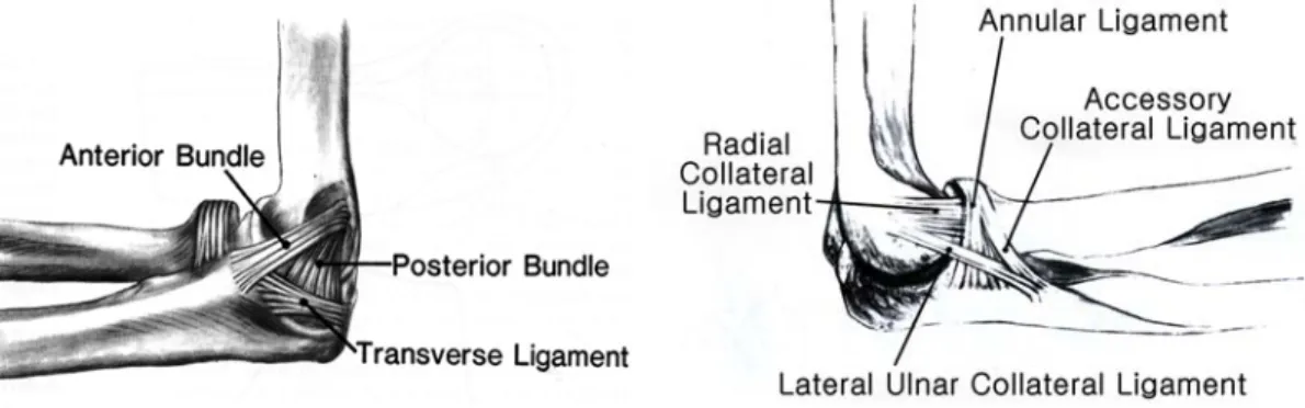
3. Lateral collateral ligament complex : varus stability9-10) radial collateral ligament
lateral ulnar collateral ligament : posterolateral stability annular ligament
accessory collateral ligament
Fig. 4. Medial and lateral collateral ligament complex.
III. Muslces
2,3)- provide dynamic stabilization and protect static constraints - 4 groups
: elbow flexor, elbow extension, forearm flexor-pronators, forearm extensors 1. Biceps : principal supinator of forearm
2. Triceps : main elbow extensor
3. Anconeus : minor role in elbow extension
dynamic constraint to varus and posterolateral rotator instability
Elbow Biomechanics
Elbow function
- link in the lever arm system - fulcrum of the forearm lever - loading-carrying joint
I. Kinematics
1. Flexion-Extension11,12) : hinge type - normal ROM : 0o - 140o
- a range of 30o to 130o required for most activities of daily living - axis of motion
: in line with the anteroinferior aspect of the medial epicondyle, the center of the trochlea, and the center projection of the capitellum onto a parasagittal plane.
: is oriented at approximately 3o to 5o of internal rotation in relation to the plane of the medial and lateral epicondyles and in 4o to 8o of valgus relative to the long axis of the humerus.
Fig. 5. Average maximum varus-valgus laxity after repair of
collateral ligaments with intact coronoid, and simulated coronoid fractures. Coronoid 1 = 10% of bone removed from coronoid tip;coronoid 2 = 50% removed; coronoid 3 = 90% removed. There was significant laxity after 50% of the coronoid was removed.
2. Pronation-Supination13)
- radiocapitellar joint & proximal radioulnar joint - normal ROM : pronation 75o, supination 85o
- 50o of pronation and 50o supination required for most activities of daily living - normal axis of forearm rotation
: runs from the center of the radial head to the center of the distal ulna
3. Carrying angle
: formed by the long axis of the humerus and the long axis of the ulna and is most evident when the elbow is straight and the forearm is fully supinated.
- man (average : 10o), women (average : 13o) - cubitus valgus : > 15o
- cubitus varus : < 5o
II. Osseous Stability
- congruent articulation of the ulnohumeral joint is responsible for as much as 50% of the stability of the elbow
1. Coronoid process14-16)
- key role in stabilization of the elbow
- pathognomonic for an episode of elbow instability - more than 50% involved fractures
: significantly increase varus-valgus laxity, even in the setting of repaired collateral ligament.
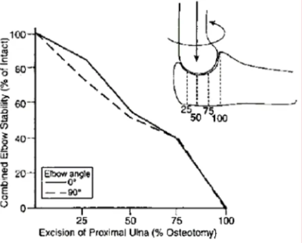
2. Olecranon17,18)
- 80% of the olecranon could be removed without compromising elbow stability.
- but significant increases in joint pressure with excision of 50% of the olecranon, which over time may contribute to elbow pain and arthritis.
Fig. 6. Successive resection of the proximal ulna
showed a linear decrease in elbow stability in both full extension and 90° flexion.3. Proximal radius2,19)
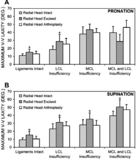
- important secondary valgus stabilizer of the elbow
- radial head is responsible for approximately 30% of valgus stability
- become more important for valgus stability in the presence of MCL deficiency
- radial head excision increases varus-valgus laxity, regardless of whether the collateral ligaments are intact.
Fig. 7. Maximum varus-valgus laxity plotted
for intact and insufficient collateral ligaments with radial head intact, excised, and replaced.Significant increases in laxity after radial head excision are denoted by asterisks.
III. Soft Tissue Stability
1. Medial collateral ligament complex3,7-9)
- consists of two main components, neither of which originates on the axis of rotation of the elbow.
- anterior bundle has been further subdivided into an anterior band that is taut in extension and a posterior band that is taut in flexion.
2. Lateral collateral ligament complex3,9,10)
- LCL lying on the axis of rotation will assume a rather uniform tension, regardless of elbow motion - lateral ulnar collateral ligament
: insert on the ulna and help to stabilize the lateral ulnohumeral joint : major component on the varus and posterolateral rotatory stability : essential to control the pivot shift maneuver
Fig. 8. Mean internal-external elbow rotation during the pivot
shift test is plotted for the intact elbow and after sectioning components of lateral collateral ligament. The only significant difference occurred after sectioning of the complete LCL.IV. Joint Forces
2,3,20)- with extension and axial loading, the distribution of stress is 40% across the ulnohumeral joint and 60% across the radiohumeral joint.
- force transmission through the radial head
: radial head forces were greatest from 0o to 30o flexion and always higher in pronation
Fig. 9. Greater force transmission across the
radial head with pronation, suggesting proximal migration of radial head with pronation.- When the elbow is extended, the overall force on the ulnohumeral joint is more concentrated at the coronoid; as the elbow is flexed, the force moves toward the olecranon.
Fig. 10. Concentration of the force at the ulno-
humeral joint varies with flexion and extension of the elbow. When the elbow is flexed at 90° (solid line), force is concentrated at the olecranon. When the elbow is extended (dashed line) the forced is concentrated at the coronoid. The olecranon fracture (a) and coronoid fracture (b) are shown.REFERENCES
1. O’Driscoll SW, Jupiter JB, King GJ, et al.: The unstable elbow. Instr Course Lect 2001;50:89-102.
2. Bryce CD, Armstrong AD: Anatomy and biomechanics of the elbow. Orthop Clin North Am 2008;39:141-154.
3. Fornalski S, Gupta R, Lee TQ: Anatomy and Biomechanics of the elbow joint. Tech Hand Upper Extremity Surg 2003;7:168-178.
4. Miyasaka KC: Anatomy of the elbow. Orthop Clin North Am 1999;30:1-13.
5. King GJ, Zarzour ZD, Patterson SD, et al.: An anthropometric study of the radial head: implications in the design of a prosthesis. J Arthroplasty 2001;16:112-116.
6. Gallay SH, Richards RR, O’Driscoll SW: Intraarticular pressure and capacity and compliance of stiff and normal elbows. Arthroscopy 1993;9:9-13.
7. Safran MR, Baillargeon D: Soft-tissue stabilizers of the elbow. 2005;14:179-185.
8. Callaway GH, Field LD, Deng XH, et al.: Biomechanical evaluation of the medial collateral ligament of the elbow. J Bone Joint Surg Am 1997;79:1223-1231.
9. Morrey BF, An KN: Functional anatomy of the ligaments of the elbow. Clin Orthop Relat Res 1985;201:84-90.
10. O’Driscoll SW, Bell DF, Morrey BF: Posterolateral rotator instability of the elbow. J Bone Joint Surg Am 1991;73:440-446.
11. Morrey BF, Askew LJ, Chao EY: A biomechanical study of normal functional elbow motion. J Bone Joint Surg Am 1981;63:872-877.
12. Bottlang M, Madey SM, Steyers CM, et al.: Assessment of elbow joint kinematics in passive motion by electromagnetic motion tracking. J Orthop Res 2000;18:195-202.
13. Kapandji A: Biomechanics of pronation and supination of the forearm. Hand Clin 2001;17:111-122.
14. Beingessner DM, Dunning CE, Stacpoole RA, et al.: The effect of coronoid fractures on elbow kinematics and stability. Clin Biomech 2007;22:183-190.
15. Morrey BF, An KN: Articular and ligamentous contributions to the stability of the elbow joint. Am J Sports Med 1983;11:315-319.
16. Hull JR, Owen JR, Fern SE, et al.: Role of the coronoid process in varus osteoarticular stability of the elbow. J Shoulder Elbow Surg 2005;14:441-446.
17. McKeever F, Buck R: Fracture of the olecranon process of the ulna. JAMA 1947;135:1-5.
18. Moed BR, Ede DE, Brown TD: Fractures of the olecranon: an in vitro study of elbow joint stresses after tension-band wire fixation versus proximal fracture fragment excision. J Trauma 2002;53:1088-1093.
19. Beingessner DM, Dunning CE, Gordon KD, et al: The effect of radial head excision and arthroplasty on elbow kinematics and stability. J Bone Joint Surg Am 2004:86:1730-1739.
20. Wake H, Hashizume H, Nishida K, et al.: Biomechanical analysis of the mechanism of elbow fracture-dislocations by compression force. J Orthop Sci 2004;9:44-50.