171
<원례보저>
랫드 두개골 결손부에서 단삼 수용성 추출물의 골형성 효과
심경미
1·김세은
2·강성수
2,*
1남부대학교 방사선학과, 2전남대학교 수의과대학 (게재승인: 2010년 7월 9일)
Effect of water extract of Danshen on bone regeneration of rat calvarial defect model
Kyung Mi Shim
1, Se Eun Kim
2, Seong Soo Kang
2,*
1Department of Radiology, Nambu University, Gwangju 500-706, Korea
2College of Veterinary Medicine, Chonnam National University, Gwangju 500-757, Korea
(Accepted: July 9, 2010)
Abstract :
The purpose of this study was to evaluate the osteogenic capacity of water extract of danshen (Salvia miltiorrhiza
Bunge). We have established in rat critical-sized calvarial defect model using the combination with collagen scaffold and danshen hydrophilic extract. All rats were extinguished at 8 weeks after bone graft surgery, and the bone regeneration ability of bone grafting sides was evaluated by plain radiography and micro-CT. These results revealed water extract of danshen had the potential to promote osteogenesis especially continuous oral administration with local treatment compared to one-shot local treatment. This compound may provide a new alternative agent for growth factors to promote bone healing and bone regeneration. In conclusion, these results suggest that danshen hydrophilic extract have the potential to promote osteogenesis in bone defects. Further studies about fusion technology with salvianolic acid B, peptides, growth factors, and scaffolds using of the combination of tissue engineering, cell engineering and mechanical engineering are needed.Keywords :
danshen, osteogenesis, rat, salvianolic acid B서 론
골이식은주로정형외과수술시골결손부를채우거 나부전유합및지연유합을치료하기위해 [11, 13, 22],
심하게복잡골절된골절부위를치유시키기위해 [28],
척추를융합시키기위해 [25], 광범위한종양절제부위를
채우기위해 [23], 관절고정술이나사지구제술을실시하
는경우 [12, 26]에적용된다. 그리고치과영역에서치
주질환에의한골흡수와하악골절부위를채워골재생
을돕기위해 [7, 29, 30], 치과용임플란트가안착하기
에치조골이부족한경우 [6, 32]에실시된다. 골이식은
1668년 Job van Meekeren [8]에의해최초로시행된후,
끊임없는시도와연구를거쳐현재는혈액다음으로가
장빈번히 이식되는 조직으로 발전했다 [2]. 1999년
Shors [31]는미국에서만연간 500,000건의골이식이시
행되고있고, 나머지나라에서그두배인 1,000,000건
이시행되고있다고보고하였다.
현재동종골과이종골및합성골의골재생능을증진 시키기위해다양한연구가진행되고있으며, 주로기계
적강도를유지하면서골전도능을향상시키기위한기 질(matrix)의크기와다공성(porosity)에대한연구 [1, 10],
bone morphogenetic protein과같이골유도능을갖는성
장인자에대한연구 [19, 27], mesenchymal stem cell과 같이골형성능을갖는세포에대한연구가끊임없이진
행중에있다 [7, 21]. 그리고실제임상상황에서, 생체
에서채취한골형성과관련된세포를일련의배양과정
*Corresponding author: Seong Soo Kang
College of Veterinary Medicine, Chonnam National University, Gwangju 500-757, Korea
[Tel: +82-62-530-2877, Fax: +82-62-530-2809, E-mail: [email protected]]
을통하여증식시킨후이들세포의활동을촉진시키는 성장인자와세포의성장을보존시킬수있는지지체를 인위적으로제작하여이를통하여새로운골조직을형 성하는과정인골조직공학(bone tissue engineering)이요 구되고있다.
골조직공학에서는지지체, 세포이외에도세포의활 성을증가시키는생물학적인자의사용이고려되고있
다. 다수의식물추출물(natural products)에대한골형성
촉진효과는 여러연구에 의해밝혀져 있으며, 이들은 국소또는전신투여되어골형성을증가시키거나골흡 수를감소시켜그효과를발휘한다. 대표적인골형성촉
진천연물에는칡, 두충, 단삼, 골쇄보등이있으며, 이 들은생체내에서골유도능을발휘한다. 이들의대표적 인약리효과로 isoflavones나 flavones와같은식물성에
스트로겐(phytoestrogens)을다량함유하여칼슘의장내
흡수를증가시키고칼슘의뇨배출을감소시킴으로써칼 슘이용률을높이는능력을들수있다. 이외에광물화
및골모세포활성을촉진시키고파골세포(osteoclast)의 활성을억제시키는등의효과도가지고있다 [34]. 단삼
(Salvia miltiorrhiza Bunge)은동서양에서심혈관계질환
의치료및예방을목적으로널리사용되는약제로혈 류를개선시키고창상치유를촉진시키는약리효과를
갖는다 [4, 5, 36]. 그리고파골세포의활성을억제하고
[16, 18] 신생골형성을촉진시킴으로써골형성에기여
할 수 있다. 특히 단삼의 수용성 성분 중 하나인
salvianolic acid B는그함량이높으며, 혈관신생작용
(angiogenesis) 및골수세포의 alkaline phosphatase(ALP)
활성을증강시키는약리작용을갖는다 [9, 20, 33].
본연구는 salvianolic acid B를주성분으로하는단
삼수용성추출물의임상적적용가능성을확인하고자 실시되었다. 본연구진은예비실험을통하여 salvianolic
acid B의골모세포활성촉진효과를확인하였다. 그러
나단삼에서순수한 1가지특정성분인 salvianolic acid
B의분리를위한적절한추출조건을찾기위해서는시
간및비용 면에서매우불리한 경우가많다. 그리고
특정약리효과를나타내는데 있어한가지성분만작 용하지않고, 여러성분이서로작용하여그효과를증
강시키기도한다. 따라서 salvianolic acid B만을임상에
적용하기에는비용적부담이너무크므로, 단삼으로부 터수용성성분을추출하여임상실험에적용하고자하
였다. 그리고추출과정에서 salvianolic acid B의구조
변화를막고, 추출되는 양및용해성을최대화시키기 위해다양한추출조건을 비교, 분석하고자 하였다. 그
리고랫드두개골결손부에서단삼수용성추출물의골
재생효과를단순방사선및 Micro CT를이용하여평
가하고자하였다.
재료 및 방법
단삼으로부터수용성성분의추출
환류 및 저온 초음파 추출법의 비교: 5 g의단삼파우
더를 50 mL의 70% 메탄올을용매로 하여끓는점에서
환류시키면서추출하거나, 물을용매로하여 20oC에서
30 Hz 초음파로 1시간동안추출하였다. 그리고추출결
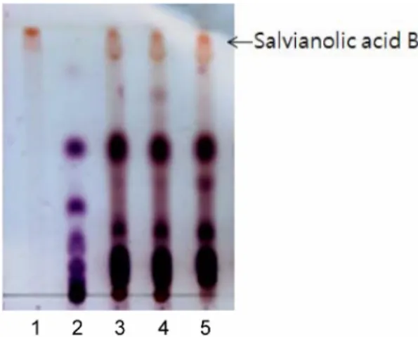
과물의 salvianolic acid B의양을 thin-layer chromatogram (TLC) 상에서표준물질(Ivy Fine Chemicals, USA)과비 교하였다. TLC는실온에서 silica gel 60 F254 TLC plates (Merck, USA)에서시행되었다. 시료 1µL를 silica gel plate에점적한후, ethyl acetate-acetic acid-water(3 : 1 : 1,
v/v/v)의용액을전개용매로사용하였고, 건조하였다. 그
후, 생성된산물들은 0.3%(w/v) N-(1-naphthyl)-ethylene- diamine과 5%(v/v) H2SO4를함유하고있는메탄올에담 그고, 말린후 121oC 오븐에서 10분간구워주고, 나타나 는물질을표준물질과비교하여확인하였다.
추출 용매에 따른 추출 효과 비교: 단삼의수용성성 분추출을위한저온초음파추출시가장효과적인용
매를평가하기위해물, 10% 메탄올, 70% 메탄올을용
매로하여 20oC에서 30 Hz 초음파로 1시간동안추출하
였다. 추출물을 12,000 rpm에서 10분간원심분리하여상
층액을얻은후, TLC 상에서의스팟을 NIH 프로그램으
로정량하여 salvianolic acid B의양을 비교분석하였
다. 동물실험에사용할시료는저압상태와저온의동결 건조기에미리−80oC에서동결한샘플을넣고, 액상을
거치지않고직접수증기로승화시킨후건조하는방법 을이용하여샘플을동결건조하였다.
단삼수용성추출물의골재생효과확인
실험동물: 체중 200 g 전후의건강한 6주령수컷랫드
(SD rat; Samtaco, Korea) 30마리를실험환경에적응시
키기위하여 1주일의적응기를거친후, 실험전기간 동안사육환경을온도 23±2oC, 상대습도 60±10%로
유지하고 12시간의명암주기가유지되는실내에서사료 와식수를자유로이공급하였다. 동물의사육과실험은 전남대학교동물실험지침에따라수행되었다.
실험방법: 무작위로 6마리씩임계결손부군(대조군), 콜
라겐기질(TERUPLUG; Termo, Japan) 이식군, 콜라겐
기질에단삼수용성추출물국소이식군및국소이식 후실험전기간동안수용성단삼추출물을경구투여 한군으로나누었다. 랫드두개골결손부형성을위해서
ketamine 40 mg/kg과 xylazine 10 mg/kg을복강주사하여
전신마취한후랫드두개골부위를전모하고, 포비돈으 로소독하였다. 두개정중부 피부및골막을 U자형으 로절개한후, 골막하박리를통하여두개골을 노출시
켰다. 생리식염수 관주 하에 8 mm trephin bur와 #2
round bur를이용하여뇌경막이손상되지않도록두개
골중앙부에 원형결손부를 형성하였다. 이원형결손
부에임계결손부군은 아무처리도하지않고(대조군),
콜라겐기질이식군에는준비된직경 8 mm, 두께 2 mm
의 TERUPLUG를, 나머지두군에는 TERUPLUG를단
삼수용성추출물(salvianolic acid B 5 mg/mL)에 15분 동안담궜다 이식하였다. 골막은 4-0 흡수성봉합사 (Surgisorb; Samyang, Korea)로연속봉합하였고, 피부는
3-0 비흡수성봉합사(Black Silk; Ailee, Korea)로단순결 절봉합하였다. 단삼수용성추출물국소이식및경구
투여군에는실험전 기간에걸쳐 1일체중 100 g 당
salvianolic acid B 1 mg을음수에첨가하여공급하였다.
골이식부에대한방사선학적평가
골이식후 8주째에실험동물에이산화탄소를흡입시
켜희생시킨후, diamond disc로골결손부위를포함하여
골조직을채취한후 Port-X portable X-ray (Genoray, Korea)
를이용하여, 70 kVp, 3 mA, 0.03 sec, 20 cm FFD의조 건으로일반방사선촬영을실시하였다. 획득된영상은 Digi-X intra oral digital sensor(한진덴탈, 한국)를통해 저장되었고, 각군별로방사선밀도의변화를영상분석 프로그램을이용하여비교및평가하였다.
Micro CT촬영및골형성량측정
채취된골조직을 Skyscan 1172 Desktop X-ray Microto- mograph (Skyscan, Belgium)를이용하여 50 kVp의관전 압과 200µA의노출조건으로컴퓨터단층촬영하였다.
획득된영상에대해 Skyscan CTan ver. 1.5.0 및 Shortcut
to ANT (Skyscan, Belgium) 영상분석프로그램을이용
해골밀도(bone mineral density; BMD)와골부피(bone
volume; BV)를측정하고, 삼차원영상으로재구성하였다.
결 과
단삼으로부터수용성성분의추출
환류 및 저온 초음파 추출법의 비교: 단삼의수용성 성분추출시효과적인추출법을평가하기위해메탄올 을용매로한고온환류추출법과물을용매로한저온 초음파추출법을비교하였다. 수율및성분의안정성은
TLC 상에서 salvianolic acid B를기준으로평가하였다.
TLC 상에서의 스팟을 NIH 프로그램으로 정량하여
salvianolic acid B의양을 비교 분석하였다. 그결과
salvianolic acid B의수율면에서고온환류추출법및
저온초음파 추출법이차이가없었다. 그리고 고온환 류 추출법의 경우 더다양한 성분들이 추출되었고,
salvianolic acid B의구조가변형되어스팟이분리된것
을확인하였다(Fig. 1).
용매에 따른 추출 효과 비교: 저온초음파추출시효
과적인추출용매를찾기위해물, 10% 메탄올, 70% 메
탄올을용매로하여 salvianolic acid B의수율을평가하
고, 이를물에대한수율과비교하였다. 그결과 10% 메
탄올이물의 106.7%, 70% 메탄올이 96.8%로 10% 메탄
올, 물, 70% 메탄올순으로추출되는 salvianolic acid B
의양이줄어들었다(Fig. 2). 추후시료의용해성을고려
하여용매를물로선택하여 5 g의단삼파우더를 50 mL
의물로초음파추출하여원심분리한결과 30 mL의추 출용액을얻었고, 이를저온동결건조한결과 1,400 mg
의파우더를획득할수있었다. 그리고총 1,400 mg의
파우더에서 salvianolic acid B가차지하는양은 80.5 mg
이었다.
단삼수용성추출물의골재생효과확인
골이식부에 대한 방사선학적 평가 결과: 콜라겐기질 과단삼의수용성추출물이랫드두개골결손부의골형 성에미치는영향을평가하기위해골이식후 8주째에 실험동물을희생시켜채취된골조직을일반방사선촬영 하였다. 임계결손부군은대부분의개체에서골형성이미 미하여결손부의방사선투과도가높았다. 결손부가장 자리로부터의신생골형성으로부분적인방사선비투과
Fig. 1.
Thin-layer chromatogram of Danshen extract. Lane 1, authentic salvianolic acid B; Lane 2, Danshen extract with water by temperature controlled ultrasonography; Lane 3, Danshen extract with 70% MeOH by reflux. One µL of the sample was spotted onto a lane on a silica gel plate, and the plaste was developed with a solvent mixture of ethyl acetate-acetic acid-water (3 : 1 : 1, v/v/v). The developed plate was then dried and visualized.도를보였으나, 신생골부위의방사선밀도는정상골에
비해낮았다. 콜라겐기질만이식한군에서도신생골형 성이관찰되었으며, 이는임계결손부군에비해양및질
적으로훨씬우수한것으로확인되었다. 콜라겐기질을 단삼의수용성추출물에적셔이식한군및실험전기 간동안단삼의수용성추출물을경구투여한군에서도 다량의신생골형성이관찰되었으며, 그양이콜라겐기 질만을이식한군보다더많음을확인할수있었다.
신생골형성에의한각군간의방사선밀도의변화 를비교하기위해영상분석프로그램을이용해골결손 부의 OD 값을측정하였다. 콜라겐기질과단삼수용성
추출물국소이식군(p< 0.05) 및경구투여를병행한군
(p< 0.01)에서임계결손부군에비해신생골형성이통계
적유의성을띠며증가하였고, 특히단삼의지속적인경 구투여가단회국소적용보다효과가큰것으로확인되 었다(Table 1).
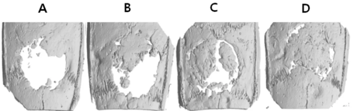
Micro CT 촬영 및 골형성량 측정 결과: 채취된골조 직을 Skyscan 1172 Desktop X-ray Microtomograph를이
용하여 50 kVp의관전압과 200µA의노출조건으로컴
퓨터단층촬영하여골단면의영상을획득하고 3차원영 상으로재구성하였다. 그리고골결손부양쪽의정상골
및가장자리, 골결손부중앙의단면상에대해신생골형 성을평가하였다. 그결과임계결손부군에서는골결손 부가장자리에서만소량의얇은골신생을확인할수있
었다. 콜라겐기질을이식한군에서는약간의골신생이 관찰되었으나, 단삼수용성추출물을함께이식한군에
비해새로형성된골의양이적었다. 그리고단삼수용 성추출물을단회국소적용한군보다이식후지속적으 로경구투여한군에서신생골형성이현저히많았다(Fig.
3). 초기골결손형성부를측정영역으로설정하여골밀
도(BMD) 및골부피(BV)를측정하였다. 그결과모든
군에서임계결손부에비해골밀도가유의적으로높았고
(p< 0.01), 단삼수용성추출물을국소및경구투여한군
은콜라겐만이식한군에비해서도골밀도가높았다(p
< 0.05) (Table 2). 골부피또한단삼수용성추출물국소
Fig. 2.
Thin-layer chromatogram of Danshen extracted by temperature controlled ultrasonography. Lane 1, authentic salvianolic acid B; Lane 2, isomalto oligosaccharide; Lane 3, Danshen extract with water; Lane 4, Danshen extract with 10% MeOH; Lane 5, Danshen extract with 70%MeOH. One µL of the sample was spotted onto a lane on a silica gel plate, and the plaste was developed with a solvent mixture of ethyl acetate-acetic acid-water (3 : 1 : 1,
Table 1.
Bone regeneration of water extract of Danshen (WED) on rat calvarial defect model (mean±SD)Group OD
Critical Defect (control) 1.51 ± 0.13
Collagen 1.71 ± 0.12
Collagen with WED local administration 1.78 ± 0.02* Collagen with WED local and oral
administration 1.88 ± 0.17**
*p< 0.05 as compared with the control (critical defect) group.
**p< 0.01 as compared with the control (critical defect) group. Values are expressed in mean±SD (n = 5).
Table 2.
Effect of WED on bone mineral density (BMD) of rat calvarial defect modelGroup BMD
Critical Defect (control) 0.148 ± 0.022
Collagen 0.204 ± 0.014**
Collagen with WED local administration 0.217 ± 0.008**
Collagen with WED local and oral
administration 0.242 ± 0.003*,**
*p< 0.05 as compared with the collagen graft group. **p<
0.01 as compared with the control (critical defect) group.
Values are expressed in mean±SD (n = 5).
Table 3.
Effect of WED on bone volume (BV) of rat calvarial defect modelGroup BV
Critical Defect (control) 99.6800 ± 0.035
Collagen 11.9267 ± 1.194
Collagen with WED local administration 14.5000 ± 0.504 Collagen with WED local and oral
administration 16.7833 ± 0.777*
*p< 0.05 as compared with the control (critical defect) group.
Values are expressed in mean±SD (n = 5).
및경구투여군에서유의적으로높게 측정되었다(p<
0.05) (Table 3).
고 찰
단삼은꿀풀과(Labiate)에속하는다년생약용식물로,
이들의건조한뿌리는신체기능을증진시키기위해일 반적으로사용되는 전통약제이며, 적삼, 자단삼, 대홍
포, 화혈근이라고도불리며, 붉은색을띠고있다. 뿌리 는특이한냄새가나고, 약간쓴맛이나며, 한방에서는
약제로사용하는데부인의생리불순, 생리통, 산후복통 에쓰인다. 또한, 어혈성의심복부동통과타박상을치 료하고, 불면증, 피부발진등에도사용된다. 그리고 동
양뿐아니라서양에서도혈류개선효과를보여심혈관 계질환의치료및예방을위해널리사용되며, 창상치 유를촉진시키는효과도보고되었다 [4, 5, 35, 36].
단삼은크게두가지서로다른성분인수용성과지 용성성분으로구성되고, 이중수용성인페놀성화합물
(phenolic compounds)과지용성인디터펜(diterpenes)이
많은약리활성을갖는다. 1930년대초반부터단삼의화 학성분에대한연구가시행되었고, 초반에는주로지용 성성분에대한연구가이루어졌으나, 최근에는수용성
성분에대한연구가더활발히진행되고있다 [16, 36].
단삼의알려진수용성유효성분으로는 salvianolic acid A와 B, danshensu, caffeic acid, protocatechualdehyde, rosmarinic acid 등이있다 [36]. 특히 salvianolic acid B
는혈관내피세포에서산화질소(nitric oxide) 합성을증 가시키고 [24], 안지오텐신전환효소(angiotensin converting
enzyme)를방해하여혈관을이완시켜혈압을낮추고 [4,
5, 15], 뇌및심장과같은주요장기의허혈-재관류손상
을예방하는것으로알려져있다 [33]. 그리고골모세포
인 MC3T3-E1 세포및마우스골수세포의 ALP 합성을
증가시키고 [9, 20], murine SVR 내피세포주에서 VEGF,
VEGF-R2 유전자발현을증가시켜혈관신생을촉진시
킨다는보고도있다 [14]. 그리고다양한동물실험에서
단삼추출물의골형성촉진효과가보고되었다 [3, 16,
18, 33]. Salvianolic acid B의골모세포활성촉진효과
가증명된바, 이의임상적적용가능성을확인하기위
해랫드두개골결손부에서의골재생효과를평가하고 자하였다. 천연물에서순수한 1가지특정성분을분리 해내는것은매우어려우며, 적절한추출조건을찾기
위해서는시간및비용면에서매우불리한경우가많
다. 따라서 salvianolic acid B만을임상에적용하기에는
비용적부담이너무크므로, 단삼으로부터수용성성분
을추출하여임상실험에적용하였다. 그리고추출과정
에서 salvianolic acid B의구조변화를막고, 추출되는양
및용해성을최대화시키기위해다양한추출조건을시 도하여결과적으로물을용매로한저온초음파추출법 을확립하였다. 이렇게추출된단삼수용성성분을콜라
겐기질을지지체로사용하여랫드두개골의 8 mm 임
계결손부에이식하였다. 그리고단삼수용성추출물의 경구투여효과를확인하기위해수술후 2개월동안지 속적으로경구투여한군과단회국소처치군의골형성 정도를비교하였다. 신생골형성은골이식후 8주째에 실험동물을희생시켜골편을채취한후일반방사선촬 영하여획득된방사선밀도차이를영상분석프로그램 을이용하여정량적으로평가하였다. 일반방사선사진 상에서콜라겐만이식한군에서도약간의신생골형성 이관찰되었고, 단삼수용성추출물을처리하여이식한
군에서는현저한신생골형성이관찰되었다. 특히단삼 수용성추출물을지속적으로경구투여한군에서가장
높은방사선밀도를보였다. Micro CT 상의결과도일
Fig. 3.
2D and 3D micro CT images of 8 mm critical-size calvarial defects at 8 weeks postsurgery (A; critical defect, B; collagen, C; collagen with water extract of Danshen (WED) local administration, D; collagen with WED local and oral administration). In collagen with WED local and oral administration groups, the defect almost completely repaired by the graft materials after 8 weeks of postsurgery.반방사선촬영상의결과와동일하였고, CT 단면상을 3
차원으로재구성한결과신생골형성정도를정확히확 인할수있었다. 본연구를통하여단삼으로부터수용성
성분을추출하기위한추출법으로물을용매로한저온 초음파추출법이매우유용하였다. 그리고콜라겐기질
이신생골형성을위한지지체로적합하며, 단삼수용성 추출물의운반체로도 유용함을확인할수있었다. 또
salvianolic acid B를포함하고있는단삼수용성추출물
의단회국소처치가생체내에서골재생효과가뛰어나 며, 특히골이식후단삼수용성추출물의지속적인경 구투여로골재생효과를증가시킬수있음이확인되었 다. 따라서단삼수용성추출물의단회국소처치에더하 여지속적인경구투여가골재생효과를극대화시킬수 있을것으로생각된다.
현재까지의연구를통하여골이식에서골유도체로서 천연물인단삼수용성추출물의이용가능성을확인할 수있었다. 이의사용은부가적인수술부담및합병증
의감소, 고비용에대한부담의감소, 합성약제의고용 량사용에의한잠재적인독성유발가능성의감소등 다양한측면에서임상적상황에의적용에유리할것으 로생각된다. 앞으로골형성능을갖는세포와단삼수용 성추출물및이들의운반체로적절한지지체의스크리 닝이필요할것으로생각되며, 이들의적절한융합을위
한골조직공학적연구가더필요할것으로생각된다.
감사의 글
이논문은 2007년정부(교육과학기술부)의재원으로
한국연구재단의지원을받아수행된연구임(KRF-2007- 331-E00269).
참고문헌
1.
Bolder SB, Schreurs BW, Verdonschot N, van Unen JM, Gardeniers JW, Slooff TJ.
Particle size of bone graft and method of impaction affect initial stability of cemented cups: human cadaveric and synthetic pelvic specimen studies. Acta Orthop Scand 2003,74
, 652- 657.2.
Boyce T, Edwards J, Scarborough N.
Allograft bone.The influence of processing on safety and performance.
Orthop Clin North Am 1999,
30
, 571-581.3.
Chae HJ, Chae SW, Yun DH, Keum KS, Yoo SK, Kim HR.
Prevention of bone loss in ovariectomized rats: the effect of Salvia miltiorrhiza extracts.Immunopharmacol Immunotoxicol 2004,
26
, 135-144.4.
Chang PN, Mao JC, Huang SH, Ning L, Wang ZJ, On T, Duan W, Zhu YZ.
Analysis of cardioprotective effects using purified Salvia miltiorrhiza extract on isolated rat hearts. J Pharmacol Sci 2006,101
, 245-249.5.
Cheng TO.
Cardiovascular effects of Danshen. Int J Cardiol 2007,121
, 9-22.6.
Clayman L.
Implant reconstruction of the bone-grafted maxilla: review of the literature and presentation of 8 cases. J Oral Maxillofac Surg 2006,64
, 674-682.7.
Clokie CM, Sndor GK.
Reconstruction of 10 major mandibular defects using bioimplants containing BMP- 7. J Can Dent Assoc 2008,74
, 67-72.8.
de Boer HH.
The history of bone grafts. Clin Orthop Relat Res 1988,226
, 292-298.9.
Ding Y, Soma S, Takano-Yamamoto T, Matsumoto S, Sakuda M.
Effects of salvia miltiorrhiza bunge (SMB) on MC3T3-E1 cells. J Osaka Univ Dent Sch 1995,35
, 21-27.10.
Dunlop DG, Brewster NT, Madabhushi SP, Usmani AS, Pankaj P, Howie CR.
Techniques to improve the shear strength of impacted bone graft: the effect of particle size and washing of the graft. J Bone Joint Surg Am 2003,85-A
, 639-646.11.
Goldberg VM, Stevenson S.
The biology of bone grafts. Semin Arthroplasty 1993,4
, 58-63.12.
Hayden RJ, Jebson PJ.
Wrist arthrodesis. Hand Clin 2005,21
, 631-640.13.
Hernigou P, Poignard A, Manicom O, Mathieu G, Rouard H.
The use of percutaneous autologous bone marrow transplantation in nonunion and avascular necrosis of bone. J Bone Joint Surg Br 2005,87
,896- 902.14.
Huang X, Jiang H, Liu D, Zhou Z, Wang L.
Implantation of calcium phosphate cement/Danshen drug delivery system for avascular necrosis of femoral head. Zhongguo Xiu Fu Chong Jian Wai Ke Za Zhi 2008,
22
, 307-310.15.
Kang DG, Oh H, Chung HT, Lee HS.
Inhibition of angiotensin converting enzyme by lithospermic acid B isolated from Radix Salviae miltiorrhiza Bunge.Phytother Res 2003,
17
, 917-920.16.
Kim HK, Woo ER, Lee HW, Park HR, Kim HN, Jung YK, Choi JY, Chae SW, Kim HR, Chae HJ.
The correlation of Salvia miltiorrhiza extract-induced regulation of osteoclastogenesis with the amount of components tanshinone I, tanshinone IIA, cryptotan- shinone, and dihydrotanshinone. Immunopharmacol
Immunotoxicol 2008,
30
, 347-364.17.
Kraus KH, Kirker-Head C.
Mesenchymal stem cells and bone regeneration. Vet Surg 2006,35
, 232-242.18.
Kwak HB, Yang D, Ha H, Lee JH, Kim HN, Woo ER, Lee S, Kim HH, Lee ZH.
Tanshinone IIA inhibits osteoclast differentiation through down-regulation of c- Fos and NFATc1. Exp Mol Med 2006,38
, 256-264.19.
Linkhart TA, Mohan S, Baylink DJ.
Growth factors for bone growth and repair: IGF, TGF beta and BMP.Bone 1996,
19
(Suppl), 1S-12S.20.
Liu YR, Qu SX, Maitz MF, Tan R, Weng J.
The effect of the major components of Salvia Miltiorrhiza Bunge on bone marrow cells. J Ethnopharmacol 2007,111
, 573-583.21.
Meijer GJ, de Bruijn JD, Koole R, van Blitterswijk CA.
Cell-based bone tissue engineering. PLoS Med 2007,4
, e9.22.
Munk B, Larsen CF.
Bone grafting the scaphoid nonunion: a systematic review of 147 publications including 5,246 cases of scaphoid nonunion. Acta Orthop Scand 2004,75
, 618-629.23.
Nishida J, Shimamura T.
Methods of reconstruction for bone defect after tumor excision: a review of alternatives. Med Sci Monit2008,14
, RA107-113.24.
O K, Cheung F, Sung FL, Zhu DY, Siow YL.
Effect of magnesium tanshinoate B on the production of nitric oxide in endothelial cells. Mol Cell Biochem 2000,207
, 35-39.25.
Pilitsis JG, Lucas DR, Rengachary SS.
Bone healing and spinal fusion. Neurosurg Focus 2002,13
, e1.26.
Safran O, Iannotti JP.
Arthrodesis of the shoulder. J Am Acad Orthop Surg 2006,14
, 145-153.27.
Sakou T.
Bone morphogenetic proteins: from basic studies to clinical approaches. Bone1998,22
, 591-603.28.
Sauer HD, Schoettle H.
The stability of osteosyntheses bridging defects. Arch Orthop Trauma Surg 1979,95
, 27-30.29.
Shi H, Ma J, Zhao N, Chen Y, Liao Y.
Periodontal regeneration in experimentally-induced alveolar bone dehiscence by an improved porous biphasic calcium phosphate ceramic in beagle dogs. J Mater Sci Mater Med 2008,19
, 3515-3524.30.
Shim KM, Kim SE, Yoo KH, Bae CS, Choi SH, Kang SS.
Case studies of repair of pathological mandibular fracture due to periodontal disease in dogs.J Vet Clin 2007,
24
, 653-657.31.
Shors EC.
Coralline bone graft substitutes. Orthop Clin North Am 1999,30
, 599-613.32.
Tiwana PS, Kushner GM, Haug RH.
Maxillary sinus augmentation. Dent Clin North Am 2006,50
, 409-424.33.
Wong RW, Rabie AB.
Effect of Salvia miltiorrhiza extract on bone formation. J Biomed Mater Res A 2008,85
, 506-512.34.
Wong RW, Rabie AB.
Traditional Chinese medicines and bone formation°™a review. J Oral Maxillofac Surg 2006,64
, 828-837.35.