http://dx.doi.org/10.14405/kjvr.2015.55.4.247
247
<원례보저>
말의 운동 스트레스에 관한 새로운 생물지표
이승우·오다영·김명철·김용백·류덕영*
서울대학교 수의과대학, BK21수의과학연구인력양성사업단, 수의과학연구소 (접수: 2015년 10월 27일, 수정: 2015년 12월 16일, 게재승인: 2015년 12월 24일)
A novel biomarker of exercise-induced stress in horses
Seungwoo Lee, Dayoung Oh, Myung-Chul Kim, Yongbaek Kim, Doug-Young Ryu*
College of Veterinary Medicine, BK21plus Program for Creative Veterinary Science Research, and Research Institute for Veterinary Science, Seoul National University, Seoul 08826, Korea.
(Received: October 27, 2015; Revised: December 16, 2015; Accepted: December 24, 2015)
Abstract : Exercise is one of the most common stressors in horses. Although various physiological parameters such as cortisol respond to exercise, there is no reliable parameter for the measurement of exercise-induced stress in sport horses. This study was performed to discover a new biomarker with high sensitivity for exercise-induced stress.
The expression of fos mRNA was increased more than 10-fold in horse blood samples collected after an hour of exercise, as compared with before the exercise. The plasma cortisol levels were also increased after the exercise, but only by about two-fold. The fos mRNA levels were well-correlated with plasma cortisol concentrations. These findings suggest that fos mRNA expression in blood may be useful for the measurement of exercise-induced stress in horses.
Keywords : biomarker, exercise, fos, horse, stress
서 론
말은 수송, 운동, 파행, 그리고 온도와 습도 등의 변화 때 문에 스트레스(stress)를 받을 수 있다 [8]. 스트레스는 다양 한 질병을 유발하고 건강 유지에 악영향을 미치므로 말의 건 강 관리와 경기력 향상에 관련된 요인으로 중요하게 다루어 지고 있다 [2, 8, 22, 30, 31]. 운동에 의한 스트레스로 유 발되는 생화학적 변화 등에 대한 연구는 주로 경주용 말에 서 광범위하게 이루어지고 있다 [23].
말의 운동에 의한 스트레스 정도를 측정하기 위해서 심박 수, 체중, 체온, 혈중 젖산, 카테콜아민(cathecolamine), 베타 엔도르핀(beta-endorphin), 코르티솔(cortisol) 등 다양한 생리 학적 인자들을 이용하고 있다 [8, 18]. 그중 코르티솔은 신장 의 부신피질에서 분비되는 하이드로코르티손(hydrocortisone) 이라 알려진 스테로이드 호르몬으로써 운동에 의한 스트레 스 정도를 평가하기 위해 가장 빈번히 사용되고 있다 [7, 9, 11, 12, 16, 17, 33]. 최근에는 운동 스트레스에 의한 유전
자 발현을 포함한 생화학적 변화에 대한 여러 연구가 많이 수행되고 있다 [2, 21, 24, 25].
Fos는 종양 형성 유전자로서 류신(leucine) 지퍼구조를 매 개로 하여 c-Jun과 결합, 이합체(heterodimer)를 형성하여 전 사조절인자의 역할을 한다. 주로 세포증식, 분화, 세포사멸 등과 같은 다양한 생물학적 과정과 관련된 기능이 밝혀졌다 [1, 15]. 또한 fos는 허혈, 저산소증, 고체온증과 같은 대사성 스트레스와 연관되어 있다. 스트레스 상황에서 마우스, 랫드 를 이용한 in vivo 동물실험과 in vitro 실험을 통해 fos 발 현 변화가 나타난다고 보고된 바 있다 [4, 6, 20, 32]. 또한 최근 운동 스트레스에 의해 말의 근육 조직 내에서 fos의 발 현이 증가한다고 보고되었다 [21, 24]. 하지만 운동 스트레스 에 대한 생체지표로서의 활용 가능성을 위한 혈액 내 fos 발 현 연구는 미미한 상황이다. 본 연구에서는 말의 운동 후 혈 중 fos 발현 변화를 분석하여 운동에 따른 스트레스 평가 지 표로서의 가치를 평가하고자 하였다.
*Corresponding author
Tel: +82-2-880-1253, Fax: +82-2-878-2360 E-mail: [email protected]
재료 및 방법
말의 운동실험
본 연구에 사용된 말은 약 600 kg의 8–16세 Warmblood (Equus caballus) 거세마 5두로 각 개체에 대한 구체적인 정 보는 Table 1에 요약하였다. 각 개체는 운동 전까지 마구간 에서 관리되었으며 야간 절식 후 영하 약 4oC의 기온 아래 오전 10시경부터 실내 모래 트랙(sand track)에서 약 200–
300 m/min의 속도로 1시간 동안 속보를 하였다. 동물실험과 정은 서울대학교 동물실험윤리위원회의 사전검토 및 승인(승 인번호: SNU-131218-1) 하에 시행되었다.
채혈과 혈장 분리
운동 전 혈액/혈장 샘플은 운동 10분 전 말의 경정맥으로 부터 약 14 mL의 혈액을 채혈하여 2개의 BD Vacutainer EDTA-K2 10.8 mL(BD Biosciences, USA) 튜브에 7 mL씩 분주하였다. 첫 번째 튜브의 혈액 6 mL은 Trizol 용액 (Invitrogen, USA)과 1 : 4의 비율로 혼합하여 4oC에서 4시간 동안 보관한 후 RNA 추출에 사용하였다. 그리고 나머지 혈 액 1 mL은 4oC에서 4시간 동안 보관한 후 혈액학적 검사에 사용하였다. 두 번째 튜브의 혈액 7 mL은 10분간 300 × g로 원심분리하여 혈장을 분리하였다. 분리된 혈장은 4oC에서 4 시간 동안 보관한 후 혈청학적 검사에 사용하였다.
운동 직후의 혈액/혈장 샘플도 운동 전과 동일한 과정 및 방법으로 분석을 진행하였다. 혈액학적, 혈청학적 검사는 ADVIA 120 혈액검사기기(Siemens Healthcare, USA)와 FUJI DRY-CHEM 3500i(Fujifilm, Japan)를 사용하여 시행 하였다.
RNA 추출과 cDNA 합성
혈액 및 Trizol 혼합액에 Trizol 용액 750 µL 당 Chloro- form(Sigma, USA) 200 µL를 넣고, 15초 동안 흔들어 실온 에서 3분 동안 반응시켰다. 4oC에서 15분간 10,000 × g의 속 도로 원심분리하였다. 원심분리 후 약 500 µL의 상층액을 확 보하여 Isopropylalcohol(Sigma)과 1 : 1 비율로 혼합하였다.
실온에서 10분간 반응시킨 후 4oC에서 10분간 10,000 × g의 속도로 원심분리하였다. 원심분리 후 RNA 침전물을 확인하 고, 상층액을 모두 제거하였다. 그리고 RNA 침전물을 세척 하기 위해 70% EtOH(Sigma) 1 mL을 첨가한 후 4oC에서 5분간 5,800 × g의 속도로 원심분리하였다. 원심분리 후 상층 액을 모두 제거하고 실온에서 튜브 뚜껑을 열어놓은 채
RNA 침전물을 완전히 말렸다. 그리고 RNase가 포함되지 않 은 20–100 µL의 HPLC 수(Sigma)를 첨가하여 RNA 용출 과정을 진행하였다.
각 개체로부터 추출된 운동 전후 혈액 내 RNA는 Nano drop(Thermo Scientific, USA)으로 농도와 순도를 측정하였 다. 그리고 cDNA를 합성하기 위해 1 µg의 RNA에 10 mM deoxynucleotide, 10ρmol 무작위서열 프라이머(random primer) 정방향 및 역방향 프라이머, 0.1 M dithiothreitol, 40 U RNase inhibitor, 200 U reverse transcriptase, 5배 반 응용 완충용액(reaction buffer), 증류수를 혼합하였다. 이 혼 합물은 중합효소 연쇄반응 장비(PCR machine)를 사용하여 42oC에서 60분, 95oC에서 5분간 반응시켰다. 이후 합성된 cDNA는 실시간 중합효소 연쇄반응(Real-time PCR)과 역전 사 중합효소 연쇄반응(RT-PCR) 전까지 −21oC에서 보관하였다.
Real-time PCR과 RT-PCR
Real-time PCR은 합성된 cDNA와 2배 SYBR Master Mix, fos나 β-actin에 대한 정방향 프라이머(fos: 5'- AAGCGGAGACAGACCAACTAGAAGATG-3', β-actin: 5'- ATCGTGCGTGACATCAAGGAGAAGCT-3'), 역방향 프라 이머(fos: 5'-ACTCAGATCAAGGGAAGCCACAGACAT-3', β-actin: 5'-TTCATGATGGAGTTGAAGGTAGTTTCGT-3'), 증류수를 넣은 혼합액으로 진행하였다. Fos와 β-actin 염기서 열은 NCBI(National Center for Biotechnology Information, USA)의 유전자 번호 XM_001491972.3과 NM_001081838.1 서열을 참고하여 각각 사용하였다. 혼합물은 Real-time PCR machine(Qiagen, USA)을 사용하여 총 45 Cycle(95oC에서 30초, 65oC에서 20초) 동안 반응시켰으며, Rotor-Gene Q Series Software(ver. 2.1.0; Qiagen) 프로그램을 사용하여 데 이터 분석을 수행하였다.
RT-PCR은 합성된 cDNA와 2 mM deoxynucleotide, 10배 Taq buffer, Taq DNA 중합효소, Real-time PCR에 사용한 fos나 β-actin에 대한 정방향 및 역방향 프라이머, 증류수를 넣어 혼합액으로 진행하였다. 혼합물은 PCR기를 사용하여 총 25 Cycle(95oC에서 30초, 65oC에서 30초, 72oC에서 20 초) 동안 반응시켰다. 각 PCR 최종산물은 1% agarose gel 을 사용하여 전기영동을 시행하였다. Gel 상의 PCR 최종산 물의 농도에 대한 계측 정량화는 Image J Software(ver.
1.47; National Institute of Health, USA) 프로그램을 사용 하여 분석하였다.
코르티솔 농도 측정
혈액으로부터 분리한 혈장 샘플 100 µL를 바코드화된 시 료 컵에 담은 후 로드 플랫폼(load platform)에 장착하고 Immulite 1000 장비(Siemens Healthcare, Germany)를 사용 하여 코르티솔의 농도를 측정하였다.
통계 분석
데이터 통계 처리는 SPSS Package(ver. 23.0; IBM, USA) Table 1. Five horses used in this study
Number Type Age, yr (date of birth) Gender 1 Warmblood 11 (June 1st 2003) Gelding 2 Warmblood 12 (July 5th 2002) Gelding 3 Warmblood 8 (June 12th 2006) Gelding 4 Warmblood 13 (February 3rd 2001) Gelding 5 Warmblood 16 (February 6th 1998) Gelding
를 활용하여 실시하였다. 말 운동 전후 fos 발현량에 대한 Real-time PCR 결과 및 코르티솔 농도 결과 분석은 Wilcoxon signed-rank test를 이용하였고, fos와 코르티솔의 상관관계 분석은 Spearman’s rank correlation을 이용하였다.
통계학적 유의성은 p < 0.05 또는 p < 0.001 수준에서 판정하 였다.
결 과
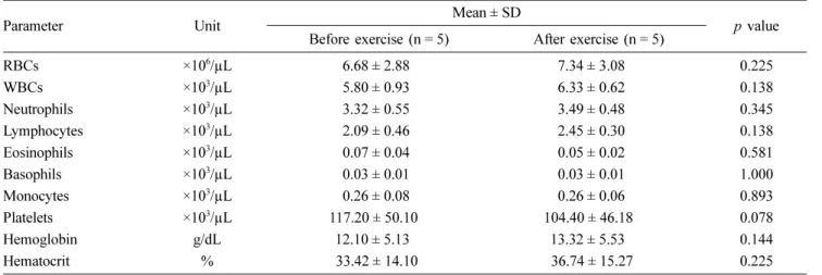
혈액 및 혈청학적 검사
운동 전후 시행한 혈액학적 검사 결과는 Table 2에 제시되 었다. 운동 전과 비교하였을 때 적혈구, 백혈구, 림프구, 호중 구, 호산구, 호염구, 단핵구, 혈소판, 헤모글로빈(hemoglobin), 헤마토크릿(hemtocrit) 수치는 운동 후에 의미 있는 변화가 관찰되지 않았다.
운동 전후 시행한 혈청학적 검사 결과(Table 3), 운동 전 과 비교하였을 때 마그네슘, 나트륨/염화이온의 혈중 농도는 운동 후에 의미 있는 변화가 관찰되지 않았다. 그러나 철의 혈중 농도는 약 1.19배 증가하였다(p = 0.042). 그리고 무기 인의 혈중 농도도 운동 후 약 1.18배 증가하였다(p = 0.042).
혈장 내 코르티솔 농도 변화
운동 전후 스트레스 정도를 측정하기 위해서 코르티솔의
혈장 내 농도를 측정하였다(Fig. 1). Figure 1과 같이 혈장 내 코르티솔의 농도는 말의 운동 전보다 운동 후 약 1.98배 증가하였다(p = 0.043).
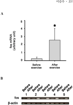
Fos mRNA 발현량 변화
Real-time PCR 결과, 운동 전 fos의 발현량은 housekeeping gene인 β-actin에 대해 약 0.25배를 나타내었다(Fig. 2A). 반 면 운동 후 fos의 발현량은 2.56배를 나타내었다. 이처럼 fos의 상대적 발현량은 운동 전보다 운동 후 약 10.22배 증 가하였다(p = 0.043).
또한 RT-PCR 결과, 모든 개체에서 β-actin의 발현량이 운 Table 2. Hematologic analyses of five horses before and after exercise
Parameter Unit Mean ± SD
p value Before exercise (n = 5) After exercise (n = 5)
RBCs ×106/µL 6.68 ± 2.88 7.34 ± 3.08 0.225
WBCs ×103/µL 5.80 ± 0.93 6.33 ± 0.62 0.138
Neutrophils ×103/µL 3.32 ± 0.55 3.49 ± 0.48 0.345
Lymphocytes ×103/µL 2.09 ± 0.46 2.45 ± 0.30 0.138
Eosinophils ×103/µL 0.07 ± 0.04 0.05 ± 0.02 0.581
Basophils ×103/µL 0.03 ± 0.01 0.03 ± 0.01 1.000
Monocytes ×103/µL 0.26 ± 0.08 0.26 ± 0.06 0.893
Platelets ×103/µL 117.20 ± 50.10 104.40 ± 46.18 0.078
Hemoglobin g/dL 12.10 ± 5.13 13.32 ± 5.53 0.144
Hematocrit % 33.42 ± 14.10 36.74 ± 15.27 0.225
RBC, red blood cell; WBC, white blood cell.
Table 3. Serologic analyses of five horses before and after exercise
Parameter Unit Mean ± SD
p value Before exercise (n = 5) After exercise (n = 5)
Magnesium mg/dL 1.28 ± 0.08 1.18 ± 0.08 0.059
Inorganic Phosphorus mg/dL 2.34 ± 0.19 2.76 ± 0.25* 0.042
Fe µg/dL 30.80 ± 3.56 36.60 ± 5.59* 0.042
Na+ mmol/L 121.20 ± 8.44 128.80 ± 9.65 0.345
Cl− mmol/L 86.80 ± 3.42 90.40 ± 7.16 0.225
*vs. before exercise at p < 0.05.
Fig. 1. Cortisol concentrations in plasma of five horses before and after exercise (*p < 0.05, Wilcoxon signed ranks test).
동 전후 유사한 가운데 fos의 발현량은 운동 후 의미 있는 수준의 증가세를 보였음을 agarose gel 상에서 확인할 수 있 었다(Fig. 2B).
Fos 발현량과 코르티솔 농도 변화의 상관관계 분석 운동 전후 코르티솔 농도와 fos 발현량 변화 수준의 의미 있는 상관관계가 확인되었다(Fig. 3; Spearman’s rank correlation coefficient [rs] = 0.927, p < 0.001).
고 찰
기존 보고에 따르면 운동 후 말에서 혈중 적혈구, 백혈구,
호중구, 단핵구, 헤모글로빈, 헤마토크릿의 수치는 상승했지 만 림프구, 호산구, 혈소판의 수치는 감소한다 [3, 13, 14, 27, 29]. 그러나, 본 연구에서 해당 수치는 정상범위에 속한 가운데, 운동에 의한 의미 있는 변화가 관찰되지 않았다. 이 는 말의 운동 방법, 운동 강도와 시간, 품종 및 개체 간의 차이 등에 의한 것으로 판단된다.
운동에 의한 스트레스는 혈중 전해질 농도에도 영향을 줄 수 있다. 기존 연구에서 말의 운동에 의한 혈중 나트륨, 무 기인산의 농도는 증가하였고, 철의 농도는 감소하는 것으로 보고된 바 있다 [3, 19]. 본 연구에서의 결과는 기존 연구와 다소 상반되었는데, 운동 때문에 증가한 혈중 철의 농도는 혈액순환의 스트레스(circulatory stress)에 의한 일부 적혈구 파괴에 의한 것일 수 있다 [5, 13, 14, 26]. 운동에 의한 무기인산의 농도 상승은 근육수축 과정에서 adenosine triphosphate가 가수 분해되면서 혈액으로 무기인산이 방출됨 에 따라 혈중 농도가 증가한 것으로 판단된다 [28].
운동 스트레스의 정도를 측정하기 위해서 여러 생체지표 가 사용되고 있으나 가장 흔히 사용되고 있는 생체지표는 코 르티솔로 사람, 마우스, 말 등에서 보고된 바 있다. 사람의 경우 운동 1시간 동안 혈장 내 코르티솔의 농도가 2배 증가 한다 [33]. 말의 경우 점프, 속보, 달리기, 지구력 훈련 등을 새들브레드(saddlebred), 스탠더드브레드(standardbred), 서러 브레드(thoroughbred) 종에서 실시한 결과 혈장 내 코르티솔 의 농도가 1.5–2.5배 증가하는 것이 관찰되었다 [16]. 또한 말의 다른 연구에서도 운동 시간이 증가함에 따라 혈장 내 코르티솔 농도는 증가하였으며, 운동 30분 후 1.8배 증가하 는 것으로 보고되었다 [9]. 본 연구에서도 이전 연구와 유사 하게 운동 후 혈장 내 코르티솔의 농도는 정상범위(2.9–6.6 µg/dL) 내에서 약 2배 증가하였다. 이처럼 운동 스트레스에 의한 혈중 코르티솔의 증가가 확인되었지만, 2배 정도의 변 화는 큰 변동 폭이 아니어서 생물학적 지표로서의 가치가 높 다고 보기 어렵다 [1]. 또 생체주기, 사료, 섭취, 환경, 온도, 습도, 관리, 생리학적 상태 등 스트레스 이외의 다른 여러 요인에 의해서 혈중 코르티솔 농도가 변화할 수 있으므로 특 이성이라는 면에서도 문제가 지적될 수 있다 [10]. 그러므로 운동에 의한 스트레스를 측정하는 데 있어서 민감도와 특이 성이 높으며 코르티솔과 상호보완적으로 이용 가능한 새로 운 생체지표가 필요하다.
다양한 세포와 동물 실험에서 허혈, 저산소증, 고체온증과 같은 대사성 스트레스 상황에서 fos mRNA 및 단백질의 발 현이 증가한다 [4, 6, 20, 32]. 또 말의 운동 후 근육조직에 서 fos의 발현이 증가한다 [21, 24]. 본 연구에서 말의 운동 후 혈중 fos의 발현량이 운동 후 약 10배 증가하여 코르티 솔보다 예민하게 변화됨을 알 수 있었다. 혈중 fos mRNA 는 주로 혈구세포에 존재할 것으로 추정된다.
코르티솔에 비하여 약 5배 높은 민감도는 fos 발현이 운동 스트레스에 대한 생물학적 지표로서의 가치가 상대적으로 높 음을 시사하고 있다. 이러한 결과를 토대로 혈중 fos의 발현 변화를 말의 건강관리 시스템에 활용할 수 있을 것으로 생 Fig. 2. Fos mRNA expression by real-time PCR (A) and RT-
PCR (B) in blood samples of five horses before and after exercise (*p < 0.05, Wilcoxon signed ranks test). β-actin was used as an internal control.
Fig. 3. A correlation of fos mRNA expression in blood and plasma cortisol concentrations in five horses before and after exercise. rs, Spearman’s rank correlation coefficient.
각한다. 추가로 fos 단백질에 대한 항체를 사용하여 혈액 내 fos 단백질을 이용한 스트레스 측정기법도 미래 연구에서 다 루어질 가치가 있을 것으로 본다.
감사의 글
본 연구는 농림축산식품부 농림수산식품기술기획평가원 생 명산업기술개발사업(과제번호: 1111594)의 지원에 의하여 수 행되었습니다.
References
1. Cacioppo JT, Berntson GG. Integrative neuroscience for the behavioral sciences: implications for inductive inference. In:
Handbook of Neuroscience for the Behavioral Science. pp. 3- 11. John Wiley & sons, New Jersey, 2009.
2. Capomaccio S, Vitulo N, Verini-Supplizi A, Barcaccia G, Albiero A, D’Angelo M, Campagna D, Valle G, Felicetti M, Silvestrelli M, Cappelli K. RNA sequencing of the exercise transcriptome in equine athletes. PLoS One 2013, 8, e83504.
3. Carlson GP. Hematology and body fluids in the equine athlete: a review. In: Gillespie JR, Robinson NE (eds.). Equine Exercise Physiology 2. pp. 393-425. ICEEP publications, Davis, 1987.
4. Cho S, Park EM, Kim Y, Liu N, Gal J, Volpe BT, Joh TH.
Early c-fos induction after cerebral ischemia: a possible neuroprotective role. J Cereb Blood Flow Metab 2001, 21, 550-556.
5. Couëtil LL, Sojka JE, Nachreiner RF. Primary hypo- parathyroidism in a horse. J Vet Intern Med 1998, 12, 45-49.
6. Cummings M. Increased c-fos expression associated with hyperthermia-induced apoptosis of a Burkitt lymphoma cell line. Int J Radiat Biol 1995, 68, 687-692.
7. Ferlazzo A, Medica P, Cravana C, Fazio E. Endocrine changes after experimental showjumping. Comp Exerc Phys 2009, 6, 59-66.
8. Foreman JH, Ferlazzo A. Physiological responses to stress in the horse. Pferdeheilkunde 1996, 12, 401-404.
9. Freestone JF, Wolfsheimer KJ, Kamerling SG, Church G, Hamra J, Bagwell C. Exercise induced hormonal and metabolic changes in Thoroughbred horses: effects of conditioning and acepromazine. Equine Vet J 1991, 23, 219- 223.
10. González-de-la-Vara MdelR, Valdez RA, Lemus-Ramirez V, Vázquez-Chagoyán JC, Villa-Godoy A, Romano MC.
Effects of adrenocorticotropic hormone challenge and age on hair cortisol concentrations in dairy cattle. Can J Vet Res 2011, 75, 216-121.
11. Gordon ME, McKeever KH, Betros CL, Manso Filho HC.
Exercise-induced alterations in plasma concentrations of ghrelin, adiponectin, leptin, glucose, insulin, and cortisol in horses. Vet J 2007, 173, 532-540.
12. Hoffman-Goetz L, Thorne RJ, Houston ME. Splenic immune responses following treadmill exercise in mice. Can J Physiol Pharmacol 1988, 66, 1415-1419.
13. Johansson AM, Skidell J, Lilliehöök I, Tvedten HW.
Chronic granulocytic leukemia in a horse. J Vet Intern Med
2007, 21, 1126-1129.
14. Larsson J, Pilborg PH, Johansen M, Christophersen MT, Holte A, Roepstorff L, Olsen LH, Harrison AP. Physiological parameters of endurance horses pre- compared to post-race, correlated with performance: a two race study from scandinavia. ISRN Vet Sci 2013, 2013, 684353.
15. Lee WB, Kwon SH, Kim HM. Effect of hypoxia-ischemia on c-fos expression in the neonatal rat brain. Korean J Pediatr 2000, 43, 386-394.
16. Linden A, Art T, Amory H, Desmecht D, Lekeux P. Effect of 5 different types of exercise, transportation and ACTH administration on plasma cortisol concentration in sport horses. In: Persson SGB, Lindholm A, Jeffcott LB (eds.).
Equine Exercise Physiology 3. pp. 391-396, ICEEP publications, Davis, 1991.
17. Malinowski K, Shock EJ, Rochelle P, Kearns CF, Guirnalda PD, McKeever KH. Plasma beta-endorphin, cortisol and immune responses to acute exercise are altered by age and exercise training in horses. Equine Vet J Suppl 2006, 36, 267-273.
18. Marc M, Parvizi N, Ellendorff F, Kallweit E, Elsaesser F.
Plasma cortisol and ACTH concentrations in the warmblood horse in response to a standardized treadmill exercise test as physiological markers for evaluation of training status. J Anim Sci 2000, 78, 1936-1946.
19. Marlin DJ, Fenn K, Smith N, Deaton CD, Roberts CA, Harris PA, Dunster C, Kelly FJ. Changes in circulatory antioxidant status in horses during prolonged exercise. J Nutr 2002, 132 (Suppl 2), 1622S-1627S.
20. McGivney BA, Eivers SS, MacHugh DE, MacLeod JN, O’Gorman GM, Park SDE, Katz LM, Hill EW.
Transcriptional adaptations following exercise in thoroughbred horse skeletal muscle highlights molecular mechanisms that lead to muscle hypertrophy. BMC Genomics 2009, 10, 638.
21. McGivney BA, McGettigan PA, Browne JA, Evans AC, Fonseca RG, Loftus BJ, Lohan A, MacHugh DE, Murphy BA, Katz LM, Hill EW. Characterization of the equine skeletal muscle transcriptome identifies novel functional responses to exercise training. BMC Genomics 2010, 11, 398.
22. McGorum BC, Anderson RA. Biomarkers of exposure to cyanogens in horses with grass sickness. Vet Rec 2002, 151, 442-445.
23. Müller JM, Krauss B, Kaltschmidt C, Baeuerle PA, Rupec RA. Hypoxia induces c-fos transcription via a mitogen- activated protein kinase-dependent pathway. J Biol Chem 1997, 272, 23435-23439.
24. Park KD, Park J, Ko J, Kim BC, Kim HS, Ahn K, Do KT, Choi H, Kim HM, Song S, Lee S, Jho S, Kong HS, Yang YM, Jhun BH, Kim C, Kim TH, Hwang S, Bhak J, Lee HK, Cho BW. Whole transcriptome analyses of six thoroughbred horses before and after exercise using RNA- Seq. BMC Genomics 2012, 13, 473.
25. Park W, Kim J, Kim HJ, Choi J, Park JW, Cho HW, Kim BW, Park MH, Shin TS, Cho SK, Park JK, Kim H, Hwang JY, Lee CK, Lee HK, Cho S, Cho BW.
Investigation of de novo unique differentially expressed genes related to evolution in exercise response during domestication in thoroughbred race horses. PLoS One 2014, 9, e91418.
26. Peeling P, Dawson B, Goodman C, Landers G, Wiegerinck ET, Swinkels DW, Trinder D. Effects of exercise on
hepcidin response and iron metabolism during recovery. Int J Sport Nutr Exerc Metab 2009, 19, 583-597.
27. Piccione G, Giannetto G, Fazio F, Di Mauro S, Caola G.
Haematological response to different workload in jumper horses. Bulgarian J Vet Med 2007, 10, 21-28.
28. Poole DC, Erickson HH. Exercise physiology of terrestrial animals. In: Reece WO, Erickson HH, Goff JP, Uemura EE (eds.). Dukes’ Physiology of Domestic Animals. 13th ed. pp.
443-466, Wiley-Blackwell, New Jersey, 2015.
29. Segura D, Monreal L, Espada Y, Pastor J, Mayós I, Homedes J. Assessment of a platelet function analyser in horses: reference range and influence of a platelet aggregation inhibitor. Vet J 2005, 170, 108-112.
30. Sellon DC, Long MT. Epidemiology of equine infectious
disease. In: Equine Infectious Diseases. 2nd ed. pp. 515-529, Saunders Elsevier, St. Louis, 2014.
31. Songer JG, Trinh HT, Dial SM, Brazier JS, Glock RD.
Equine colitis X associated with infection by Clostridium difficile NAP1/027. J Vet Diagn Invest 2009, 21, 377-380.
32. Veening JG, Bouwknecht JA, Joosten HJ, Dederen PJ, Zethof TJ, Groenink L, van der Gugten J, Olivier B.
Stress-induced hyperthermia in the mouse: c-fos expression, corticosterone and temperature changes. Prog Neuropsycho- pharmacol Biol Psychiatry 2004, 28, 699-707.
33. Wellhoener P, Born J, Fehm HL, Dodt C. Elevated resting and exercise-induced cortisol levels after mineralocorticoid receptor blockade with canrenoate in healthy humans. J Clin Endocrinol Metab 2004, 89, 5048-5052.