β-Ionone Enhances TRAIL-Induced Apoptosis in
Hepatocellular Carcinoma Cells through Sp1-Dependent Upregulation of DR5 and Downregulation of NF-κB Activity
Mun-Ock Kim1, Dong-Oh Moon1, Chang-Hee Kang1, Taeg Kyu Kwon2, Yung Hyun Choi3, and Gi-Young Kim1
Abstract
β-Ionone (ION), an end-ring analogue of β-carotenoid, has been known to inhibit tumor cell growth and induce apoptosis in various types of cancer cells. Nevertheless, its apoptosis-related molecular me- chanisms remain unclear. Here, we first investigated the molecular mechanisms by which ION sensitizes cancer cells to the therapeutic potential of tumor necrosis factor–related apoptosis-inducing ligand (TRAIL). Notably, treatment with subtoxic concentrations of ION and TRAIL effectively inhibited cell viability in the hepatocellular carcinoma cell line Hep3B and other cancer cell lines such as colon carcino- ma cell line HCT116 and leukemia cell line U937. Combined treatment with ION and TRAIL was also more effective in inducing DR5 expression, caspase activities, and apoptosis than treatment with either agent alone. ION-mediated sensitization to TRAIL was efficiently reduced by treatment with a chimeric blocking antibody or small interfering RNA specific for DR5. Electrophoretic mobility shift assay and a chro- matin immunoprecipitation assay confirmed that ION treatment upregulates the binding of transcription factor Sp1 to its putative site within the DR5 promoter region, suggesting that Sp1 is an ION-responsive transcription factor. In addition, ION significantly increased hepatocellular carcinoma cell sensitivity to TRAIL by abrogating TRAIL-induced NF-κB activation and decreasing the expression of antiapoptotic proteins such as XIAP and IAP-1/2. Taken together, these data suggest that ION is a useful agent for TRAIL-based cancer treatments.Mol Cancer Ther; 9(4); 833–43. ©2010 AACR.
Introduction
Tumor necrosis factor (TNF)–related apoptosis-induc- ing ligand (TRAIL), a member of the tumor necrosis fac- tor superfamily, is considered a promising anticancer agent because it can induce apoptosis in various cell types. Moreover, the cytotoxic activity of TRAIL is selec- tive for human tumor cells and leaves normal cells un- harmed (1, 2). Thus, TRAIL is a promising anticancer cytokine (3). TRAIL induces apoptosis in various types of tumor cells through the death receptor pathway by using a mechanism that resembles one involving TNF- α (2). TRAIL reacts with the death receptor DR4 or DR5, leading to the aggregation of the receptors, recruitment of the adaptor molecule Fas-associated death domain
protein, and activation of initiator caspase-8 (4). Activat- ed caspase-8 is released into the cytoplasm and initiates a protease cascade that activates effector caspases such as caspase-3 and caspase-7 (4). Therefore, agents that can sensitize cells to TRAIL-mediated apoptosis are good candidates for chemotherapy against various types of cancer.
β-Ionone (ION), an end-ring analogue of β-carotenoid, has been known for in vivo and in vitro protection against various types of cancer cells (5, 6). It is also well estab- lished that ION suppresses the proliferation of melanoma (7, 8), leukemia (9), and meningioma cells (10). Despite many previous studies, the exact mechanisms of ION re- main unclear. Therefore, we investigated the usefulness of ION as a potent sensitizer of TRAIL-resistant human hepatocellular carcinoma cells for TRAIL-induced apo- ptosis. This report provides the first evidence that sub- toxic doses of ION increase the expression of DR5 by upregulating the binding of Sp1 to its DR5 promoter, leading to a rapid induction of TRAIL-mediated signal- ing and cell death. Additionally, TRAIL-induced NF-κB activation is a major factor that prevents TRAIL-induced apoptosis; this is achieved through the expression of anti- apoptotic proteins (11). We also found that ION strongly abrogates TRAIL-induced NF-κB activation and potenti- ates TRAIL-mediated apoptosis in TRAIL-resistant hepa- tocellular carcinoma cells.
Authors' Affiliations:1Laboratory of Immunobiology, Department of Marine Life Sciences, Jeju National University, Jeju, Republic of Korea;
2Department of Immunology, School of Medicine, Keimyung University, Taegu, Republic of Korea; and3Department of Biochemistry, Dongeui University College of Oriental Medicine, Busan, Republic of Korea Note: G.-Y. Kim and Y.H. Choi contributed equally as experimental designers to this work.
Corresponding Author: Gi-Young Kim, Department of Marine Life Sciences, Jeju National University, Jeju 690-756, Republic of Korea.
Phone: 82-64-754-3427; Fax: 82-64-756-3493. E-mail: immunkim@
jejunu.ac.kr
doi: 10.1158/1535-7163.MCT-09-0610
©2010 American Association for Cancer Research.
Therapeutics
Here, we show that ION increases the sensitivity of can- cer cells to TRAIL by enhancing DR5 expression, and not DR4 expression, and by activating Sp1 in the DR5 promoter and inhibiting TRAIL-mediated NF-κB activation.
Materials and Methods Reagents and antibodies
Antibodies against Sp1, nucleolin, p50, p65, caspase-3, caspase-8, caspase-9, poly ADP ribose polymerase (PARP), DR4, DR5, IAP-1, IAP-2, XIAP, Bcl-2, and Bid were purchased from Santa Cruz Biotechnology. The an- tibody againstβ-actin was purchased from Sigma. Perox- idase-labeled donkey anti-rabbit and sheep anti-mouse immunoglobulin, recombinant human TRAIL/Apo2 li- gand (the nontagged 19-kDa protein, amino acids 114–
281), anti-Fas antibody, and TNF-α were purchased from KOMA Biotechnology. The blocking antibody against DR5 was purchased from R&D Systems. 3,3-PDTC, MG132, and PS341 were purchased from Sigma. ION was also purchased from Sigma and dissolved in DMSO (vehicle). z-VAD-fmk was purchased from Calbiochem.
Cell culture and viability assay
Human hepatocellular carcinoma cell lines Hep3B and HepG2, human colon cancer cell line HCT116, and hu- man leukemia cell line U937 were obtained from the American Type Culture Collection. Cells were cultured at 37°C in a 5% CO2-humidified incubator and main- tained in RPMI 1640 culture medium containing 10%
heat-inactivated fetal bovine serum (Life Technologies Bethesda Research Laboratories and 1% penicillin-strep- tomycin (Sigma). The cells were seeded (4 × 10⁴cells/mL), grown for 24 h, and then incubated for up to 24 h with ION and/or TRAIL. MTT assays were done to assess cell viability.
Flow cytometric analysis
Cells were fixed in 1 U/mL of RNase A (DNase free) and 10μg/mL of propidium iodide (Sigma) overnight in the dark at room temperature. To assess whether apopto- sis had occurred, the cells were incubated with Annexin V (R&D Systems). A FACSCalibur flow cytometer (Bec- ton Dickinson) was used to determine the number of ap- optotic cells, i.e., cells with sub-G1 DNA that were Annexin V positive.
Determination of mitochondrial membrane potential
The mitochondrial membrane potential was monitored by measuring the uptake of dihexyloxacarbocyanine io- dide (DiOC6; Molecular Probes). Briefly, the cells were har- vested, loaded with 50 nmol/L DiOC6at 37°C for 30 min in the dark, and then analyzed using a flow cytometer.
In vitro caspase activity assay
A caspase activation kit was used according to the manufacturer's instructions (R&D Systems) to measure the activity of caspase-like proteases.
DNA fragmentation
Cells were treated under various conditions and then lysed on ice in a buffer containing 10 mmol/L Tris-HCl (pH 7.4), 150 mmol/L NaCl, 5 mmol/L EDTA, and 0.5% Triton X-100 for 30 min. Lysates were vortexed and cleared by centrifugation at 10,000 g for 20 min. Frag- mented DNA in the supernatant was extracted with an equal volume of neutral phenol/chloroform/isoamylal- cohol (25:24:1, v/v/v) and was analyzed electrophoreti- cally on a 1.5% agarose gel containing ethidium bromide.
Western blot
Total cell extracts were prepared using the PRO-PREP protein extraction solution (iNtRON Biotechnology).
Cytoplasmic and nuclear extracts were prepared using NE-PER nuclear and cytosolic extraction reagents (Pierce), respectively. Total cell extracts were separated by PAGE and standard procedures were then used to transfer them to nitrocellulose membranes. The mem- branes were developed using an enhanced chemilumi- nescence (Amersham) reagent (Amersham).
RNA extraction and reverse transcription-PCR Total RNA was extracted using the Trizol reagent (Invi- trogen) and reverse transcription-PCR was conducted.
The sense primer 5-GTC TGC TCT GAT CAC CCA AC-3 and the antisense primer 5-CTG CAA CTG TGA CTC CTA TG-3 were used to amplify human DR5 mRNA. For glyc- eraldehyde-3-phosphate dehydrogenase, the sense primer 5-CGT CTT CAC CAT GGA GA-3 and the antisense prim- er 5-CGG CCA TCA CGC CCA CAG TTT-3 were used.
Gel mobile shift assays (electrophoretic mobility shift assay)
DNA-protein binding assays were carried out with the nuclear extract. Synthetic complementary NF-κB– and Sp1-binding oligonucleotides (5-AGT TGA GGG GAC TTT CCC AGG C-3 and 5-ATT CGA TCG GGG CGG GGC GAG C-3, respectively) were 3-biotinylated using a bi- otin 3-end DNA labeling kit (Pierce) according to the manufacturer's instructions. Assays were done using a Lightshift electrophoretic mobility shift assay (EMSA) Optimization kit (Pierce) according to the manufacturer's protocol.
Chromatin immunoprecipitation assay
A chromatin immunoprecipitation assay was done with a EZ-Chip assay kit used according to the manufacturer's in- structions (Upstate Biotechnology). The primers used to am- plify the Sp1-binding site in the DR5 promoter region were 5-GCC AGG GCG AAG GTT A-3 (sense) and 5-GGG CAT CGT CGG TGT AT-3 (antisense; a 276-bp DNA product).
Plasmids, transfections, and luciferase gene assays The pDR5/SacI plasmid [containing a DR5 promoter se- quence (-2500/+3)], pDR5/-605 plasmid [containing a DR5 promoter sequence (-605/+3)], and point-mutated plasmids containing sequences with the Sp1-binding sites Kim et al.
Published OnlineFirst March 30, 2010; DOI: 10.1158/1535-7163.MCT-09-0610
in the DR5 promoter region were gifted by Dr. Sakai T.
(Kyoto Prefectural University, Kyoto, Japan). Plasmid transfection was done using the FuGENE 6 Transfection Reagent (Roche Molecular Biochemicals) according to the manufacturer's instructions. Cells were plated onto 6- well plates at a density of 4 × 10⁴cells per well and grown overnight. The cells were then cotransfected with 1μg of various plasmid constructs and 1μg of the pCMV-β-galac- tosidase plasmid for 24 h. After transfection, the cells were cultured in 10% FCS medium with a vehicle or with drugs for 24 h. Luciferase andβ-galactosidase activities were as- sayed according to the manufacturer's protocol (Promega).
Luciferase activity was normalized withβ-galactosidase activity in the cell lysate and was expressed as an average of the results of three independent experiments.
Small interfering RNA
The 25-nucleotide small interfering RNA (siRNA) duplexes used in this study were purchased from
Santa Cruz Biotechnology. Cells were transfected with the siRNA oligonucleotides by using the FuGENE 6 Transfection Reagent according to the manufacturer's recommendations.
Statistical analysis
All data from the MTT assays, caspase activity, lucif- erase activity, and fluorescence-activated cell sorting analysis were derived from at least three independent experiments. The images were visualized with Chemi- Smart 2000 (Vilber Lourmat). Images were captured using Chemi-Capt (Vilber Lourmat) and transported in- to Photoshop. All bands were quantified by the Scion Imaging software (12). Statistical analyses were con- ducted using the SigmaPlot software, and values were presented as mean ± SD. The significance of the differ- ences between the groups was determined using the unpaired Student's t test. A value of P < 0.05 was considered statistically significant.
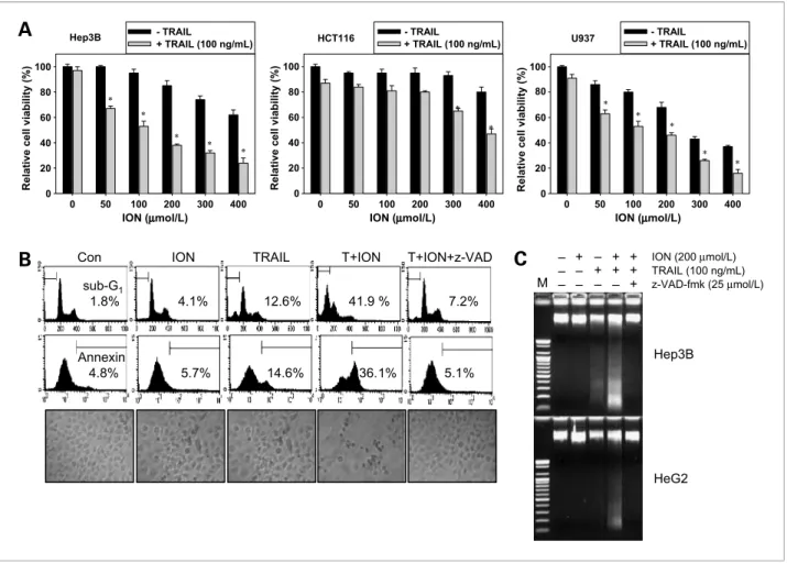
Figure 1.Effect of ION and/or TRAIL on cell viability. A, hepatocellular carcinoma Hep3B cells, colon cancer HCT116 cells, and leukemia U937 cells were cotreated with the indicated concentrations of ION in the presence of 100 ng/mL TRAIL for 24 h. Cell viability was measured by an MTT assay. B, to examine the effect of caspase inhibition, Hep3B cells were pretreated with 25μmol/L z-VAD-fmk for 30 min and then treated with ION/TRAIL for 24 h.
DNA content (top) and Annexin V-FITC–positive cells (middle) were analyzed by flow cytometry. Morphology of cells was examined under light microscopy (×400; bottom). C, a DNA fragmentation assay was done for the Hep3B and HepG2 cells. After the Hep3B and HepG2 cells were treated as indicated for 24 h, fragmented DNAs were extracted from the treated cells and analyzed on a 1.5% agarose gel. Columns, mean from three independent experiments;
bars, SD. Statistical significance was determined by the Student's t test (*, P < 0.05 versus vehicle control).
Results
ION sensitizes various types of cancer cells to TRAIL-induced apoptosis
We first investigated the cytotoxic effect of ION alone or in combination with TRAIL in various types of cancer cell lines. Treatment with TRAIL (100 ng/mL) alone re- sulted in the death of only a few Hep3B cells and HCT116 cells; no >10% cells had died after 24 hours
(Fig. 1A). This observation suggests that these cell lines are quite resistant to TRAIL-induced apoptosis.
Treatment with ION alone decreased cell viability in a dose-dependent manner up to 400μmol/L. However, treatment with both ION (increasing concentrations) and TRAIL (100 ng/mL) synergistically reduced cell via- bility in a dose-dependent manner. This effect was slight in HCT-116 cells and more drastic in U937 cells than in Hep3B cells. To further investigate whether the combined
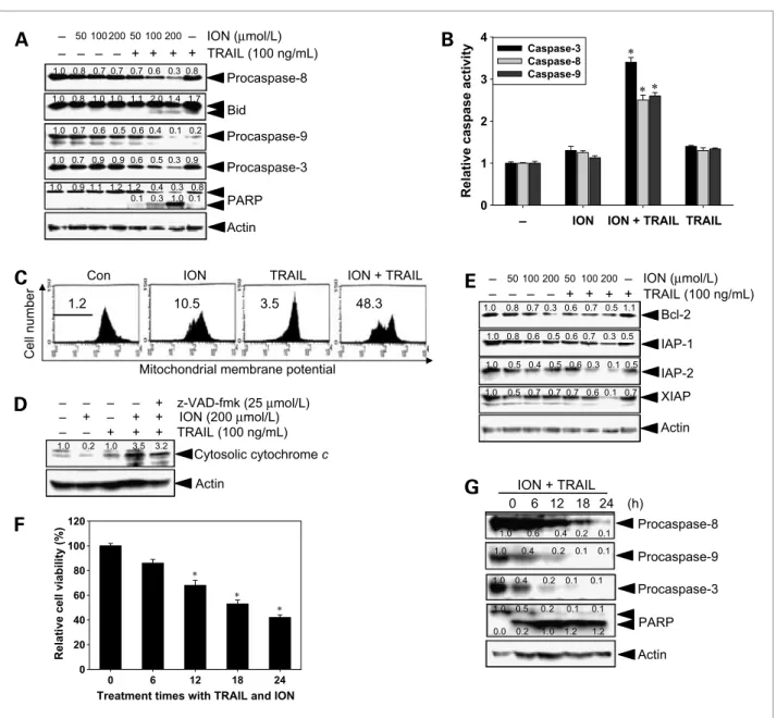
Figure 2.Effect of ION and/or TRAIL on the expression of caspases and various intracellular regulators of apoptosis. Hep3B cells were treated with 100 ng/mL TRAIL alone, ION alone, or a combination of both for 24 h. Cell extracts were prepared for Western blot analysis by using specific antibodies.
A, caspases-8, Bid, caspase-9, caspase-3, and PARP; E, Bcl-2, IAP-1, IAP-2, and XIAP. B, caspase-3, caspase-8, and caspase-9 activities were determined by doing the analysis according to the manufacturer's protocol, after cells were treated with 100 ng/mL TRAIL alone, 100μmol/L ION alone, or a combination of both for 24 h. C, mitochondria potential was measured by flow cytometry using DiOC6dye. D, the translocation of cytochrome c was analyzed with cytosolic extracts by Western blot analysis. F and G, Hep3B cells were cotreated with the indicated concentrations of ION (100μmol/L) in the presence of TRAIL (100 ng/mL) for the indicated periods. Cell viability was measured by an MTT assay (F) and cell lysates were subjected to Western blot for detecting the procaspase-8, procaspase-9, procaspase-3, and the cleavage of PARP (G). Columns, mean from three independent experiments are expressed; bars, SD. Statistical significance was determined by the Student's t test (*, P < 0.05 versus vehicle control).
Kim et al.
Published OnlineFirst March 30, 2010; DOI: 10.1158/1535-7163.MCT-09-0610
treatment triggered cell death through caspases, we used a caspase-8 inhibitor, z-VAD-fmk. Flow cytometric anal- yses revealed cells exhibiting apoptotic shrinkage, con- firming that the combined treatment had a synergistic effect on apoptosis in the sub-G1population and Annex- in V–positive cells (Fig. 1B). However, z-VAD-fmk effec- tively blocked the apoptosis induced by the combined treatment, indicating that ION sensitizes Hep3B cells to TRAIL-induced apoptosis in a caspase-dependent man- ner. Additionally, the DNA fragmentation assay revealed typical DNA fragmentation in Hep3B cells treated with a combination of ION and TRAIL; DNA fragmentation was rarely seen in cells treated with ION or TRAIL alone (Fig. 1C). Pretreatment with z-VAD-fmk significantly blocked DNA fragmentation induced by the combined
treatment of ION and TRAIL. Furthermore, these results were also observed in another hepatocellular carcinoma cell line, HepG2, which was treated identically. The re- sults indicate that ION sensitizes cancer cells to TRAIL's actions through a caspase-dependent mechanism.
ION enhances TRAIL-mediated signaling by engaging the mitochondrial death pathway
To assess whether ION induces apoptosis by triggering the mitochondrial apoptotic pathway, we investigated the changes that are responsible for resistance to TRAIL-induced apoptosis. The changes we studied in- cluded the activation of caspases, mitochondrial perme- ability, cytochrome c release, expression of the Bcl-2 family proteins that contribute to increased mitochondrial
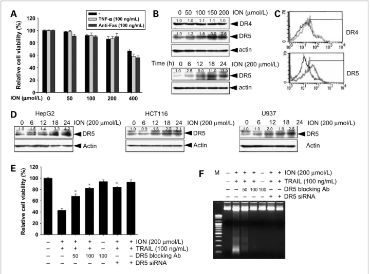
Figure 3.Effect of ION on DR4 and DR5 expression. A, HepG2 cells were treated with the indicated concentrations of ION with or without 100 ng/mL TNF-α or 100 ng/mL anti-Fas antibody for 24 h. Cellular viabilities were analyzed by MTT assays. B, Hep3B cells were treated with increasing concentrations of ION for 24 h (top) and 200μmol/L ION for the indicated times, and Western blot analysis was done for DR5 and DR4. β-Actin was used as the loading control. C, surface-expression levels of DR5 and DR4 were analyzed using flow cytometry. X axis, fluorescence intensity; Y axis, relative number of cells. Black lines, nontreated cells; gray lines, treated cells with ION. D, human hepatocellular carcinoma HepG2, leukemia U937, and colon cancer HCT116 cells were treated with 200μmol/L ION for up to 24 h, and cell extracts were prepared for Western blot analysis of DR5. E and F, Hep3B cells were pretreated with or without 200μmol/L ION or 100 ng/mL TRAIL for 24 h in the presence of indicated concentrations of the DR5-specific blocking chimera antibody (Ab) or siRNA duplexes against DR5 mRNA. Cell viability was assessed by an MTT assay (E) and a DNA fragmentation assay (F) was done to confirm cell death. Columns, mean from three independent experiments; bars, SD. Statistical significance was determined by the Student's t test (*, P < 0.05 versus vehicle control).
permeability, and expression of the IAP family of pro- teins. Cells were treated with increasing concentrations of ION and 100 ng/mL TRAIL for 24 hours. The activa- tion status of two initiation caspases, including caspase- 8 and caspase-9, and effector caspase-3 was measured by Western blot analyses. Treatment with ION or TRAIL alone slightly decreased the expression of procaspases (Fig. 2A). However, combined treatment significantly downregulated procaspases in a dose-dependent manner.
Correspondingly, combined treatment caused the signifi- cant truncation of Bid and cleavage of PARP. To further confirm caspase activation, we examined caspase activities with a caspase activation assay. The result revealed that increase in caspase activities correlated with the ION concentration in cells cotreated with TRAIL
(Fig. 2B). Combined treatment also decreased the percent- age of cells labeled with DiOC6, although staining was slightly less in the cells treated with ION alone (Fig. 2C). After the combined treatment, the amount of cytochrome c released from the intermembrane space of the mitochondria into the cytosol increased (Fig. 2D).
However, pretreatment with z-VAD-fmk blocked this release. Because the antiapoptotic molecules Bcl-2, Bcl- xL, and IAPs can protect some tumor cell lines from TRAIL-induced apoptosis (13, 14), we investigated the expression levels of Bcl-2 and proteins belonging to the IAP family. The combined treatment induced the down- regulation of the antiapoptotic proteins Bcl-2, IAP-1, IAP- 2, and XIAP (Fig. 2E). However, the levels of antiapopto- tic Bcl-xL did not change significantly. We also examined
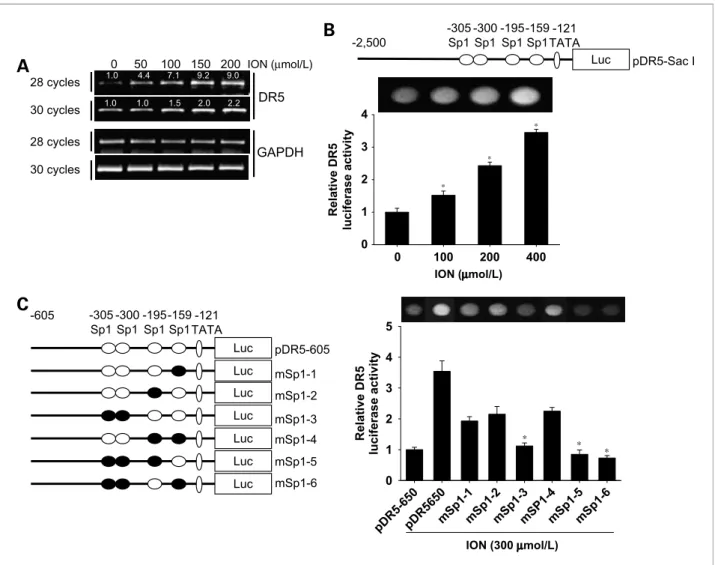
Figure 4.Effect of ION on DR5 transcriptional activity. A, Hep3B cells were treated with up to 200μmol/L ION for 24 h. Total RNA was isolated, and reverse transcription-PCR analysis for DR5 and glyceraldehyde-3-phosphate dehydrogenase (GAPDH) was done. B, top, schematic structures of the full DR5 promoter constructs used to measure luciferase activity. Bottom, Hep3B cells transfected with the reporter constructs and lysates from cells treated with or without the indicated concentrations of ION were assayed for luciferase activity. C, left, a schematic structure series of the DR5 promoter constructs mutated at the Sp1 consensus site(s). Right, cells transfected with each reporter construct, and lysates from cells treated with or without indicating concentrations of ION were assayed for luciferase activity. Columns, mean from three independent experiments; bars, SD. Statistical significance was determined by the Student's t test (*, P < 0.05 versus vehicle control).
Kim et al.
Published OnlineFirst March 30, 2010; DOI: 10.1158/1535-7163.MCT-09-0610
the time course effects of ION on TRAIL-initiated cell death and caspase cascade. Dramatic cell death was found at 12 hours after treatment with ION and TRAIL (Fig. 2F).
Notably, treatment with ION augmented the TRAIL- induced downregulation of procaspase-8, procaspase-9, and procaspase-3 as well as the cleavage of one of the caspase-3 substrated PARP (Fig. 2G). These results indicate that combined treatment with ION and TRAIL activates the mitochondrial apoptotic pathway.
DR5 upregulation is important for ION/TRAIL- induced apoptosis
Because a previous study showed that members of the TNF superfamily have protein structures similar to those of the proteins involved in death receptor–mediated apo- ptosis signaling pathways (2), we did experiments to examine whether ION could sensitize cancer cells to TNF-α– or anti-Fas antibody–mediated apoptosis. As shown in Fig. 3A, no synergistic cytotoxic effect was
observed in HepG2 cells treated with TNF-α or anti-Fas antibody in the presence of ION. These results suggest that ION selectively executes TRAIL-induced apoptosis but not TNF-α– or anti-Fas antibody–mediated apoptosis.
To elucidate the molecular mechanism underlying the enhancement of TRAIL-induced apoptosis by ION, we examined the expression of TRAIL receptors DR4 and DR5. As shown in Fig. 3B, ION treatment increased the DR5 level in dose- and time-dependent manners but did not affect the levels of DR4. Fluorescence-activated cell sorting analyses also showed that the ION-induced cell surface expression levels of DR5 but not DR4 were signif- icantly increased in Hep3B cells (Fig. 3C). ION treatment also consistently increased DR5 protein levels in other cancer cell lines such as HepG2, HCT116, and U937 (Fig. 3D). These results suggest that DR5 upregulation is a common response of cancer cells to ION treatment.
Therefore, to confirm the functional role of DR5 in cells sensitized to TRAIL-induced apoptosis by ION, we
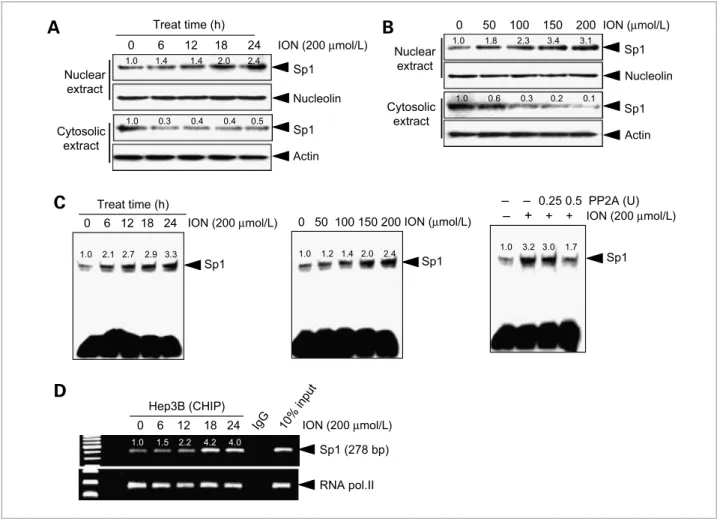
Figure 5.Effect of ION on Sp1's DNA-binding activity with regard to the DR5 promoter. Hep3B cells were incubated with 200μmol/L ION for the indicated time and with increasing concentrations of ION for 24 h. A and B, cytosolic and nucleus protein extracts were prepared, and Western blotting analysis of Sp1 was done. C, Sp1's DNA-binding activity was analyzed using the nucleus extracts (left and middle). Nuclear extracts from cells that were subjected to the combined treatment were incubated with the indicated concentrations of PP2A for 30 min in vitro, followed by an EMSA assay (right).
D, chromatin immunoprecipitation (CHIP) assay was done using antibodies against Sp1 in Hep3B cells. Rabbit IgG was used as the negative control, and 10% input was used as the positive control.
examined the effect of a dominant-negative form of the TRAIL receptor (DR5/Fc chimeric protein) on ION/
TRAIL-induced apoptosis. DR5/Fc chimeric protein sig- nificantly inhibited cell death following the combined
treatment (Fig. 3E). In addition, suppression of DR5 ex- pression by transfection of Hep3B cells with DR5 siRNA also effectively blocked combined treatment–induced ap- optosis (Fig. 3E). To confirm these results, we did a DNA
Figure 6.Effect of ION on TRAIL-induced NF-κB activation. A, Hep3B and HepG2 cells were incubated with 50 ng/mL TRAIL for 3 h with or without pretreatment with 200μmol/L ION. Nuclear extracts were then prepared and assayed for NF-κB by doing an EMSA. B, nuclear extracts from nontreated or ION-treated cells were incubated with unlabeled NF-κB oligoprobe (competitor) and then assayed for NF-κB activation by EMSA. C, cells were treated with ION and with or without 50 ng/mL TRAIL. Then, NF-κB transcriptional activity was measured by a NF-κB–responsive luciferase reporter assay.
D, Hep3B cells were treated with 200μmol/L ION for 1 h, followed by treatment with 50 ng/mL TRAIL for the indicated time intervals. Equal amounts of cytosolic and nucleus proteins (50μg) were used. Western blotting was done to check for p65, p50, p-Akt, and Akt. Nucleolin and β-actin were used as the loading controls. E, Hep3B cells were pretreated with 20μmol/L of PDTC, 20 μmol/L MG-132, or 10 μmol/L PS-341 for 2 h before treatment with 50 ng/mL TRAIL alone or 50 ng/mL TRAIL plus 200μmol/L ION for 24 h. Cellular DNA content of was analyzed by flow cytometry. Columns, mean from three independent experiments; bars, SD. Statistical significance was determined by the Student's t test (*, P < 0.05 versus vehicle control).
Kim et al.
Published OnlineFirst March 30, 2010; DOI: 10.1158/1535-7163.MCT-09-0610
fragmentation assay under identical conditions. ION/
TRAIL-induced synergistic cytotoxicity was abolished by pretreatment with DR5/Fc and by pretreatment with DR5 siRNA (Fig. 3F). These results indicate that ION en- hances TRAIL's actions through interactions between TRAIL and TRAIL receptors.
ION enhances DR5 transcription through Sp1 binding at−300/−305 in the DR5 promoter region
To determine whether ION-induced DR5 upregulation is controlled at the transcriptional level, we analyzed the expression of DR5 mRNA by subjecting ION-treated Hep3B cells to reverse transcription-PCR. As shown in Fig. 4A, ION treatment increased DR5 mRNA expression in a dose-dependent manner. Therefore, we investigated whether ION activates the DR5 promoter by using a lu- ciferase gene expression system. We first examined the effects of ION on the promoter activities of a reporter construct containing the full-length DR5 gene promoter region (Fig. 4B). Cells were transfected with a plasmid containing the wild-type construct (pDR5-Sac I), and lu- ciferase activities were assayed 24 hours after treatment with different concentrations of ION. We found that ION significantly increases DR5 promoter activity in a dose-dependent manner, supporting the idea that ION- induced DR5 upregulation is controlled at the transcrip- tional level. Previously, Yoshida et al. (15) showed that the region of the DR5 promoter spanning nucleotides
−605 to +3 contains typical transcription factor–binding sites, including four Sp1 sites and a TATA-like box site (Fig. 4C). To determine which Sp1 site(s) in the DR5 pro- moter is responsible for ION-induced DR5 upregulation, we analyzed reporter constructs with mutations at differ- ent Sp1-binding sites with luciferase assays (Fig. 4C).
Transfection with mSp1-3 (mutated at the −305 and
−300 Sp1 sites), mSp1-5 (mutated at the −305, −300, and−195 Sp1 sites), and mSp1-6 (mutated at the −305,
−300, and −159 sites; ref. 21) showed significantly de- creased ION-induced DR5 promoter activities compared with transfection with the wild-type construct (pDR5/
−605). Although all cells with constructs containing any mutated Sp1 site decreased DR5 promoter activity, these significant declines confirmed that the two putative Sp1- binding sites present at−305/−300 play a critical role in the ION-induced enhancement of DR5 promoter activity.
ION enhances the direct binding of Sp1 on DR5 promoter
To further investigate the role of ION in the upregula- tion of Sp1 activity, Western blot analysis was done to measure Sp1 protein levels in Hep3B cells treated with ION. At 18 hours after ION treatment, the nuclear Sp1 protein levels had increased, whereas the cytosolic Sp1 le- vels had decreased (Fig. 5A). In addition, Sp1 protein translocation into the nucleus also increased in a dose- dependent manner at 24 hours (Fig. 5B). To examine the effect of ION on the constitutive DNA-binding activity of Sp1, we did an EMSA by using the nuclear extracts of
Hep3B cells treated with ION. As shown in Fig. 5C, ION treatment increased constitutive Sp1-binding activity in time- and dose-dependent manners (left and middle).
Additionally, to investigate whether ION-induced Sp1 DNA–binding activity was due to Sp1 phosphorylation, we incubated the nuclear extract with the serine-threo- nine phosphatase PP2A before the binding reaction. The increased Sp1 DNA–binding activity provoked by the ION treatment was blocked in the lanes treated with PP2A (right). We also investigated the effects of ION on the direct induction of Sp1's binding to the DR5 promoter by doing a chromatin immunoprecipitation assay (Fig. 5D). ION treatment directly induced binding of Sp1 to the DR5 promoter after 18 hours, using primers that target the Sp1 element in the DR5 promoter. RNA po- lymerase II was the positive control. These results suggest that ION increases sensitivity to TRAIL-induced apopto- sis by increasing the activity of Sp1, which in turn leads to increased DR5 promoter activity and DR5 transcription, ultimately increasing surface DR5 protein expression.
ION-induced inhibition of NF-κB activity potentiates TRAIL-mediated apoptosis
Previous studies have shown that TRAIL-induced NF- κB activation in cancer cells contributes to resistance to TRAIL-induced apoptosis (16, 17). To investigate TRAIL-induced NF-κB activation, NF-κB DNA–binding activity was measured in Hep3B and HepG2 cells by EMSA. As shown in Fig. 6A, NF-κB activity was activat- ed in both hepatocellular carcinoma cell lines from 1 hour after the treatment with 50 ng/mL TRAIL; however, treatment with ION suppressed NF-κB activity. Addition of excess unlabeled NF-κB probe (competitor; 100-fold) caused complete disappearance of the NF-κB activity in- duced by TRAIL (Fig. 6B). NF-κB transcriptional activity was also measured with a NF-κB–responsive luciferase reporter assay. In keeping with the EMSA results, data showed an increase in NF-κB luciferase activity after TRAIL stimulation, but treatment with ION abolished this activity in a dose-dependent manner (Fig. 6C). Next, we confirmed the effect of ION on TRAIL-mediated NF-κB activation in Hep3B cells by using Western blot analysis. As shown in Fig. 6D, ION treatment decreased TRAIL-induced translocation of p65 and p50 in the nuclear extract. Consistent with these observations, the decrease in translocated NF-κB protein levels correlated with decreased phosphorylation of AKT.
To further examine the role of NF-κB in ION/TRAIL- induced apoptosis, we investigated the effect of noncyto- toxic concentrations of NF-κB inhibitors, including PDTC, and proteasome inhibitors (MG-132 and PS-341) on cancer cells. Cells pretreated with NF-κB inhibitors were significantly sensitized to TRAIL-induced apoptosis following treatment with ION or TRAIL alone and after combined treatment (Fig. 6E). These results indicate that the attenuation of NF-κB activity by NF-κB inhibitors rendered the Hep3B cells more prone to ION/TRAIL- induced apoptosis. In summary, we confirmed that ION
treatment potentiates TRAIL-induced apoptosis through the inhibition of the NF-κB–mediated survival pathway.
Discussion
TRAIL has attracted public attention for its potential as an anticancer agent based on evidence of its beneficial therapeutic effects for a range of advanced cancers of he- matologic and solid tumor origin. However, there are re- ports that many cancer cells resist TRAIL-mediated apoptosis (1–3). Therefore, a TRAIL-sensitizing agent is believed to be required in chemotherapy for TRAIL- resistant cancers. Recently, several chemical compounds that sensitize cancer cells to TRAIL-induced apoptosis, including histone deacetylase inhibitors (18), cyclin- dependent kinase inhibitors (19), proteasome inhibitors (20), and some natural products (21–23), have been iden- tified. Most of these TRAIL-sensitizing compounds regu- late two death domain–containing receptors and are thought to have potential cancer chemotherapeutic appli- cations. In particular, a previous study also showed that fresh melanoma isolates are relatively resistant to TRAIL- induced apoptosis because they have low levels of TRAIL-death receptors (24). Another recent study em- phasized that DR5 may play a significantly more promi- nent role than DR4 in TRAIL-induced apoptosis (25). Our study is the first investigation into the ability of ION to potentiate TRAIL-induced apoptosis in hepatocellular carci- noma cells through DR5 upregulation. Additionally, because ION did not enhance TNF-α- or anti-Fas antibody–
induced apoptosis, other mechanisms that participate in TRAIL-specific sensitization, including upregulation of DR5 expression, remain to be clarified.
Sp1 is known to bind to G-rich elements such as the GC-box (GGGGCGGGG) and GT-box (GGTGTGGGG;
ref. 26). Recently, it has been shown that the binding of Sp1 to promoter regions tightly regulates DR5 transcrip- tion in a variety of cancer cells. Further, the required spe- cific Sp1 site is mainly found−195/−159 bp from the transcription start site (27, 28). In this study, reporter gene analysis showed two regions (−305 and −300) of the DR5 promoter that seem to be important in the ION regulation of DR5 promoter activity. Additionally, Sp1 is known to participate in inducible gene expression by posttransla- tional modifications such as phosphorylation (29, 30).
We also confirmed that the serine-threonine phosphatase PP2A reduces ION-induced Sp1-binding activity. This re- sult suggests that ION increases the binding activity and phosphorylation of Sp1. Although Sp1 is the main regula- tor of DR5 transcription, many other transcription factor binding sites exist in the DR5 promoter region, including cyclophosphamide-Adriamycin-vincristine-prednisone (Oncovin) and Yin Yang 1 (15, 31, 32). Therefore, further studies are required to verify the regulation of DR5 tran- scription by ION. Recent reports have also shown that several other factors could modulate the death signal of the TRAIL pathway, including mitogen-activated protein kinase (33, 34). c-Jun-NH2-kinase 1/2 may directly
phosphorylate Sp1, thereby altering its DNA-binding ac- tivity to upregulate DR5 expression (35). However, com- bined treatment with ION and TRAIL did not affect mitogen-activated protein kinase activation in this study (data not shown). These data indicate that TRAIL-sensi- tizing agents enhance different pathways to induce sensi- tivity to TRAIL-induced apoptosis. Other studies also reported that Egr-1 may play a cooperative role with Sp1 in gene induction (36). These data indicate that tran- scriptional factors or protein kinases need to be character- ized in greater detail to elucidate the mechanism underlying Sp1-dependent TRAIL-induced apoptosis.
Resistance to cell death occurs as a result of the abnor- mal activation of intracellular antiapoptotic pathways, in- cluding those involving NF-κB, in many cancer cells (37).
Ravi et al. (38) have recently reported that Rel-A−/−mouse fibroblasts that are highly sensitive to TRAIL-induced ap- optosis and anti-CD40–mediated activation of NF-κB, including Rel-A, effectively blocked TRAIL-induced apoptosis. A previous study has also shown that NF-κB inhibition suppresses the transcription repressor Yin Yang 1, leading to DR5 upregulation (39). NF-κB inhibition can also lead to Bcl-xL downregulation and Bax induction, contributing to mitochondrial membrane depolymeriza- tion (40, 41). In this study, treatment with ION and TRIAL resulted in the activation of the mitochondrial apoptotic pathway, inhibition of antiapoptotic gene products, and activation of caspases resulting in apoptosis. Nevertheless, blocked NF-κB activation by mutant IκBα did not sensitize Hep3b cells to TRAIL-induced apoptosis (20). Therefore, more creditable evidence will be needed, although, at first glance, NF-κB inhibition seems to play a role in ION- mediated TRAIL-sensitization.
In conclusion, we provide the first evidence that ION sensitizes TRAIL-induced apoptosis through the upregu- lation of Sp1-dependent DR5 expression and suppression of TRAIL-mediated NF-κB activation in hepatocellular carcinoma cells. Our results imply that treatment with both ION and TRAIL is a promising strategy for enhanc- ing the susceptibility of cancer cells to TRAIL-mediated cancer therapeutics. Nevertheless, further studies are nec- essary to elucidate the exact in vivo mechanisms by which combined treatment with ION and TRAIL increases the sensitivity of cancer cells to TRAIL-mediated apoptosis.
Disclosure of Potential Conflicts of Interest
No potential conflicts of interest were disclosed.
Grant Support
Basic Science Research Program through the National Research Foundation of Korea funded by the Ministry of Education, Science and Technology (no. 2009-0068626; G.-Y. Kim).
The costs of publication of this article were defrayed in part by the payment of page charges. This article must therefore be hereby marked advertisement in accordance with 18 U.S.C. Section 1734 solely to indicate this fact.
Received 07/02/2009; revised 12/08/2009; accepted 01/31/2010;
published OnlineFirst 03/30/2010.
Kim et al.
Published OnlineFirst March 30, 2010; DOI: 10.1158/1535-7163.MCT-09-0610
References
1. Wiley SR, Schooley K, Smolak PJ, et al. Identification and character- ization of a new member of the TNF family that induces apoptosis.
Immunity 1995;3:673–82.
2. Locksley RM, Killeen N, Lenardo MJ. The TNF and TNF recep- tor superfamilies: integrating mammalian biology. Cell 2001;104:
487–501.
3. Ganten TM, Koschny R, Sykora J, et al. Preclinical differentiation be- tween apparently safe and potentially hepatotoxic applications of TRAIL either alone or in combination with chemotherapeutic drugs.
Clin Cancer Res 2006;12:2640–6.
4. Johnstone RW, Frew AJ, Smyth MJ. The TRAIL apoptotic pathway in cancer onset, progression and therapy. Nat Rev Cancer 2008;8:
782–98.
5. Janakiram NB, Cooma I, Mohammed A, Steele VE, Rao CV.β-Ionone inhibits colonic aberrant crypt foci formation in rats, suppresses cell growth, and induces retinoid X receptor-α in human colon cancer cells. Mol Cancer Ther 2008;7:181–90.
6. Liu JR, Sun XR, Dong HW, et al.β-Ionone suppresses mammary carcinogenesis, proliferative activity and induces apoptosis in the mammary gland of the Sprague-Dawley rat. Int J Cancer 2008;
122:2689–98.
7. He L, Mo H, Hadisusilo S, Qureshi AA, Elson CE. Isoprenoids sup- press the growth of murine B16 melanomas in vitro and in vivo.
J Nutr 1997;127:668–74.
8. Riebeling C, Forsea AM, Raisova M, Orfanos CE, Geilen CC. The bi- sphosphonate pamidronate induces apoptosis in human melanoma cells in vitro. Br J Cancer 2002;87:366–71.
9. Tatman D, Mo H. Volatile isoprenoid constituents of fruits, vegeta- bles and herbs cumulatively suppress the proliferation of murine B16 melanoma and human HL-60 leukemia cells. Cancer Lett 2002;175:129–39.
10. Johnson MD, Woodard A, Okediji EJ, Toms SA, Allen GS. Lovastatin is a potent inhibitor of meningioma cell proliferation: for inhibition of a mitogen associated protein kinase. J Neurooncol 2002;56:133–42.
11. Franco AV, Zhang XD, Berker EV, et al. The role of NF-κB in TNF- related apoptosis-inducing ligand (TRAIL)-induced apoptosis of mel- anoma cells. J Immunol 2001;166:5337–45.
12. http://www.scioncorp.com.
13. Uren AG, Pakusch M, Hawkins CJ, Puls KL, Vaux DL. Cloning and expression of apoptosis inhibitory protein homologs that function to inhibit apoptosis and/or bind tumor necrosis factor receptor-associ- ated factors. Proc Natl Acad Sci U S A 1996;93:4974–8.
14. LeBlanc H, Lawrence D, Varfolomeev E, et al. Tumor-cell resistance to death receptor-induced apoptosis through mutational inactivation of the proapoptotic Bcl-2 homolog Bax. Nat Med 2002;8:274–81.
15. Yoshida T, Maeda A, Tani N, Sakai T. Promoter structure and tran- scription initiation sites of the human death receptor 5/TRAIL-R2 gene. FEBS Lett 2001;507:381–5.
16. Braeuer SJ, Büneker C, Mohr A, Zwacka RM. Constitutively activated nuclear factor-κB, but not induced NF-κB, leads to TRAIL resistance by up-regulation of X-linked inhibitor of apoptosis protein in human cancer cells. Mol Cancer Res 2006;4:715–28.
17. Kim YS, Schwabe RF, Qian T, Lemasters JJ, Brenner DA. TRAIL- mediated apoptosis requires NF-κB inhibition and the mitochon- drial permeability transition in human hepatoma cells. Hepatology 2002;36:1498–508.
18. Inoue S, MacFarlane M, Harper N, et al. Histone deacetylase inhibitors potentiate TNF-related apoptosis-inducing ligand (TRAIL)-induced apoptosis in lymphoid malignancies. Cell Death Differ 2004;11:S193–206.
19. Palacios C, Yerbes R, Lopez-Rivas A. Flavopiridol induces cellular FLICE-inhibitory protein degradation by the proteasome and promotes TRAIL-induced early signaling and apoptosis in breast tumor cells. Cancer Res 2006;66:8858–69.
20. Ganten TM, Koschny R, Haas TL, et al. Proteasome inhibition sensitizes hepatocellular carcinoma cells, but not human hepatocytes, to TRAIL. Hepatology 2005;42:588–97.
21. Kim H, Kim EH, Eom YW, et al. Sulforaphane sensitizes tumor necrosis factor-related apoptosis-inducing ligand (TRAIL)-resistant
hepatoma cells to TRAIL-induced apoptosis through reactive oxygen species-mediated up-regulation of DR5. Cancer Res 2006;66:1740–50.
22. Son YG, Kim EH, Kim JY, et al. Silibinin sensitizes human glioma cells to TRAIL-mediated apoptosis via DR5 up-regulation and down-regulation of c-FLIP and survivin. Cancer Res 2007;67:
8274–84.
23. Lu M, Xia L, Hua H, Jing Y. Acetyl-keto-β-boswellic acid induces ap- optosis through a death receptor 5-mediated pathway in prostate cancer cells. Cancer Res 2008;68:1180–6.
24. Nguyen T, Zhang XD, Hersey P. Relative resistance of fresh isolates of melanoma to tumor necrosis factor-related apoptosis-inducing li- gand (TRAIL)-induced apoptosis. Clin Cancer Res 2001;7:966–73s.
25. Truneh A, Sharma S, Silverman C, et al. Temperature-sensitive dif- ferential affinity of TRAIL for its receptors. DR5 is the highest affinity receptor. J Biol Chem 2000;275:23319–25.
26. Black AR, Black JD, Azizkhan-Clifford J. Sp1 and krüppel-like factor family of transcription factors in cell growth regulation and cancer.
J Cell Physiol 2001;188:143–60.
27. Kim YH, Park JW, Lee JY, Kwon TK. Sodium butyrate sensitizes TRAIL-mediated apoptosis by induction of transcription from the DR5 gene promoter through Sp1 sites in colon cancer cells. Carci- nogenesis 2004;25:1813–20.
28. Sun M, Zhang J, Liu S, Liu Y, Zheng D. Sp1 is involved in 8-chloro- adenosine-upregulated death receptor 5 expression in human hepa- toma cells. Oncol Rep 2008;19:177–85.
29. Li L, He S, Sun JM, Davie JR. Gene regulation by Sp1 and Sp3. Bio- chem Cell Biol 2004;82:460–71.
30. Jackson SP, MacDonald JJ, Lees-Miller S, Tjian R. GC box binding induces phosphorylation of Sp1 by a DNA-dependent protein kinase.
Cell 1990;63:155–65.
31. Yamaguchi H, Wang HG. CHOP is involved in endoplasmic reticulum stress-induced apoptosis by enhancing DR5 expression in human carcinoma cells. J Biol Chem 2004;297:45495–502.
32. Xu J, Zhou JY, Wei WZ, Philipsen S, Wu GS. Sp1-mediated TRAIL induction in chemosensitization. Cancer Res 2008;68:6718–26.
33. Ortiz-Ferrón G, Tait SW, Robledo G, et al. The mitogen-activated protein kinase pathway can inhibit TRAIL-induced apoptosis by pro- hibiting association of truncated Bid with mitochondria. Cell Death Differ 2006;13:1857–65.
34. Tran SE, Holmstrom TH, Ahonen M, Kahari VM, Eriksson JE. MAPK/
ERK overrides the apoptotic signaling from Fas, TNF, and TRAIL re- ceptors. J Biol Chem 2001;276:16484–90.
35. Higuchi H, Grambihler A, Canbay A, Bronk SF, Gores GJ. Bile acids up-regulate death receptor 5/TRAIL-receptor 2 expression via a c- Jun N-terminal kinase-dependent pathway involving Sp1. J Biol Chem 2004;279:51–60.
36. Droin NM, Pinkoski MJ, Dejardin E, Green DR. Egr family members regulate nonlymphoid expression of Fas ligand, TRAIL, and tumor necrosis factor during immune responses. Mol Cell Biol 2003;23:
7638–47.
37. Barkett M, Gilmore TD. Control of apoptosis by Rel/NF-κB transcrip- tion factors. Oncogene 1999;18:6910–24.
38. Ravi R, Bedi GC, Engstrom LW, et al. Regulation of death receptor expression and TRAIL/Apo2L-induced apoptosis by NF-κB. Nat Cell Biol 2001;3:409–16.
39. Baritaki S, Huerta-Yepez S, Sakai T, Spandidos DA, Bonavida B.
Chemotherapeutic drugs sensitize cancer cells to TRAIL-mediated apoptosis: up-regulation of DR5 and inhibition of Yin Yang 1. Mol Cancer Ther 2007;6:1387–99.
40. Vega MI, Jazirehi AR, Huerta-Yepez S, Bonavida B. Rituximab-in- duced inhibition of YY1 and Bcl-xL expression in Ramos non-Hodg- kin's lymphoma cell line via inhibition of NF-κB activity: role of YY1 and Bcl-xL in Fas resistance and chemoresistance, respectively.
J Immunol 2005;175:2174–83.
41. Baritaki S, Suzuki E, Umezawa K, et al. Inhibition of Yin Yang 1- dependent repressor activity of DR5 transcription and expression by the novel proteasome inhibitor NPI-0052 contributes to its TRAIL- enhanced apoptosis in cancer cells. J Immunol 2008;180:6199–210.
2010;9:833-843. Published OnlineFirst March 30, 2010.
Mol Cancer Ther
Mun-Ock Kim, Dong-Oh Moon, Chang-Hee Kang, et al.
κB Activity
and Downregulation of NF-
Carcinoma Cells through Sp1-Dependent Upregulation of DR5 -Ionone Enhances TRAIL-Induced Apoptosis in Hepatocellular β
Updated version
10.1158/1535-7163.MCT-09-0610 doi:
Access the most recent version of this article at:
Cited articles
http://mct.aacrjournals.org/content/9/4/833.full.html#ref-list-1 This article cites 40 articles, 19 of which you can access for free at:
E-mail alerts Sign up to receive free email-alerts related to this article or journal.
Subscriptions
Reprints and
. [email protected] Department at
To order reprints of this article or to subscribe to the journal, contact the AACR Publications
Permissions
. [email protected] Department at
To request permission to re-use all or part of this article, contact the AACR Publications Published OnlineFirst March 30, 2010; DOI: 10.1158/1535-7163.MCT-09-0610