https://doi.org/10.3340/jkns.2015.0506.004 pISSN 2005-3711 eISSN 1598-7876
Comparative Analysis between Total Disc Replacement and Posterior Foraminotomy for Posterolateral Soft Disc Herniation with Unilateral Radiculopathy : Clinical and Biomechanical Results of a Minimum 5 Years Follow-up
Kyoung-Tae Kim, M.D.,
1Dae-Chul Cho, M.D.,
1Joo-Kyung Sung, M.D.,
1Young-Baeg Kim, M.D.,
2Du Hwan Kim, M.D.
3 Department of Neurosurgery,1 Kyungpook National University Hospital, Daegu, KoreaDepartment of Neurosurgery,2 Chung-Ang University Hospital, Seoul, Korea Department of Rehabilitation Medicine,3 Keimyung University Hospital, Daegu, Korea
Objective : To compare the clinical outcomes and biomechanical effects of total disc replacement (TDR) and posterior cervical
foraminotomy (PCF) and to propose relative inclusion criteria.
Methods : Thirty-five patients who underwent surgery between 2006 and 2008 were included. All patients had single-level
disease and only radiculopathy. The overall sagittal balance and angle and height of a functional segmental unit (FSU; upper and lower vertebral body of the operative lesion) were assessed by preoperative and follow-up radiographs. C2-7 range of motion (ROM), FSU, and the adjacent segment were also checked.
Results : The clinical outcome of TDR (group A) was tended to be superior to that of PCF (group B) without statistical significance.
In the group A, preoperative and postoperative upper adjacent segment level motion values were 8.6±2.3 and 8.4±2.0, and lower level motion values were 8.4±2.2 and 8.3±1.9. Preoperative and postoperative FSU heights were 37.0±2.1 and 37.1±1.8. In the group B, upper level adjacent segment motion values were 8.1±2.6 and 8.2±2.8, and lower level motion values were 6.5±3.3 and 6.3±3.1.
FSU heights were 37.1±2.0 and 36.2±1.8. The postoperative FSU motion and height changes were significant (p<0.05). The patient’s satisfaction rates for surgery were 88.2% in group A and 88.8% in group B.
Conclusion : TDR and PCF have favorable outcomes in patients with unilateral soft disc herniation. However, patients have
different biomechanical backgrounds, so the patient’s biomechanical characteristics and economic status should be understood and treated using the optimal procedure.
Key Words : Total disc replacement · Foraminotomy · Cervical disc · Radiculopathy.
• Received: June 1, 2015 • Revised: October 13, 2015 • Accepted: October 14, 2015
• Address for reprints : Kyoung-Tae Kim, M.D., Ph.D.
Department of Neurosurgery, Kyungpook National University Hospital, 130 Dongdeok-ro, Jung-gu, Daegu 41944, Korea Tel : +82-53-200-5657, Fax : +82-53-423-0504, E-mail: [email protected]
This is an Open Access article distributed under the terms of the Creative Commons Attribution Non-Commercial License (http://creativecommons.org/licenses/by-nc/4.0) which permits unrestricted non-commercial use, distribution, and reproduction in any medium, provided the original work is properly cited.
INTRODUCTION
Various surgical approaches have been described to treat cervical disc disorders causing radiculopathy
1,3,4,7,19,21,27). Among these approaches, anterior cervical discectomy with bone fusion (ACDF) was developed in the 1950s to achieve direct decompression of the herniated disc fragment
6), and it has been widely adopted for treating of cervical radiculopa- thy in the past 50 years. However, fusion techniques can in- crease adjacent segment disease or degeneration after surgery.
To decrease these problems and to preserve segmental mo- tion, total disc replacement (TDR) and posterior cervical fo- raminotomy (PCF) have been developed as alternative surgi- cal techniques
8-10,12,24,28-30). Posterolateral soft disc herniation with unilateral radiculopathy is a good indication for both procedures, both of which provide good clinical outcomes.
However, there are currently no substantiated comparative biomechanical and clinical results with long-term follow-up.
This study was designed to compare the clinical outcomes and biomechanical changes after TDR and PCF with at least 5 years of follow-up.
MATERIAL AND METHODS Patients
This retrospective study was designed to evaluate surgical procedures for patients presenting with unilateral cervical radiculopathy caused by a posterolateral soft disc herniation.
We selected patients who underwent TDR or PCF between January 2006 and December 2008. All patients had single- level disease and only unilateral radiculopathy without my- elopathy. Patients with traumatic injury, neoplasm, a previ- ous cervical operation, or myelopathy were excluded.
TDR (group A) and PCF (group B) were performed in 18 and 20 patients, respectively. Complete data with a long-term follow-up evaluation were available for 35 patients (group A:
17, and group B: 18); three patients could not be contacted.
Preoperative and perioperative data were obtained by review- ing patients’ charts and radiologic examinations, and surgi-
cal outcomes were determined based on clinical outpatient follow-up with radiologic examinations. The follow-up peri- od ranged was 60–95 months (mean, 83.2±15.9 months). All patients underwent preoperative computed tomography and magnetic resonance imaging. Preoperative overall sagittal balance and functional segmental unit (FSU; upper and low- er vertebral body of operative lesion) angle and height were assessed by preoperative and follow-up static neutral lateral radiographs. C2–7 range of motion (ROM), FSU, and the ad- jacent segment were also checked by dynamic radiography.
Surgical techniques
A conventional anterior cervical approach was used in group A via a transverse incision in all cases. A complete dis- cectomy with sufficient foraminal and central decompres- sion was routinely performed. The posterior longitudinal lig- ament was routinely removed. A rasp was used to complete the endplate preparation. A rail cutter guide was used to pre- pare the implant fixation channels. A fixation channel in the endplate was drilled, and a rail punch was impacted into the disc space. The Prestige LP (Medtronics Sofamor Danek, Memphis, TN, USA) disc rails were aligned with the chan- nels on the endplates and inserted. Anterior-posterior and lateral fluoroscopy was performed to verify proper place- ment. All patients were encouraged to ambulate without neck braces immediately after surgery.
PCF was performed by using a tubular retractor (METRx
system; Medtronic Sofamor Danek, Memphis, TN, USA) and
a microscope in group B
12,18). A skin incision was made ap-
proximately 5 mm ipsilateral to the midline at the target lev-
el. The cervical fascia was incised equal to the length of the
incision using a monopolar cautery, and tubular muscle dila-
tors were serially placed. After dilation was complete, a final
working channel (16-mm or 18-mm tubular retractor) was
placed over the dilators and fixed over the laminofacet junc-
tion with a table-mounted flexible retractor arm, and the di-
lators were removed. Partial hemilaminectomy and forami-
notomy with partial facetectomy at the target level was
performed under a microscope. The proximal root was ade-
quately visualized for removal of the compressing disc mate-
rial. The patients were placed in a soft collar for 2–3 weeks postoperatively.
Assessment of clinical and radiological outcomes Preoperative and postoperative neurological status, visual analog scale (VAS) score of the neck and arm, and the neck disability index (NDI) were evaluated routinely. Surgery-re- lated complications, such as hoarseness, dysphagia, and cere- brospinal fluid leakage, were also investigated. We regarded the surgery as a success if postoperative NDI improvement was more than 15 points at the final follow-up (minimum 5 years) without repeat surgery.
The overall cervical sagittal balance and FSU angle and height were assessed on preoperative and postoperative static neutral lateral radiographs (Fig. 1). C2–7 ROM, FSU, and the adjacent segment were assessed by dynamic cervical spine radiographs (Fig. 2). Lordosis was shown as a negative value, and kyphosis was shown as a positive value. To compare
changes in disc height, we also examined FSU height com- pensated for by radiological magnification errors. The reason we measured FSU height instead of actual disc space height was that endplate milling in TDR made it difficult to com- pare disc height changes. The incidence of heterotopic ossifi- cation (HO) in group A was assessed according to the McAfee classification
22). We measured the angles with quan- titative measurement analysis software on a PACS worksta- tion (Centricity 2.0; General Electric Medical Systems, Mil- waukee, WI, USA).
Assessment of patient satisfaction and cognition for surgery
We asked the patients 1 year after surgery: “Are you satis- fied with the results of surgery?” and “Would you choose the same procedure again?” Additionally, we asked the pa- tients “What do you think was the best benefit of your pro- cedure?”
A B
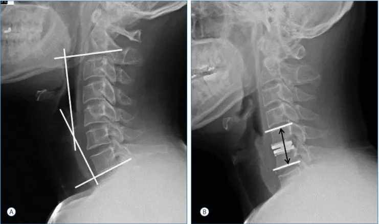
Fig. 1. A : Sagittal balance was measured as the angle between the lower margin of C2 and C7 on a static neutral lateral radiograph. B : Functional segmental unit (FSU; upper and lower endplate of the operative lesion) height was measured as the length from the upper endplate of the superior segment to the lower endplate of the inferior segment at the operated level.
Statistical analysis
Differences in the clinical and radiological results of each group were evaluated by the Wilcoxon signed-rank test with SPSS software for Windows ver. Version 18.0 (SPSS Inc., Chi- cago, IL, USA).
We applied nonparametric statistical tests exclusively, such
as the χ
2test, the Fisher’s exact test, the t-test, and the Mann- Whitney U test, as appropriate. The mean values of the groups were compared by using t-tests after performing F- tests for homogeneity of variances. A p-value<0.05 was con- sidered significant. Nonparametric correlation analyses were performed using the Spearman-rho rank-ordered correlation
Table 1. Patient characteristics
Variable Group A (17 patients) Group B (18 patients) p-value
Mean age (yr, range) 42.1±5.5 (29–51) 42.8±5.9 (33–49) 0.680
Gender (male : female) 12 : 5 11 : 7 0.725
Affected lesion 0.347
C4–5 (%) 5 (29.4) 5 (27.8)
C5–6 (%) 7 (41.2) 5 (27.8)
C6–7 (%) 5 (29.4) 5 (27.8)
C7–T1 (%) 0 3 (16.6)
Duration of symptom (mon, range) 5.1±3.4 (3–16) 4.4±3.0 (1–18) 0.483
Surgical time (min, range) 90.3±17.6 (63–119) 77.4±18.3 (52–114) 0.041
Length of hospital stay (day, range) 6.9±2.2 (5–18) 4.1±1.8 (3–15) 0.032
Follow-up period (mon, range) 82.5±15.4 (61–91) 84.1±16.1 (60–95) 0.783
Group A : total disc replacement. Group B : posterior cervical foraminotomy
A B
Fig. 2. Range of motion was measured as the difference of the angle on a simple dynamic radiograph.
coefficient and the Kendall-tau coefficient analyses to detect associations between categorical variables.
RESULTS
Clinical outcomes
Patient demographics and preoperative neurological status are summarized in Table 1, 2. No differences in these vari- ables were observed between the groups, except length of hospital stay, which may have been associated with early sur- gical complications (Table 3). The difference in early compli- cation was caused by using the anterior approach.
Clinical outcomes are summarized in Table 4. Preopera- tive NDI scores in groups A and B were 34.1±5.6 and 33.6±
8.4, and the postoperative NDI scores were 9.5±3.4 and 9.9±
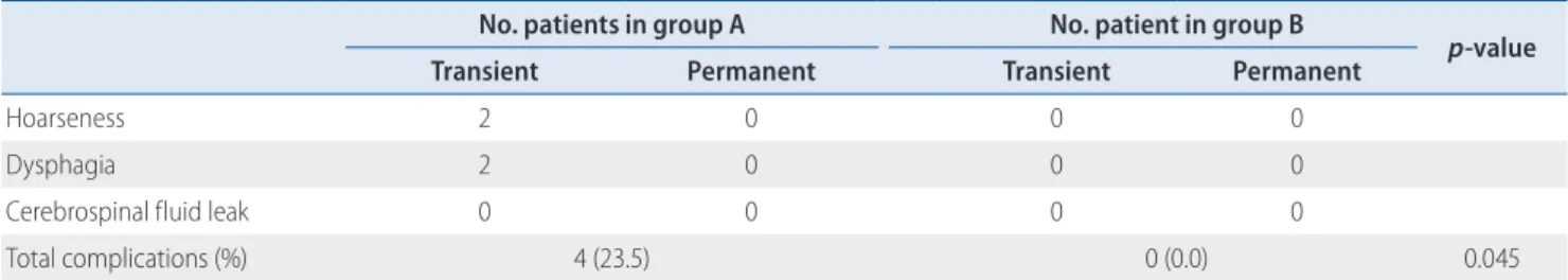
4.7, respectively. The success rate of group A was 94.1%, which was slightly superior to that of group B (88.9%), but the difference was not significant. No surgical complications were detected in group B, whereas group A had four cases of complications (23.5%), but all were transient. Operating time and hospitalization time were 90.3±17.6 min and 6.9±
2.2 d in group A, and 77.4±18.3 min and 4.1±1.8 d in group B, respectively.
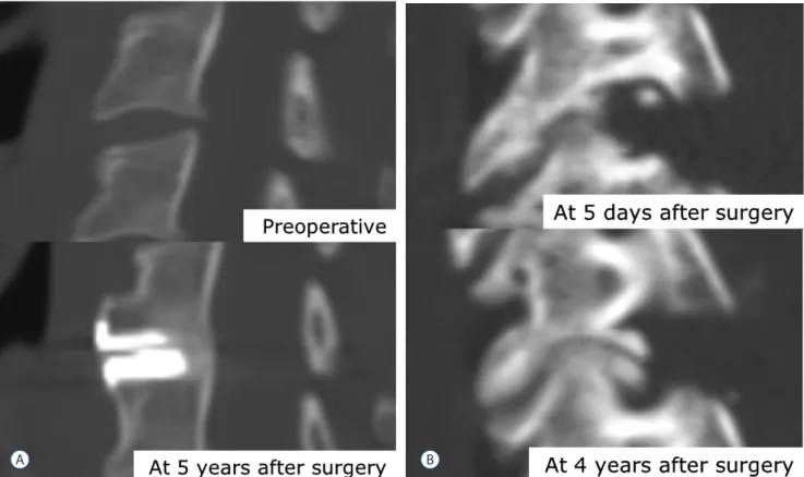
Repeat surgery was necessary for a 48-year-old male pa- tient in group B at 4 years after primary surgery. He experi- enced an ipsilateral recurrent arm pain causing progression of spondylosis (Fig. 3) and underwent anterior discectomy and fusion.
Radiologic data
The overall biomechanical data are summarized in Table 5.
The preoperative C2–7 angle and FSU in group A were -13.4±
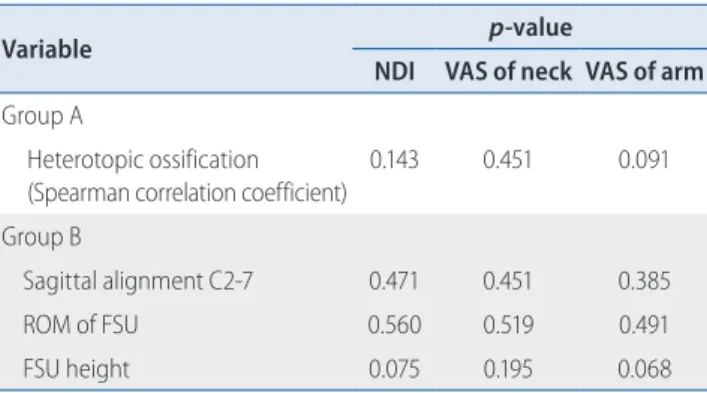
11.6 and -0.2±3.8, and the postoperative C2–7 angle and FSU were -14.0±10.4 and 0.4±3.8, respectively. Preoperative C2–7 ROM and FSU were 44.5±6.3 and 10.0±1.6, and the postoper- ative values were 44.3±5.7 and 8.4±2.1, respectively. Preopera- tive and postoperative upper level adjacent segment motion values were 8.6±2.3 and 8.4±2.0, and the lower level motion values were 8.4±2.2 and 8.3±1.9, respectively. Preoperative and postoperative FSU heights were 37.0±2.1 and 37.1±1.8, re- spectively. Postoperative FSU ROM decreased significantly final follow-up ( p=0.007), which was caused by HO (Table 6).
We also found a high rate of HO in group A (70.6%; 12/17).
In particular, severe HO (grade III+IV) was directly relevant to segmental motion (29.4%; 5/17). However, the occurrence of HO was not related with clinical outcomes (Table 7).
Preoperative C2–7 angle and FSU in group B were -14.0±
10.4 and 0.4±3.8, and postoperative C2–7 angle and FSU were -12.8±9.5 and 0.6±4.5, respectively. Preoperative C2–7 ROM and FSU were 44.9±8.3 and 10.4±1.5, and the postoperative values were 44.0±8.0 and 8.2±1.9, respectively. Preoperative and postoperative upper level adjacent segment motion val- ues were 8.1±2.6 and 8.2±2.8, and the lower level motion val- ues were 6.5±3.3 and 6.3±3.1, respectively. Preoperative and postoperative FSU heights were 37.1±2.0 and 36.2±1.8, respec-
Table 2. Preoperative neurological statuses Symptoms No. patients in
group A (%)
No. patient in
group B (%) p-value
Motor deficits 9 (52.9) 11 (61.1) 0.625
Sensory deficits 13 (76.5) 14 (77.8) 0.927
Pain only 2 (11.8) 2 (11.1) 0.952
Altered reflex 10 (56.8) 12 (66.7) 0.631
Group A : total disc replacement. Group B : posterior cervical foraminotomy
Table 3. Surgery-related complications
No. patients in group A No. patient in group B
p-value
Transient Permanent Transient Permanent
Hoarseness 2 0 0 0
Dysphagia 2 0 0 0
Cerebrospinal fluid leak 0 0 0 0
Total complications (%) 4 (23.5) 0 (0.0) 0.045
Group A : total disc replacement. Group B : posterior cervical foraminotomy
tively. Postoperative FSU motion and height changes were significant compared to those taken preoperatively ( p<0.05).
The decrease in FSU height tended to be associated with clin-
ical outcomes ( p=0.068; Table 7). Cervical foraminal stenosis was aggravated by the decrease in FSU height, and it may have increased the arm VAS and NDI scores. Additionally, we compared the degree of biomechanical changes between group A and B. The postoperative change in C2–7 sagittal alignment (group A, 0.1±0.1; group B, 1.2±0.4, p =0.043), ROM of FSU (group A, 1.6±0.5; group B, 2.2±0.6, p=0.039), and FSU height (group A, 0.1±0.0; group B, 0.9±0.1, p=0.033) were significantly higher in group B than those in group A.
Patient satisfaction and cognition for surgery We asked the patients 1 year after surgery: “Are you satis- fied with the results of surgery?” and “Would you choose the same procedure again?” The satisfaction rates of group A and B were 88.2% and 88.8%, respectively. The “yes” rates in group A and B for the second question were 82.3% and 83.3%. Additionally, we asked the patients “What do you think was the best benefit of your procedure?”, “Motion pres-
Table 4. Long-term outcomes after the operation
Variable Group A Group B p-value
NDI
Preoperative 34.1±5.6 33.6±8.4 0.854
Postoperative 9.5±3.4 9.9±4.7 0.767
VAS of neck
Preoperative 2.9±1.1 2.9±1.0 0.986
Postoperative 1.1±0.7 1.2±0.9 0.664
VAS of arm
Preoperative 7.0±1.2 6.9±1.1 0.889
Postoperative 1.3±0.8 1.6±0.8 0.278
Success* 16 (94.1) 16 (88.9) 0.581
Values are presented as mean±standard deviation or number (%). Group A : total disc replacement. Group B : posterior cervical foraminotomy.
*NDI improvement of more than 15 points at the final follow-up with no device failure or major complication. NDI : neck disability index, VAS : visual analog scale
Fig. 3. Case 1 in the total disc replacement (A). At 5 years after surgery, severe heterotophic ossification was shown on a computed tomography scan, but no clinical symptoms were observed. Case 2 in the posterior cervical foraminotomy (B). The patient complained of recurrent arm pain 4 years after the surgery, so we performed anterior cervical fusion as the revision surgery.
A B
ervation” was given by 88.2% of group A patients. However, the group B answers varied: “motion preservation” in 38.9%,
“no device use” in 27.8%, and “cost effectiveness” in 22.2%.
DISCUSSION
The primary goal of all operative treatment methods in patients with pure radiculopathy is decreased pain and sen- sorimotor deficits, as well as restore of working ability and quality of life. These goals can be reached by permanent de- compression of the compressed nerve root. Various morpho- logical causes (hard or soft disc or both) can lead to radicu- lopathy, which is treated using different techniques. These techniques differ in approach, complexity, aim, duration, and complications. We compared perioperative data and long-term outcomes of patients with pure radiculopathy treated at a single institution. These patients underwent de-
compression, with either anterior microdiscectomy and in- sertion of an artificial disc or posterior foraminotomy. Al- though both techniques helped preserve motion, they have different characteristics.
The advantages of TDR are preservation of motion and disc height, familiarity of the approach, and a relatively good long-term result including a lower incidence of adja- cent segmental disease. Peng et al.
25)reported that TDR pro- duces significant improvement in clinical outcomes after 2 years. Moreover, TDR restores segmental lordosis and pre- serves segmental motion up to 2 years postoperatively. In the present study, TDR showed good clinical outcomes, but
Table 5. Biomechanical follow-up results
Variable Group A Group B
Preoperative Postoperative p-value Preoperative Postoperative p-value Sagittal alignment (º)
C2–7 -13.4±11.6 -13.3±8.8 0.921 -14.0±10.4 -12.8±9.5 0.091
FSU -0.2±3.8 -0.2±4.0 0.977 0.4±3.8 0.6±4.5 0.775
ROM (º)
C2–7 44.5±6.3 44.3±5.7 0.904 44.9±8.3 44.0±8.0 0.133
FSU 10.0±1.6 8.4±2.1 0.007 10.4±1.5 8.2±1.9 0.003
Adjacent segment
Upper 8.6±2.3 8.4±2.0 0.340 8.1±2.6 8.2±2.8 0.753
Lower 8.4±2.2 8.3±1.9 0.718 6.5±3.3 6.3±3.1 0.268
FSU height (mm) 37.0±2.1 37.1±1.8 0.178 37.1±2.0 36.2±1.8 0.011
Values are presented as mean±standard deviation. Group A : total disc replacement. Group B : posterior cervical foraminotomy. FSU : functional segmental unit, ROM : range of motion
Table 6. Incidence of heterotopic ossification in Group A
McAfee class No. of patient (%)
0 5 (29.4)
I 4 (23.5)
II 3 (17.6)
III 2 (11.8)
IV 3 (17.6)
Group A: Total disc replacement
Table 7. Relationship between clinical outcomes and the presence of heterotopic ossification in group A and sagittal alignment C2–7, ROM, and FSU height in group B
Variable p-value
NDI VAS of neck VAS of arm Group A
Heterotopic ossification (Spearman correlation coefficient)
0.143 0.451 0.091
Group B
Sagittal alignment C2-7 0.471 0.451 0.385
ROM of FSU 0.560 0.519 0.491
FSU height 0.075 0.195 0.068
Group A : total disc replacement. Group B: posterior cervical foraminotomy.
NDI : neck disability index, VAS : visual analog scale, FSU : functional segmental unit, ROM : range of motion
whether motion preservation was questionable in some cas- es at a minimum 5-year follow-up. Moreover, TDR has dis- advantages, such as HO, implant-related complications, and risk of anterior structural injury
2,17,22,23,26,31). Quan et al. re- ported that the Bryan TDR maintains favorable clinical and radiological results, preserves movement, and leads to satis- factory clinical outcomes in the majority of cases at the 8-year follow-up
26). However, HO was evident in 48% of op- erated segments, and the incidence of HO causing restricted ROM of the prosthesis appeared to increase with time. In the same report, mean VAS score for both neck and arm pain was slightly higher in patients in whom HO developed than in those without HO. In the present study, no statisti- cally significant correlation was detected between HO and clinical outcomes, although HO was evident in 12 of 17 (70.6%) cases. However, the correlation was almost margin- al ( p=0.091), suggesting possible significance in a long-term follow-up. Many studies have examined risk factors for HO after cervical TDR, such as male sex, old age, longer post- operative period, prosthesis type, and preoperative calcifi- cation of longitudinal ligaments and osteophyte
5,15,20,34). The reason for the high incidence of HO in our study was thought to be the difference in sex ratio (male : female=12 : 5) and prosthesis type.
Implant-related complications can be a problem with TDR. Quan et al. reported a case of posterior migration of the implant, whereas Hrabálek reported no implant-related complications, such as migration, loosening, or subsid- ence
26). These results are consistent with previous studies, in which incidence of implant-related complications was very low
13,26,33). In contrast with PCF, TDR has a risk of intraoper- ative or early complications related to the anterior approach including hoarseness, dysphagia, and cerebrospinal fluid leakage. Early complication rates for TDR are 5
–30%, and dysphagia is the most common complication
2,17,23,31). In the present study, two patients (11.7%) had dysphagia after TDR, but no permanent symptoms were observed. Dysphagia af- ter the anterior approach improves gradually over time, and severe complications are rare. However, it is clear that the anterior approach results in a higher complication rate than
that of the posterior approach.
PCF also has advantages and disadvantages. The first ad- vantage is that it does not require specialized instrumenta- tion, so no instrument-related complications, such as infec- tion or instrumental failure, occur after PCF. Additionally, PCF is more cost-effective compared with TDR. The cost of PCF in Korean insurance system is about one-half to one- third that of TDR. Four patients (22.2%) in the group B chose cost-effectiveness as an important benefit of PCF, which may be related with patient’s satisfaction. Another ad- vantage of PCF is the low complication risk. Because PCF uses a posterior approach, the injury risk to anterior struc- tures, such as the esophagus, carotid artery, and recurrent la- ryngeal nerve, is eliminated. In contrast, the most common complaint during the early postoperative period is neck pain and discomfort
16,21). The extensive incision and dissection during PCF can increase neck discomfort and pain. Because of the slow recovery from this extensive incision, we used a tubular retractor system for minimally invasive surgery. We previously compared the open procedure and the tubular re- tractor assisted procedure for patients undergoing cervical radiculopathy
18). The neck pain VAS score after the tubular retractor-assisted procedure was significantly lower than that of the open procedure. The tubular retractor-assisted proce- dure also decreases the size of skin incision, length of non- steroidal anti-inflammatory drug use, and length of hospital stay. Postoperative kyphosis can also be a problem with PCF.
Risk factors associated with postoperative kyphotic deformi- ty are old age, preoperative kyphosis, and extent of laminec-
tomy
11,14,16). Jagannathan et al. reported that patients with
postoperative kyphosis had lower quality of life outcomes
14).
In contrast, postoperative kyphosis was not correlated with
postoperative clinical outcomes in the present study, which
may have been due to including patients who were relatively
young and had single-level soft disc disease. Interestingly, de-
creased FSU height was marginally correlated with the NDI
(p=0.075) and arm VAS scores (p=0.068; Table 7), possibly
because of progression of foraminal stenosis may be related
with decreased ROM and FSU height. This finding was
caused by accelerated degeneration related to injury toe the
facet, lamina, and soft tissue during surgery
14). In addition, C5 nerve root palsy can be a complication of PCF
10,14,16,19). Fortunately, C5 root palsy did not occur in the present study, possibly because only soft disc disease was treated. Yang re- ported that risk factors for C5 palsy include ossification of the posterior longitudinal ligament and foraminal stenosis
32). Soft disc herniation has a lower risk of C5 root palsy than hard disc or foraminal stenosis.
This study had some limitations. First, the data were ob- tained from a small number of patients and may be biased.
Moreover, selection of surgical method was not randomized, although preoperative characteristics were not different be- tween the two groups (Table 1). We explained the advantages and disadvantages of the two procedures to the patients, and the patients selected the surgical procedure. This factor may have influenced the patients’ preconception for the surgery.
Second, differences in the anterior and posterior approaches could have affected postoperative complications, such as dys- phagia, neck pain, and hoarseness. However, the main objec- tive of this study was the biomechanical follow-up results for motion preservation techniques. Also, PCF and TDR are the most popular motion-preservation techniques, and no com- parative study has been conducted between the two tech- niques.
CONCLUSION
TDR and PCF provided favorable clinical and radiological outcomes for patients with unilateral cervical radiculopathy caused by posterolateral soft disc herniation, However, TDR has disadvantages including HO and anterior approach-re- lated complications, and PCF has problems, including disc recurrence, progression of spondylosis, and neck pain.
Therefore, we recommend TDR for patients with risks for progressing kyphosis (neck muscle atrophy, preoperative ky- phosis, etc), posterior neck pain and re-herniation of a disc.
In contrast, we suggest PCF for patients with risks for the anterior approach (structural problems, previous neck sur- gery, etc) and concerns about cost.
References
1. Aldrich F : Posterolateral microdisectomy for cervical monoradiculopathy caused by posterolateral soft cervical disc sequestration. J Neurosurg 72 : 370-377, 1990
2. Bazaz R, Lee MJ, Yoo JU : Incidence of dysphagia after anterior cervical spine surgery: a prospective study. Spine (Phila Pa 1976) 27 : 2453- 2458, 2002
3. Beaurain J, Bernard P, Dufour T, Fuentes JM, Hovorka I, Huppert J, et al.
: Intermediate clinical and radiological results of cervical TDR (Mobi-C) with up to 2 years of follow-up. Eur Spine J 18 : 841-850, 2009 4. Cağlar YS, Bozkurt M, Kahilogullari G, Tuna H, Bakir A, Torun F, et al. :
Keyhole approach for posterior cervical discectomy: experience on 84 patients. Minim Invasive Neurosurg 50 : 7-11, 2007
5. Cho HJ, Shin MH, Huh JW, Ryu KS, Park CK : Heterotopic ossification following cervical total disc replacement: iatrogenic or constitutional?
Korean J Spine 9 : 209-214, 2012
6. Cloward RB : The anterior approach for removal of ruptured cervical disks. J Neurosurg 15 : 602-617, 1958
7. Ducker TB, Zeidman SM : The posterior operative approach for cervical radiculopathy. Neurosurg Clin N Am 4 : 61-74, 1993
8. Duggal N, Pickett GE, Mitsis DK, Keller JL : Early clinical and biomechani- cal results following cervical arthroplasty. Neurosurg Focus 17 : E9, 2004
9. Fehlings MG, Gray RJ : Posterior cervical foraminotomy for the treatment of cervical radiculopathy. J Neurosurg Spine 10 : 343-344, 2009 10. Gala VC, O'Toole JE, Voyadzis JM, Fessler RG : Posterior minimally inva-
sive approaches for the cervical spine. Orthop Clin North Am 38 : 339-349, 2007
11. Henderson CM, Hennessy RG, Henry M Jr, Shackelford GE : Posterior- lateral foraminotomy as an exclusive operative technique for cervical radiculopathy: a review of 846 consecutively operated cases. Neuro- surgery 13 : 504-512, 1983
12. Hilton DL Jr : Minimally invasive tubular access for posterior cervical foraminotomy with three-dimensional microscopic visualization and lo- calization with anterior/posterior imaging. Spine J 7 : 154-158, 2007 13. Hrabálek L, Vaverka M, Houdek M : Cervical disc arthroplasty (Prodisc-C):
analysis of 3 to 4- year follow up results. Rozhl Chir 88 : 634-641, 2009 14. Jagannathan J, Sherman JH, Szabo T, Shaffrey CI, Jane JA : The poste- rior cervical foraminotomy in the treatment of cervical disc/osteophyte disease: a single-surgeon experience with a minimum of 5 years' clinical and radiographic follow-up. J Neurosurg Spine 10 : 347-356, 2009 15. Jin YJ, Park SB, Kim MJ, Kim KJ, Kim HJ : An analysis of heterotopic os-
sification in cervical disc arthroplasty: a novel morphologic classification of an ossified mass. Spine J 13 : 408-420, 2013
16. Jödicke A, Daentzer D, Kästner S, Asamoto S, Böker DK : Risk factors for outcome and complications of dorsal foraminotomy in cervical disc herniation. Surg Neurol 60 : 124-129; discussion 129-130, 2003
17. Kang SH, Kim DK, Seo KM, Kim KT, Kim YB : Multi-level spinal fusion and postoperative prevertebral thickness increase the risk of dysphagia after anterior cervical spine surgery. J Clin Neurosci 18 : 1369-1373, 2011 18. Kim KT, Kim YB : Comparison between open procedure and tubular
retractor assisted procedure for cervical radiculopathy: results of a ran- domized controlled study. J Korean Med Sci 24 : 649-653, 2009 19. Korinth MC, Krüger A, Oertel MF, Gilsbach JM : Posterior foraminotomy
or anterior discectomy with polymethyl methacrylate interbody stabiliza- tion for cervical soft disc disease: results in 292 patients with monora- diculopathy. Spine (Phila Pa 1976) 31 : 1207-1214, 2006
20. Leung C, Casey AT, Goffin J, Kehr P, Liebig K, Lind B, et al. : Clinical sig- nificance of heterotopic ossification in cervical disc replacement: a pro- spective multicenter clinical trial. Neurosurgery 57 : 759-763, 2005 21. Lidar Z, Salame K : Minimally invasive posterior cervical discectomy for
cervical radiculopathy: technique and clinical results. J Spinal Disord Tech 24 : 521-524, 2011
22. McAfee PC, Cunningham BW, Devine J, Williams E, Yu-Yahiro J : Clas- sification of heterotopic ossification (HO) in artificial disk replacement. J Spinal Disord Tech 16 : 384-389, 2003
23. Morpeth JF, Williams MF : Vocal fold paresis after anterior cervical dis- cectomy and fusion. Laryngoscope 110 : 43-46, 2000
24. Park CK, Ryu KS, Lee KY, Lee HJ : Clinical outcome of lumbar total disc re- placement using ProDisc-L in degenerative disc disease: minimum 5-year follow-up results at a single institute. Spine (Phila Pa 1976) 37 : 672- 677, 2012
25. Peng CW, Yue WM, Basit A, Guo CM, Tow BP, Chen JL, et al. : Interme- diate results of the prestige LP cervical disc replacement: clinical and radiological analysis with minimum two-year follow-up. Spine (Phila
Pa 1976) 36 : E105-111, 2011
26. Quan GM, Vital JM, Hansen S, Pointillart V : Eight-year clinical and ra- diological follow-up of the Bryan cervical disc arthroplasty. Spine (Phila Pa 1976) 36 : 639-646, 2011
27. Rodrigues MA, Hanel RA, Prevedello DM, Antoniuk A, Araújo JC : Pos- terior approach for soft cervical disc herniation: a neglected technique?
Surg Neurol 55 : 17-22; discussion 22, 2001
28. Roh SW, Kim DH, Cardoso AC, Fessler RG : Endoscopic foraminotomy using MED system in cadaveric specimens. Spine (Phila Pa 1976) 25 : 260-264, 2000
29. Russell SM, Benjamin V : Posterior surgical approach to the cervical neu- ral foramen for intervertebral disc disease. Neurosurgery 54 : 662- 665, 2004
30. Ryu KS, Park CK, Jun SC, Huh HY : Radiological changes of the operated and adjacent segments following cervical arthroplasty after a minimum 24-month follow-up: comparison between the Bryan and Prodisc-C de- vices. J Neurosurg Spine 13 : 299-307, 2010
31. Stewart M, Johnston RA, Stewart I, Wilson JA : Swallowing performance following anterior cervical spine surgery. Br J Neurosurg 9 : 605-609, 1995
32. Yang CW, Fuh JL : C5 palsy after cervical spine decompression surgery.
J Chin Med Assoc 76 : 363-364, 2013
33. Yi S, Lim JH, Choi KS, Sheen YC, Park HK, Jang IT : Comparison of ante- rior cervical foraminotomy vs arthroplasty for unilateral cervical radicu- lopathy. Surg Neurol 71 : 677-680; discussion 680, 2009
34. Yi S, Shin DA, Kim KN, Choi G, Shin HC, Kim KS, et al. : The predispos- ing factors for the heterotopic ossification after cervical artificial disc replacement. Spine J 13 : 1048-1054, 2013