41(4), 437~442(2012) http://dx.doi.org/10.3746/jkfn.2012.41.4.437
홍국쌀(
Monascus purpureus
) 추출물의 항산화 작용권 정 숙
안동대학교 생활과학대학 식품영양학과
Antioxidant Properties of Red Yeast Rice ( Monascus purpureus ) Extracts
Chong Suk Kwon
Dept. of Food Science and Nutrition, Andong National University, Gyeongbuk 760-749, Korea
Abstract
Red yeast rice (RER) has been used in China for centuries for its medicinal properties and is an increasingly popular alternative lipid-lowering treatment. This study was carried out to estimate the antioxidant properties of RER extracts. The ethyl acetate extract exhibited the DPPH radical scavenging activity of 85% at 0.2 mg/mL and IC50 0.13 mg/mL. A significant proportion of hydroxyl radicals in a cuvette were scavenged: 44.2% at 2.5 μg/mL, 74.1% at 5.0 μg/mL, and >100% at 10 μg/mL. The HepG2cells pre-treated with RER ethyl acetate extract reduced the hydroxyl radicals significantly compared to the control cells. Oxidative DNA damage was measured using a Comet assay. The RER ethyl acetate extract did not induce any DNA damage per se, and appeared to enhance the resistance to DNA damage caused by an oxidant challenge with H2O2, whereas lovastatin increased the level of DNA damage in the cells in both the unstressed (no oxidant) and those stressed with H2O2. The relative gene expression of the antioxidant enzymes in HepG2cells were also affected by the RER ethyl acetate extract. The HepG2cells were pre-incubated with the RER ethyl acetate extract, and then stressed with H2O2
or left unstressed (no oxidant). In the unstressed cells, superoxide dismutase (Cu/Zn SOD) and glutathione perox- idase (GPx) were increased significantly 3.25-fold and 2.67-fold, respectively, whereas in the stressed cells, the catalase (CAT) level was increased by 4.64-fold and 7.0-fold at 5 μg/mL and 10 μg/mL, respectively, com- pared to those of the control. From these results, RER appears to be effective in suppressing oxidative stress.
Key words: red yeast rice, Monascus purpureus, oxidative DNA damage, antioxidant enzymes gene expression
E-mail: [email protected]
Phone: 82-54-820-5484, Fax: 82-54-820-6281
서 론
생체에서는 superoxide, hydroxyl radical 및 과산화수소 와 같은 reactive oxygen species(ROS)들이 계속해서 생성 되며 ROS의 생성에 반응하여 이들을 제거하는 항산화 시스 템도 함께 조절되고 있다. 따라서 ROS의 생성과 항산화 방 어계는 어느 정도 균형 상태를 유지하고 있는 것이 사실이 다. 그러나 항산화 방어계가 ROS의 생성에 미치지 못하는 상태, 즉 산화적 스트레스(oxidative stress) 상태가 되면 DNA를 비롯하여 세포와 조직을 구성하는 단백질, 지질 및 다른 성분들이 ROS에 의해 손상될 수 있고 이것이 여러 종 류의 암 및 노화 관련 질병들의 발생과도 관련되어 있는 것 으로 알려져 있다(1). 따라서 산화적 스트레스와 관련된 질 병들의 발생을 예방할 수 있는 항산화 물질을 함유한 기능성 식품에 대한 연구와 개발에 관심이 높아지고 있다.
찐 백미에 홍국(Monascus purpureus)을 접종하고 배양 하여 만든 홍국쌀은 ‘red Koji’ 또는 ‘Hong Qu’로도 불리며, 중국에서는 수세기 동안 식품으로서뿐 아니라 술을 비롯한
발효식품 제조에 향미 증진제, 또는 착색제로 사용되고 있으 며, 민간에서는 소화 촉진과 혈액 순환 개선에 효과가 있는 것으로 널리 알려져 왔다(2). 홍국에는 콜레스테롤 합성 저 해제인 monacolin K(lovastatin)가 함유되어 있어서 혈액 콜 레스테롤 저하 작용이 있는 것으로 알려져 있으며(3-5), 최 근에는 홍국의 발효과정에서 생성되는 이차 대사산물들의 다양한 기능성이 보고되고 있다. 홍국쌀에 함유되어 있는 azaphilone 색소들(monascin, ankaflavin, rubropunctatin, monascorburin, rubropunctamine 및 monascorburamine) 의 항염증 작용, γ-aminobutyric acid의 신경전달 및 혈압 강하 효과, dimerumic acid, tannin, phenol 및 unsaturated fatty acids의 항산화 작용이 보고되어 있으며(6-9), 알코올 성 간손상에 대한 간보호작용(10), 그리고 Alzheimer's dis- ease 발생과 관련 있는 amyloid β-peptide의 신경세포에 대 한 독성 억제 효과(11) 등도 홍국쌀의 항산화 작용에 기인한 것으로 보고되고 있다. 아울러 고지혈증 치료제인 statins의 장기간 복용으로 근육 장애, 간과 신장 기능 장애, 갑상선 기능 저하 및 당뇨병과 같은 부작용이 보고되면서 statins의
대체제로서 홍국의 사용에 대한 관심이 높아지고 있다(12).
이에 본 연구에서는 홍국쌀 추출물의 항산화 작용을 DPPH radical 및 hydroxyl radical 소거능, 간암 세포에서의 산화적 DNA 손상과 항산화 효소의 유전자 발현에 미치는 영향으로 분석하였다.
재료 및 방법 홍국쌀 추출물의 제조
홍국쌀은 한스바이오(주)(경북 영양군, 한국)로부터 제공 받은 제품을 사용하였다. 홍국쌀 100 g을 먼저 메탄올로 3회 환류 추출한 후 90oC에서 진공 농축시켜 methanol 추출물을 얻고(5.2 g), methanol 추출물로부터 hexane, butanol, water 및 ethyl acetate 추출물을 각각 연속적으로 분획하여 제조 하였으며, 분획물의 수율은 각각 6.4%, 11.0%, 71.5% 및 6.7%
로 나타났다.
세포 배양
HepG2 cells를 10%(w/v) fetal bovine serum과 1% pen- icillin-streptomycin이 첨가된 Dulbecco's modified Eagle's medium(DMEM)을 사용하여 5% CO2, 37oC incubator에서 배양하였다.
DPPH radical 소거능
1,1-Diphenyl-2-picrylhydrazyl(DPPH) radical 소거능은 Hatano 등(13)의 방법으로 측정하였다. 추출물별로 여러 농도 (0~1.0 mg/mL)의 시료를 DMSO를 용매로 제조하여 DPPH 용액(200 μM)에 첨가한 후, 실온에 30분간 방치시켰다가 mi- croplate reader(Sunrise, Tecan, Männedorf, Switzerland) 로 515 nm에서 흡광도를 측정하였다. 시료의 DPPH radical 소거능은 control에 대한 radical 소거율(%)로 나타내었으 며, 소거율 50%에서의 시료 농도(IC50)를 산출하였다.
Hydroxyl radical 소거능
Hydroxyl radical 소거능은 fluorescent probes를 사용하 여 cuvette에서(14), 그리고 HepG2cell에서(15) 각각 측정하 였다. Cuvette에서의 측정은 H2O2(1 μM), FeSO4(50 mM), fluorescent probe인 dichlorofluorescin diacetate(DCFDA, 20 μM)와 시료(butanol 및 ethyl acetate 추출물)를 각각 농 도별(0, 2.5, 5.0 및 10.0 μg/mL)로 혼합한 다음 15분 동안 dichlorofluorescin(DCF) fluorescence 생성량을 Ex 365 nm, Em 435 nm에서 형광광도계(F-4500, Hitachi, Tokyo, Ja- pan)로 측정하였다. HepG2 cell에서의 측정은 세포를 여러 농도의 홍국쌀 ethyl acetate 추출물(0, 1, 5 및 10 μg/mL)로 1시간 동안 전 처리한 후, H2O2(1 mM)에 1시간 노출시키고, DCFDA(20 μM)를 첨가하여 30분간 incubation 하고 세포를 ice-cold PBS로 2회 세척하여 수집한 후 초음파로 파쇄하고 원심분리 하여 얻은 상등액에서 DCF fluorescence를 형광 광도계로 측정하였다. 추출물과 H2O2 대신 용매와 PBS로
처리한 세포("no oxidant")를 대조로 하여 비교하였다.
DNA 손상(Comet assay)
DNA 손상 정도는 Singh 등(16)의 방법에 따라 Comet assay로 분석하였다. HepG2 cells를 여러 농도(1, 5 및 10 μg/mL)의 홍국쌀 ethyl acetate 추출물 또는 lovastatin(1 μg/mL)으로 3시간 동안 전처리하고 수집하여 PBS에 현탁 시킨다. Slide에 0.65% agarose를 깔고 냉장고에서 고형화 시킨 후, 그 위에 세포 현탁액(약 20,000개 세포)과 0.65%
low melting point agarose를 고루 펴지게 덮고 냉장고에서 고형화시킨다. Slide를 PBS(“no oxidant”) 또는 H2O2(25 μM)에 5분 동안 담근 후 lysis buffer(4oC for 1 hr)로 lysis시 키고, unwinding buffer에 20분간 방치한 후 4oC에서 20분간 전기영동(25 V, 300 mA)한다. 전기영동이 끝나면 중화시킨 후 ethidium bromide로 염색하여 형광현미경(DMLB, Leica, Wetzlar, Germany)으로 DNA 손상 정도를 관찰한다. 시료 마다 2개씩의 slide, 한 slide에 50개씩의 cells에 대해 Komet image analysis system(Version 5.0, Kinetic Imaging, Nottingham, UK)을 사용하여 DNA 손상 정도를 측정하고, 결과는 Olive tail moment(OTM)[(Tailmean-Headmean)×Tail density(%)/100]로 나타내었다.
항산화 효소의 유전자 발현
홍국쌀 ethyl acetate 추출물이 HepG2 cell에서 항산화 효 소의 유전자 발현에 미치는 영향을 semi-quantitative real time PCR(RT-PCR)로 측정하였다. HepG2 cells를 여러 농 도의 홍국쌀 ethyl acetate 추출물(0, 1, 5 및 10 μg/mL)로 12시간 동안 전처리한 후, PBS(no oxidant) 또는 H2O2(1 mM)에 1시간 동안 노출시켰다. 세포를 수거하여 RNA 추출 용 kit(Quiagen, Valencia, CA, USA)로 RNA를 추출하고 SuperScript II reverse transcriptase(Life Technologies, Paisley, UK)를 사용하여 cDNA를 합성한 다음 mRNA 발현 정도를 Exicycler(Bioneer Co., Daejeon, Korea)로 분석하였 다. 항산화 효소 primer는 Bioneer Co.에 주문 제작하였으며, primer sequence는 Table 1과 같고 internal control로 β- actin을 사용하였다. 유전자 발현 정도는 측정된 Ct(thresh- old cycle) 값을 사용하여 the comparative CT(2-ΔΔCT) method(17)로 계산하였고 control에 대한 상대적 발현비로 나타내었다.
통계처리
자료 분석은 SPSS 18.0(statistical package for the social science, Chicago, IL, USA) PC package를 이용하였다. 결과 는 평균±표준편차로 나타내었으며, 각 변수에 대해 일원배 치분산분석(one-way ANOVA)을 실시하였고, 사후검정으 로는 Duncan's multiple range test를 적용하였으며 α=0.05 수준에서 유의성을 검정하였다. 두 그룹 간에는 Student's t-test로 유의성을 검정하였다.
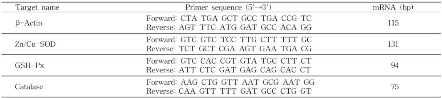
Table 1. Nucleotide sequences of PCR Primers used for semiquantitative RT-PCR of the antioxidant enzymes
Target name Primer sequence (5'→3') mRNA (bp)
β-Actin Forward: CTA TGA GCT GCC TGA CCG TC
Reverse: AGT TTC ATG GAT GCC ACA GG 115
Zn/Cu-SOD Forward: GTC GTC TCC TTG CTT TTT GC
Reverse: TCT GCT CGA AGT GAA TGA CG 131
GSH-Px Forward: GTC CAC CGT GTA TGC CTT CT
Reverse: ATT CTC GAT GAG CAG CAC CT 94
Catalase Forward: AAG CTG GTT AAT GCG AAT GG
Reverse: CAA GTT TTT GAT GCC CTG GT 75
0 20 40 60 80 100 120
0 0.2 0.4 0.6 0.8 1
Concentration (mg/mL)
DPPH radical scavenging (%) .
Ethyl acetate ext.
Methanol ext.
Butanol ext.
Hexane ext.
Fig. 1. DPPH radical scavenging activity of the RER extracts.
Table 2. IC50of DPPH radical scavenging activity of the RER extracts
Sample IC50 (mg/mL)
Ethyl acetate extract Methanol extract Butanol extract Hexane extract Water extract Vitamin C
0.13±0.011) 0.43±0.03 0.62±0.05
>1.0
>1.0 0.04±0.005
1)Values are mean±SD.
결과 및 고찰 DPPH radical 소거능
홍국쌀 추출물의 DPPH radical 소거능은 Fig. 1과 같다.
DPPH radical은 안정한 분자로부터 전자 또는 수소를 취하 는 유리기로 작용하므로 DPPH radical 소거능이 큰 물질일 수록 항산화력이 크다고 할 수 있다. 홍국쌀 ethyl acetate 추출물은 0.2 mg/mL에서 DPPH radical을 85% 소거하였고, 0.5 mg/mL에서 ethyl acetate 추출물은 100% 소거율을 나 타내었으며, methanol, butanol 및 hexane 추출물은 각각 54%, 42% 및 25% 소거하는 것으로 나타났다. DPPH radical 소거율 50%에 해당하는 추출물의 농도(IC50)를 계산한 결과 (Table 2), ethyl acetate 추출물이 0.13 mg/mL로 항산화력 이 가장 높았고, methanol 추출물(0.43 mg/mL), butanol 추 출물(0.62 mg/mL) 순으로 나타났다. 한편, vitamin C의 DPPH 소거능은 0.1 mg/mL에서 99.4%(IC50는 0.04 mg/mL)
였다. Yang 등(18)도 홍국쌀 methanol 추출물의 DPPH rad- ical 소거능이 1 mg/mL에서 88.6%였으며, vitamin C는 0.1 mg/mL에서 90∼96%, methanol 추출물의 IC50는 0.59 mg/
mL라고 하여 본 실험과 비슷한 결과를 보고하였다. Lee 등 (11)도 홍국쌀 ethanol 추출물 0.1 mg/mL의 DPPH radical 소거능이 200 μM vitamin C와 같다고 보고하였다. 홍국쌀에 는 항산화 물질로 dimerumic acid, tannin, phenol, unsatur- ated fatty acid 및 기타 대사산물들이 함유되어 있는 것으로 알려져 있다(19,20).
이상의 DPPH radical 소거능 측정 결과로부터 홍국쌀 추 출물 시료 중 항산화능이 가장 우수한 ethyl acetate 추출물 을 주 시료로 하여 다음 실험을 시행하였다.
Hydroxyl radical 소거능
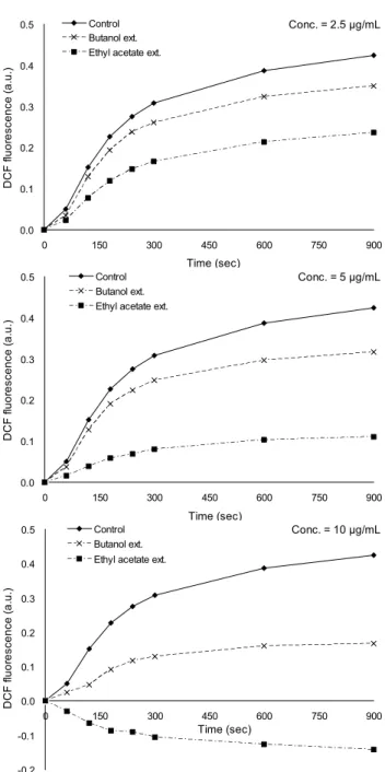
Hydroxyl radical은 반응성이 높아 생성된 장소에서 매우 강한 공격성을 나타내는 radical로 알려져 있다(21). 홍국쌀 butanol 및 ethyl acetate 추출물의 hydroxyl radical 소거능 을 cuvette에서 측정한 결과를 Fig. 2에 제시하였다. Cuvette 에서 H2O2/FeSO4의 Fenton 반응으로 hydroxyl radical을 생 성하고 fluorescent probe인 DCFDA를 사용하여 hydroxyl radical로 인해 생성된 DCF fluorescence를 15분 동안 측정 하였다. 홍국쌀 ethyl acetate 및 butanol 추출물(0, 2.5, 5 및 10 μg/mL)의 DCF fluorescence 생성 저해율을 측정한 결과, 2.5 μg/mL에서 control에 비해 각각 44.2%와 17.7%, 5.0 μg/
mL에서 각각 74.1%와 25.5%, 10 μg/mL에서 각각 >100%와 60.4%의 생성저해율을 나타내었다.
HepG2 cell에서 홍국쌀 추출물의 hydroxyl radical 소거능 을 측정한 결과는 Fig. 3과 같다. 세포에서 DCFDA는 세포막 을 쉽게 통과하여 세포 내로 들어가고 세포의 esterase에 의해 DCFH(nonfluorescent)로 전환되어 세포 내에 갇히게 되는데 세포 내에 존재하는 ROS, 특히 hydrogen peroxide (H2O2)와 hydroxyl radical에 의해 신속히 산화되어 DCF (highly fluorescent)로 전환된다. 따라서 DCF fluorescence 의 강도는 세포의 hydrogen peroxide 또는 hydroxyl radical 생성량에 비례한다. HepG2cell을 1 mM H2O2에 1시간 동안 노출시킨 세포(0 μg/mL)의 DCF fluorescence가 H2O2에 노 출시키지 않은 세포(no oxidant)에 비해 34.5% 유의적으로 증가하였다. 홍국쌀 ethyl acetate 추출물(0. 1, 5 및 10 μg/
Conc. = 2.5 μg/mL
0.0 0.1 0.2 0.3 0.4 0.5
0 150 300 450 600 750 900
Time (sec)
DCF fluorescence (a.u.) .
Control Butanol ext.
Ethyl acetate ext.
Conc. = 5 μg/mL
0.0 0.1 0.2 0.3 0.4 0.5
0 150 300 450 600 750 900
Time (sec)
DCF fluorescence (a.u.) .
Control Butanol ext.
Ethyl acetate ext.
Conc. = 10 μg/mL
-0.2 -0.1 0.0 0.1 0.2 0.3 0.4 0.5
0 150 300 450 600 750 900
Time (sec)
DCF fluorescence (a.u.) .
Control Butanol ext.
Ethyl acetate ext.
Fig. 2. The hydroxyl radical scavenging activity of the RER extracts.
a
b
a
a a
0 20 40 60 80 100 120 140 160
0 (no oxidant)
0 1 5 10
Concentration (μg/mL)
DCF Fluorescence (%) .
Fig. 3. The hydroxyl radical scavenging activity of the RER ethyl acetate extract in H2O2-treated HepG2 cells. Different letters indicate significant differences from one another (p<0.05).
a a a
a
b
A A
B
A
C
0 0.1 0.2 0.3 0.4 0.5 0.6 0.7
0 1 5 10 1 (Lovastatin)
Concentration (μg/mL)
Olive Tail Moment (a.u.) .
no oxidant H2O2 (25 uM)
* * *
*
ns
Fig. 4. Effect of the RER ethyl acetate extract on DNA dam- age in HepG2 cells treated with or without H2O2 by comet assay. Different letters indicate significant differences from one another (p<0.05).*p<0.05 by Student's t-test between the un- stressed (no oxidant) cells and the stressed (H2O2) cells. ns: not significant.
mL)로 1시간 동안 전처리하고 H2O2에 노출시킨 세포의 DCF fluorescence가 추출물로 전처리하지 않고 H2O2에 노출시킨 세포(0 μg/mL)에 비해 유의적으로 감소하였고, 추출물의 농 도가 높을수록 더 많이 감소하는 경향을 보였으나 농도 간에 는 유의적인 차이가 없었다.
DNA 손상 정도(Comet assay)
HepG2 cell의 산화 스트레스로 인한 DNA 손상 정도를 Comet assay로 측정한 결과를 Fig. 4에 제시하였다. 세포를 3시간 동안 홍국쌀 ethyl acetate 추출물 농도별(0, 1, 5 및 10 μg/mL) 및 lovastatin(1 μg/mL)으로 각각 전처리한 후, phosphate buffer(no oxidant) 또는 25 μM H2O2에 각각 5분
간 노출시켜 산화 스트레스를 가한 후 DNA 손상 정도를 Comet assay로 분석하였다. No oxidant 세포에서 보듯이, 홍국쌀 ethyl acetate 추출물 자체로 인한 DNA 손상은 관찰 되지 않았으며 오히려 추출물 농도가 증가할수록 DNA 손상 이 감소하는 경향을 보였으나 유의성은 없었다. 하지만 추출 물 대신에 lovastatin을 처리한 세포는 추출물(10 μg/mL)로 전처리한 세포(0.18±0.07)에 비해 DNA 손상이 211%(0.38
±0.09) 유의적으로 증가하였다. H2O2(25 μM)로 산화 스트레 스를 가한 세포(0.30±0.07)는 no oxidant 세포(0.24±0.08)에 비해 DNA 손상이 유의적으로 증가하였다(p<0.05). 추출물 5 μg/mL로 전처리하고 H2O2로 산화 스트레스를 가한 세포 (0.20±0.07)와 산화 스트레스를 가하지 않은(no oxidant) 세 포(0.22±0.08)의 DNA 손상에서 유의적인 차이가 없는 것으 로 보아 H2O2로 인한 DNA 손상으로부터 세포가 보호된 것 으로 보인다. Lovastatin을 적용한 세포(0.50±0.10)는 추출 물로 전처리한 세포들보다 DNA 손상이 유의적으로 증가한 것으로 나타났다. 장기간에 걸친 산화적 DNA 손상은 여러
No oxidant
b
a a
B B
A ns
0 1 2 3 4 5 6 7 8 9
0 5 10
Concentration (μg/mL) Relative mRNA expression . SOD(Cu/Zn)GPx
CAT
H2O2 (1 mM)
ns ns a
a
b
c
0 1 2 3 4 5 6 7 8 9
0 1 5 10
Concentration (μg/mL)
Relative mRNA expression .
SOD(Cu/Zn) GPx CAT
Fig. 5. Effect of the RER ethyl acetate extract on relative mRNA expression of the antioxidant enzymes in HepG2cells treated with or without H2O2. Different letters indicate sig- nificant differences from one another (p<0.05). ns: not significant.
종류의 암, 대장암, 전립선암, 직장암 및 유방암, 그리고 퇴행 성 질환의 발병 원인이 되며, 따라서 산화적 DNA 손상을 억 제시키는 식이, 기능성 물질 또는 치료제는 암 발생을 지연 또는 예방할 수 있는 것으로 알려져 있다(22-25). 홍국쌀의 monascorubrin 색소가 mouse의 피부암 세포의 증식을 억제 하며(26), 홍국쌀의 색소가 풍부한 분획에서 대장암 세포의 증식 억제 활성이 보고된 바 있다(27). 본 실험에서 홍국쌀 ethyl acetate 추출물은 간암 세포인 HepG2의 산화적 DNA 손상에 대해 5 μg/mL에서는 억제뿐 아니라 예방 효과도 있 었으나, 더 높은 농도인 10 μg/mL에서는 억제 효과가 나타 나지 않았으며 이에 대해서는 추후 연구가 필요한 것으로 생각된다. Lovastatin은 1 μg/mL에서 DNA 손상을 증가시 키는 것으로 나타났다.
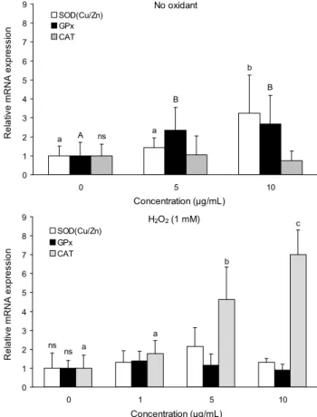
항산화 효소 유전자의 상대적 발현 정도
홍국쌀 ethyl acetate 추출물이 항산화 효소의 유전자 발 현에 미치는 영향을 HepG2에서 조사한 결과를 Fig. 5에 제시 하였다. 산화 스트레스 없이 ethyl acetate 추출물에 12시간 동안 전처리한 경우, 10 μg/mL에서 Cu/Zn superoxide dis- mutase(Cu/Zn SOD)와 glutathione peroxidase(GPx)의 mRNA 발현이 control(0 μg/mL)에 비해 각각 3.25배와 2.67 배 증가하였으며, catalase(CAT)는 변화가 없었다. 추출물
로 12시간 전처리하고 H2O2(1 mM)에 1시간 처리하여 산화 스트레스를 가한 세포의 경우, control에 비해 CAT는 5 μg/
mL에서 4.64배, 10 μg/mL에서 7.0배 유의적으로 증가하였으 며, Cu/Zn SOD는 5 μg/mL에서 증가하는 경향을 보였으나 유의성이 없었고 GPx는 변화가 없었다. 실험동물에 고콜레 스테롤식이와 홍국쌀을 혼합하여 공급한 후 혈청과 간에서 항산화 효소 활성을 측정한 연구에서도 홍국쌀을 섭취한 군 에서 대조군에 비해 SOD, GPx 및 CAT 모두 활성이 증가하 였고 아울러 지질과산화물의 농도는 감소하였다고 보고한 바 있다(28). Lee 등은 고콜레스테롤 식이를 섭취한 hamster 의 간 SOD와 CAT 활성이 control에 비해 감소하였으나 홍 국쌀을 고콜레스테롤 식이와 함께 섭취한 군에서 SOD와 CAT 모두 유의적으로 증가하였고 지질과산화물의 농도는 감소하였다고 보고하였다(29). Mouse에서 알코올성 간질환 에 대한 홍국쌀의 보호작용을 연구한 Cheng과 Pan(10)도 간의 SOD, GPx 및 CAT 활성이 알코올 투여군에서는 con- trol군에 비해 감소했으나 알코올과 홍국쌀을 함께 투여한 군에서는 알코올 투여군에 비해 각각 1.57배, 1.35배 및 2.58 배 유의적으로 증가하였다고 하였다. 알코올 섭취로 인한 세포 손상의 원인 중 하나가 알코올 대사과정에 생성되는 reactive oxygen species(ROS)로 알려져 있으며(30), 홍국 쌀은 항산화 효소를 통한 ROS의 제거를 촉진하여 알코올로 인한 세포 손상을 경감시킬 수 있다고 하였다.
요 약
홍국쌀 추출물의 항산화 작용을 DPPH radical 및 hy- droxyl radical 소거능, 간암 세포에서의 산화적 DNA 손상 과 항산화 효소의 유전자 발현에 미치는 영향으로 분석하였 다. 홍국쌀 추출물의 DPPH radical 소거능은 ethyl acetate 추출물, methanol 추출물, butanol 추출물 순이었으며, ethyl acetate 추출물의 DPPH radical 소거능은 0.2 mg/mL에서 85%, IC50는 0.13 mg/mL로 나타났다. Ethyl acetate 추출물 의 hydroxyl radical 소거능을 DCF fluorescence로 측정한 결과, cuvette에서는 2.5 μg/mL에서 44.2%, 5.0 μg/mL 74.1%, 10.0 μg/mL >100%로 나타났고, HepG2 cell에서는 ethyl acetate 추출물로 전처리한 세포의 radical이 H2O2로만 처리한 세포에 비해 유의적으로 감소하였다. 홍국쌀 ethyl acetate로 전처리한 세포의 DNA 손상이 H2O2로만 처리한 세포에 비해 유의적으로 낮았으며, 추출물 대신 lovastatin 을 처리한 세포는 DNA 손상이 증가하는 것으로 나타났다.
항산화 효소 유전자의 상대적 발현 정도를 측정한 결과, 산 화 스트레스 없이 ethyl acetate 추출물로 전처리한 세포에 서는 SOD와 GPx가 control에 비해 각각 3.25배, 2.67배 유의 적으로 증가하였으며, 추출물로 전처리한 후 산화 스트레스 에 노출시킨 세포에서는 CAT가 control에 비해 5 μg/mL에 서 4.64배, 10 μg/mL에서 7.0배 유의적으로 증가하였다.
문 헌
1. Halliwell B, Gutteridge JMC, Cross CE. 1992. Free radicals, antioxidants, and human disease: Where are we now? J Lab Clin Med 119: 598-620.
2. Ma J, Li Y, Ye Q, Li J, Hua Y, Ju D, Zhang D, Cooper R, Chang M. 2000. Constituents of red yeast rice, a traditional chinese food and medicine.J Agric Food Chem 48: 5220- 5225.
3. Wang J, Lu Z, Chi J. 1997. A multi-center clinical trial of the serum lipid lowering effects of aMonascus purpureus (red yeast) rice preparation from traditional Chinese medicine. Curr Ther Res 58: 964-978.
4. Li CL, Zhu Y, Wang Y. 1998.Monascus purpureus-fer- mented rice (red yeast rice): a natural food product that lowers blood cholesterol in animal models of hyperchole- sterolemia. Nutr Res 18: 71-81.
5. Heber D, Yip I, Ashley JM. 1999. Cholesterol-lowering ef- fects of a proprietary Chinese red-yeast-rice dietary sup- plement. Am J Clin Nutr 69: 231-236.
6. Martinkova L, Patakova-Juzlova P, Krent V, Kucerova Z, Havlicek V, Olsovsky P. 1999. Biological activities of oligo- ketide pigments of Monascus purpureus. Food Addit Contam 16: 15-24.
7. Akihisa T, Tokuda H, Yasukawa K, Ukiya M, Kiyota A, Sakamoto N. 2005. Azaphilones, furanoisophthalides, and amino acids from the extracts of Monascus pilosus-fer- mented rice (red-mold rice) and their chemopreventive effects. J Agric Food Chem 53: 562-565.
8. Taira J, Miyagi C, Aniya Y. 2002. Dimerumic acid as an antioxidant from the mold, Monascus anka: the inhibition mechanisms against lipid peroxidation and hemeprotein- mediated oxidation. Biochem Pharmacol 63: 1019-1026.
9. Lee CL, Wang JJ, Kuo SKL, Pan TM. 2006.Monascusfer- mentation of dioscorea for increasing the production of cho- lesterol-lowering agent-monacolin K and anti-inflammation agent-monascin.Appl Microbiol Biotechnol72: 1254-1262.
10. Cheng C, Pan TM. 2011. Protective effect ofMonascus-fer- mented red mold rice against alcoholic liver disease by at- tenuating oxidative stress and inflammatory response. J Agric Food Chem 59: 9950-9957.
11. Lee CL, Wang JJ, Pan TM. 2008. Red mold rice extract represses amyloid beta peptide-induced neurotoxicity via potent synergism of anti-inflammatory and antioxidative effect. Appl Microbiol Cell Physiol 79: 829-841.
12. Iskra B, Zivko M, Kes P. 2005. Rhabdomyolysis as a side effect of simvastatin treatment. Acta Med Croatica 59:
325-328.
13. Hatano T, Kagawa H, Yasuhara T, Okuda T. 1988. Two new flavonoids and other constituents in licorice root: their relative astringency and radical scavenging effects. Chem Pharm Bull 36: 2090-2097.
14. Shao ZH, Li CQ, Vanden Hoek TL, Becker LB, Schumacker PT, Wu JA, Attele AS, Yuan CS. 1999. Extract fromScu- tellaria baicalensis Georgi attenuates oxidant stress in
cardiomyocytes. J Mol Cell Cardiol 31: 1885-1895.
15. Royall JA, Ischiropoulos H. 1993. Evaluation of 2,7-dichloro- fluorescin and dihydrorhodamine 123 as fluorescent probes for intracellular H2O2 in cultured endothelial cells. Arch Biochem Biophys301: 348-355.
16. Singh NP, McCoy MT, Tice RR, Schneider EL. 1998. A simple technique for quantitation of low levels of DNA damage in individual cells. Exp Cell Res 175: 184-191.
17. Livak KJ, Schmittgen TD. 2001. Analysis of relative gene expression data using real-time quantitative PCR and the 2-ΔΔCT method. Methods 25: 402-408.
18. Yang JH, Tseng YH, Lee YL, Mau JL. 2006. Antioxidant properties of methanolic extracts from monascal rice.LWT- Food Sci Technol 39: 740-747.
19. Juzlova P, Martinkova L, Kren V. 1996. Secondary metabo- lites of the fungusMonascus: a review.J Ind Microbiol 16: 163-170.
20. Rhyu MR, Kim DK, Kim HY, Kim BK. 2000. Nitric oxide- mediated endothelium-dependent relaxation of rat thoracic aorta induced by aqueous extract of red rice fermented with Monascus ruber. J Ethnopharmacol 70: 29-34.
21. Pastor N, Weinstein H, Jamison E, Brenowitz M. 2000. A detailed interpretation of OH radical footprints in a TBP-DNA complex revels the role of dynamics in the mechanism of sequence-specific binding.J Mol Biol 304:
55-68.
22. Halliwell B. 2002. Effect of diet on cancer development: is oxidative DNA damage a biomarker?Free Radic Biol Med 32: 968-974.
23. Totter JR. 1980. Spontaneous cancer and its possible rela- tionship to oxygen metabolism.Proc Natl Acad Sci USA 77: 1763-1767.
24. Ames BN, Shigenaga MK, Hagen TM. 1993. Oxidants, an- tioxidants and the degenerative disease of aging.Proc Natl Acad Sci USA90: 7915-7922.
25. Thompson HJ, Heimendinger J, Haegele A, Sedlack SM, Gillette C, O’Neill C, Wolfe P, Conry C. 1999. Effect of in- creased vegetable and fruit consumption on markers of oxi- dative cellular damage. Carcinogenesis 20: 2261-2266.
26. Yasukawa K, Takahashi M, Yamanouchi S, Takido M.
1996. Inhibitory effect of oral administration of Monascus pigment on tumor promotion in two-stage carcinogenesis in mouse skin. Oncology 53: 247-249.
27. Hong MY, Seeram NP, Shang Y, Heber D. 2008. Anticancer effects of Chinese red yeast rice versus monacolin K alone on colon cancer cells. J Nutr Biochem19: 448-458.
28. Mohan-Kumari HP, Dhale MA, Akhilender Naidu K, Vijayalakshmi G. 2011. Antioxidant effect of red mould rice in hypercholesterolemic Wistar male rats. Cell Biochem Funct 29: 597-602.
29. Lee CL, Hung HK, Wang JJ, Pan TM. 2007. Red mold dio- scorea has greater hypolipidemic and antiatherosclerotic effect than traditional red mold rice and unfermented dio- scorea in hamsters. J Agric Food Chem 55: 7162-7169.
30. Dey A, Cederbaum AI. 2006. Alcohol and oxidative liver injury. Hepatology43: s63-s74.
(2012년 1월 6일 접수; 2012년 2월 2일 채택)