A retrospective analysis of etiology and outcomes of hemophagocytic lymphohistiocytosis in children and adults
Abraham Kwak
1, Nani Jung
1, Ye Jee Shim
1, Heung Sik Kim
2, Hyun Ji Lim
3, Jae Min Lee
4, Mi Hwa Heo
5, Young Rok Do
51
Department of Pediatrics, Keimyung University Dongsan Hospital, Keimyung University School of Medicine, Daegu, Korea
2
Department of Pediatrics, Keimyung University Daegu Dongsan Hospital, Daegu, Korea
3
Department of Pediatrics, Yeungnam University Hospital, Daegu, Korea
4
Department of Pediatrics, Yeungnam University College of Medicine, Daegu, Korea
5
Department of Internal Medicine, Keimyung University Dongsan Hospital, Keimyung University School of Medicine, Daegu, Korea
Background: Hemophagocytic lymphohistiocytosis (HLH) is a rare but severe, life-threatening in- flammatory condition if untreated. We aimed to investigate the etiologies, outcomes, and risk factors for death in children and adults with HLH.
Methods: The medical records of patients who met the HLH criteria of two regional university hospitals in Korea between January 2001 and December 2019 were retrospectively investigated.
Results: Sixty patients with HLH (35 children and 25 adults) were included. The median age at diagnosis was 7.0 years (range, 0.1–83 years), and the median follow-up duration was 8.5 months (range, 0–204 months). Four patients had primary HLH, 48 patients had secondary HLH (20 infection-associated, 18 neoplasm-associated, and 10 autoimmune-associated HLH), and eight patients had HLH of unknown cause. Infection was the most common cause in children (14/35, 40.0%), whereas neoplasia was the most common cause in adults (13/25, 52.0%). Twen- ty-eight patients were treated with HLH-2004/94 immunochemotherapy. The 5-year overall sur- vival (OS) rate for all HLH patients was 59.9%. The 5-year OS rates for patients with primary, in- fection-associated, neoplasm-associated, autoimmune-associated, and unknown cause HLH were 25.0%, 85.0%, 26.7%, 87.5%, and 62.5%, respectively. Using multivariate analysis, neoplasm-in- duced HLH (p=0.001) and a platelet count <50×10
9/L (p=0.008) were identified as indepen- dent risk factors for poor prognosis in patients with HLH.
Conclusion: Infection was the most common cause of HLH in children, while it was neoplasia in adults. The 5-year OS rate for all HLH patients was 59.9%. HLH caused by an underlying neo- plasm or a low platelet count at the time of diagnosis were risk factors for poor prognosis.
Keywords: Hemophagocytic lymphohistiocytosis; Human herpesvirus 4; Lymphoproliferative disor- ders; Neoplasms; Survival
Yeungnam Univ J Med 2021;38(3):208-218 https://doi.org/10.12701/yujm.2020.00591
Received: July 11, 2020 Revised: September 19, 2020 Accepted: September 25, 2020 Corresponding author:
Ye Jee Shim, MD, PhD
Department of Pediatrics, Keimyung University Dongsan Hospital, Keimyung University School of Medicine, 1095 Dalgubeol-daero, Dalseo-gu, Daegu 42601, Korea Tel: +82-53-258-7824 Fax: +82-53-258-4875 E-mail: [email protected]
Copyright © 2021 Yeungnam University College of Medicine
This is an Open Access article distributed under the terms of the Creative Commons Attribution Non-Commercial License (http://creativecommons.org/licenses/by-nc/4.0/)
which permits unrestricted non-commercial use, distribution, and reproduction in any medium, provided the original work is properly cited.
Introduction
Hemophagocytic lymphohistiocytosis (HLH) is a rare, rapidly progressive, and life-threatening hematologic disorder with exces- sive immune activation [1]. HLH is a group of disorders character- ized by accumulation of lymphocytes and macrophages leading to
‘hemophagocytosis’ (phagocytosis of hematologic cells by macro- phages) [2]. If HLH is left untreated, most patients survive for only a few months with severe progressive multiple organ failure [1]. In 1983, the long-term survival in patients with HLH was reportedly only 4% [3]. HLH can be divided into two major categories; pri- mary and secondary [4]. Primary HLH includes familial HLH (FHL), X-linked lymphoproliferative disease type 1 (XLP1), and other primary immunodeficiencies associated with HLH and par- tial albinism [5,6]. However, secondary HLH can develop because of various triggers, including infections, neoplasms, and autoim- mune conditions [4]. HLH most frequently affects infants aged 0 to 18 months; however, the condition is also observed in children, adolescents, and adults of all ages [7].
In 1991, the Histiocyte Society first presented the diagnostic guidelines for HLH based on the common clinical, laboratory, and histopathological findings of HLH [8]. Subsequently, in 1994, the prospective international treatment protocol, HLH-94, based on etoposide, corticosteroids, and cyclosporine A (CSA), was intro- duced [9]. The HLH-94 protocol dramatically increased the sur- vival rate of patients with HLH to 54% with a median 6-year fol- low-up [1,10]. Thereafter, a modified treatment protocol, HLH- 2004, with revised diagnostic criteria, was introduced in 2004 [11]. In HLH-2004, CSA was incorporated into induction thera- py; however, the survival outcomes of patients with HLH were not different between the two studies [11,12]. Although secondary HLH is more common, both HLH-94 and HLH-2004 protocols mainly focused on pediatric patients with primary HLH [1].
Therefore, implementing the same treatment regimen in all HLH subtypes has been questioned [13].
Although primary and secondary HLH exhibit common clinical features in the beginning, the distribution of each subtype and their respective prognoses vary globally [14,15]. According to a na- tionwide study on pediatric patients with HLH by the Korea His- tiocytosis Working Party from the Korean Society of Hematology, the most common genetic cause of primary HLH was FHL type 3 with UNC13D variants [13]. Moreover, secondary HLH induced by Epstein-Barr virus (EBV) infection had a relatively high inci- dence, and the 5-year overall survival (OS) rate was 68% in 251 Korean children with HLH [13]. According to the reports on HLH in adults, neoplasms as triggers are more common in adults than in children [16]. Additionally, secondary HLH is the most
common HLH subtype in adults [17]. However, to date, there have been no data regarding HLH in adult patients or a compari- son of HLH in pediatric and adult patients in Korea. Therefore, we aimed to retrospectively investigate the causes and characteristics of HLH in both pediatric and adult patients with different HLH subtypes in this study. We also investigated the survival rates of and risk factors for poor prognosis in children and adults with HLH.
Materials and methods
1. Subjects and ethical statement
In this study, patients diagnosed with HLH at Keimyung Universi- ty Dongsan Hospital and Yeungnam University Medical Center in Daegu, Korea between January 2001 and December 2019 were in- vestigated. The medical records of patients with HLH were retro- spectively reviewed for age at diagnosis, sex, HLH etiology, treat- ment regimen, and death.
2. Definition and methods
HLH was diagnosed according to the diagnostic criteria presented by the Histiocyte Society in 1991 and updated in 2004 [8,11]. Ac- cording to the HLH-2004 guideline, at least five of the eight listed items must be met for the diagnosis of HLH: (1) fever ≥38.5°C;
(2) splenomegaly; (3) bicytopenia affecting ≥2 cell lines (hemo- globin <90 g/L [hemoglobin <100 g/L in infants <4 weeks], platelet <100×10
9/L, and neutrophil <1.0×10
9/L); (4) hyper- ferritinemia (ferritin >500 μg/L); (5) hypertriglyceridemia (fast- ing triglyceride >3.0 mmol/L [265 mg/dL]) and/or hyperfibrin- ogenemia (fibrinogen, <1.5 g/L); (6) hemophagocytosis in the bone marrow (BM), lymph node, spleen, or liver without evidence of malignancy; (7) elevated levels of soluble CD25 (also called in- terleukin-2 receptor) >2,400 U/mL; and (8) low or absent natu- ral killer (NK) cell activity [5,11]. In this study, NK cell activity could not be tested using flow cytometry; instead, interferon-γ was measured by enzyme-linked immunosorbent assay using the NK Vue kit (ATgen, Seongnam, Korea) through Seoul Clinical Labora- tories (Yongin, Korea; http://www.scllab.co.kr) [18]. Since this ex- ternal test method was not performed daily, the results of this test were available after several days. Therefore, it was not used directly as a diagnostic criterion but was referenced in previously diag- nosed HLH patients. Additionally, mutations in the genes of typi- cal FHL (PRF1, UNC13D, STX11, STXBP2), XLP1 (SH2D1A), and XLP2 (BIRC4) are sufficient to establish a diagnosis of HLH regardless of the number of fulfilled criteria of HLH-2004 [5,6,11].
3. Statistical analysis
The variables are described as median values and ranges. The
Mann-Whitney U-test was used to compare the variables between the two groups. The chi-square test was used to compare the ratio between the two groups. The 5-year OS rates between the two groups were compared using the Kaplan-Meier method with log-rank test and post hoc pairwise comparison. The 95% confidence intervals (CIs) were es- timated using the means and standard errors. The Cox proportional hazards model was used for multivariate analysis. The p-values of
<0.05 were considered significant. For all statistical analyses, we used IBM SPSS ver. 23.0 (IBM Corp., Armonk, NY, USA).
Results
1. Baseline characteristics of patients with HLH
During the study period, 60 patients (35 males and 25 females) fulfilled the criteria for HLH. The median age at diagnosis of HLH was 7.0 years (range, 0.1–83 years). In the study population, 35 pa- tients were children (aged <18 years) and 25 patients were adults (aged ≥18 years). Although HLH occurred in all age groups, more than 50% of the patients (34/60, 56.7%) developed HLH under 10 years of age (Fig. 1A). The median follow-up duration was 8.5
40 35 30 25 20 15 10 5 0
Age (yr) 0-9
34
2
4 3
5
1 2 2
7
10-19 20-29 30-39 40-49 50-59 60-69 70-79 80-89
No. of patients No. of patients
4
3
2
1
0
2001 2002 2003 2004 2005 2006 2007 2008 2009 2010 2011 2012 203 2014 2015 2016 2017 2018 2019■Children 3 3 2 3 1 1 1 2 2 0 2 1 1 2 4 1 2 3 1
■Adults 0 0 0 0 1 0 1 2 0 2 2 0 0 2 3 3 2 4 3
Fig. 1. (A) Age distribution of patients with hemophagocytic lymphohistiocytosis (HLH). (B) Number of patients diagnosed with HLH per year.
B
A
Table 1. Baseline characteristics of patients with HLH
Characteristic Data
Patient 60
Age at diagnosis of HLH (yr) 7 (0.1–83)
Child, < 18 yr 35 (58.3)
Adult, ≥ 18 yr 25 (41.7)
Sex (male:female) 35:25
Classification
Primary HLH 4 (6.7)
Familial HLH type 3 (UNC13D) 2
Familial HLH type 2 (PRF1) 1
X-linked lymphoproliferative disease 1 (SH2D1A) 1
Secondary HLH 48 (80.0)
Infection-associated 20
Epstein–Barr virus 6
Cytomegalovirus 3
SFTS virus 3
Mycoplasma pneumoniae 2
Enterococcus (urinary tract infection) 2
Adenovirus 1
Streptococcus agalactiae (meningitis) 1
Achromobacter xylosoxidans 1
Unknown organism (infectious colitis) 1
Neoplasm-associated 18
Lymphoma 10
Acute leukemia 3
Castleman disease 2
Myelodysplastic syndrome 1
Hepatocellular carcinoma 1
Pancreatic ductal adenocarcinoma 1
Autoimmune-associated 10
Kawasaki disease 5
Kikuchi disease 2
Rheumatoid arthritis 1
Systemic lupus erythematosus 1
Steven-Johnson syndrome 1
Uncertain cause 8 (13.3)
Treatment regimen
HLH-2004 21 (35)
HLH-94 7 (11.7)
Others 32 (53.3)
Corticosteroid 7
Antibiotics 5
Chemotherapy for underlying neoplasm 3
Corticosteroid+antibiotics 4
Corticosteroid+chemotherapy 3
Corticosteroid+IgG+antibiotics 2
IgG+antibiotics 2
Corticosteroid+IgG 1
Corticosteroid+IgG+cyclosporin A 1
Corticosteroid+IgG+cyclosporin A+antibiotics 1
Corticosteroid+plasma exchange 1
No treatment 2
Values are presented as number (%), median (range), or number only.
HLH, hemophagocytic lymphohistiocytosis; SFTS, severe fever with thrombocytopenia syndrome; IgG, immunoglobulin G.
months (range, 0–204 months). On dividing the patients accord- ing to age, it was observed that the number of adult patients with HLH increased each year from 2001 to 2019 (Fig. 1B).
The baseline characteristics of the HLH patients are presented in Table 1. Among 60 patients, 15 underwent genetic testing for HLH, four of whom were confirmed as having primary HLH. Out of the four patients with primary HLH, three patients had FLH while one had XLP1. Forty-eight patients had secondary HLH; 20 patients with infection-associated HLH, 18 with neoplasm-associ- ated HLH, and 10 with autoimmune-associated HLH. In the re- maining eight patients, the cause of HLH was unknown. In sec- ondary HLH, EBV was the most common cause of infection-asso- ciated HLH (6/20, 30.0%), lymphoma was the most common of the neoplasm-associated HLH (10/18, 55.6%), and Kawasaki dis- ease was the most common cause of autoimmune-associated HLH (5/10, 50.0%). With respect to the treatment regimen, 28 patients were treated with HLH-2004- or HLH-94-based immu- nochemotherapy, and the remaining 32 patients were treated with other regimens; corticosteroid was the commonly used medica- tion (20/32, 62.5%).
Each of the 60 HLH patients in this study underwent BM aspi- ration and biopsy. None of them underwent biopsy of organs (lymph node, spleen, or liver) other than BM. Among 60 patients, hemophagocytosis was found in the BM of 56 patients, but not in that of four patients. There were 26 patients who were included in the BM study, despite not meeting the five diagnostic criteria for HLH; the median hemoglobin level in these patients was 87 g/L (range, 51–157 g/L), the median neutrophil count was 1.95×10
9/ L (range, 0.18–14.09×10
9/L), and the median platelet count was 70×10
9/L (range, 15–108×10
9/L). The blood test results of the remaining 34 patients were as follows: the median hemoglobin lev- el was 96 g/L (range, 65–142 g/L), the median neutrophil count was 0.85 ×10
9/L (range, 0.06–7.71 ×10
9/L), and the median platelet count was 59 ×10
9/L (range, 23–384 ×10
9/L). There were no statistically significant differences in hemoglobin, neutro- phil count, and platelet count between the two groups (p=0.200, p=0.051, and p=0.617, respectively).
2. Comparison of the causes of HLH according to the age
The underlying pathophysiology of HLH according to the age
group is shown in Fig. 2. In children with HLH, infection was the
most common cause (14/35, 40.0%), followed by autoimmune
diseases (6/35, 17.1%), uncertain causes (7/35, 20.0%), neoplasm
(5/35, 14.3%), and primary HLH (3/35, 8.6%). In adults with
HLH, neoplasm was the most common cause (13/25, 52.0%), fol-
lowed by infection (6/25, 24.0%), autoimmune diseases (4/25,
16.0%), and primary HLH or uncertain cause (both, 1/25, 4.0%).
A higher proportion of children had infection as the cause of HLH (40.0%) than that of adults (24.0%) (p=0.029). Moreover, neo- plasm-associated HLH was more common in adults (52.0%) than in children (14.3%) (p=0.004). Conversely, the difference in the prevalence of autoimmune diseases, uncertain causes, and primary HLH was not significant between children and adults (p=1.000, p=0.123, and p=0.634, respectively).
3. Comparison of the variables between pediatric and adult patients with HLH
A comparison of the variables of clinical manifestations, laboratory tests, and treatment regimens between pediatric and adult patients with HLH is presented in Table 2. In the case of clinical manifesta- tions, the prevalence of splenomegaly was higher in adults (24/25, 96.0%) than in children (24/35, 68.6%) (p=0.010). Conversely, the prevalence of hepatomegaly was higher in children (28/35, 80.0%) than in adults (10/25, 40.0%) (p=0.003). The levels of the liver enzyme alanine aminotransferase were also higher in chil- dren (median, 232 U/L; range, 14–1,230 U/L) than in adults (me- dian, 120 U/L; range, 8–2,019 U/L) (p=0.024). With respect to the treatment regimen, the proportion of patients treated with HLH-2004/94 protocol was higher among children (28/35, 80.0%) than that in adults (0/25, 0%) (p<0.001). Differences in the variables for the rest of the clinical features and test findings be- tween children and adults were not significant.
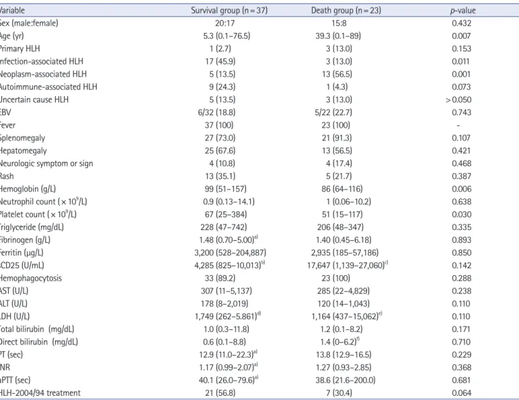
4. Comparison of the variables between survival and death groups of patients with HLH
A comparison of the variables between survival and death groups
is presented in Table 3. The median age at diagnosis was lower in the survival group (median, 5.3 years; range, 0.1–76.5 years) than that in the death group (median, 39.3 years; range, 0.1–89 years) (p=0.007). The prevalence of neoplasm-induced HLHs in the survival group (5/37, 13.5%) was lower than that in the death group (13/23, 56.5%) (p=0.001). The hemoglobin levels were higher in the survival group (median, 99 g/L; range, 51–157 g/L) than in the death group (median, 86 g/L; range, 64–116 g/L) (p=0.006). The platelet levels were higher in the survival group (median, 67 ×10
9/L; range, 25–384 ×10
9/L) than in the death group (median, 51×10
9/L; range, 15–117×10
9/L) (p=0.030).
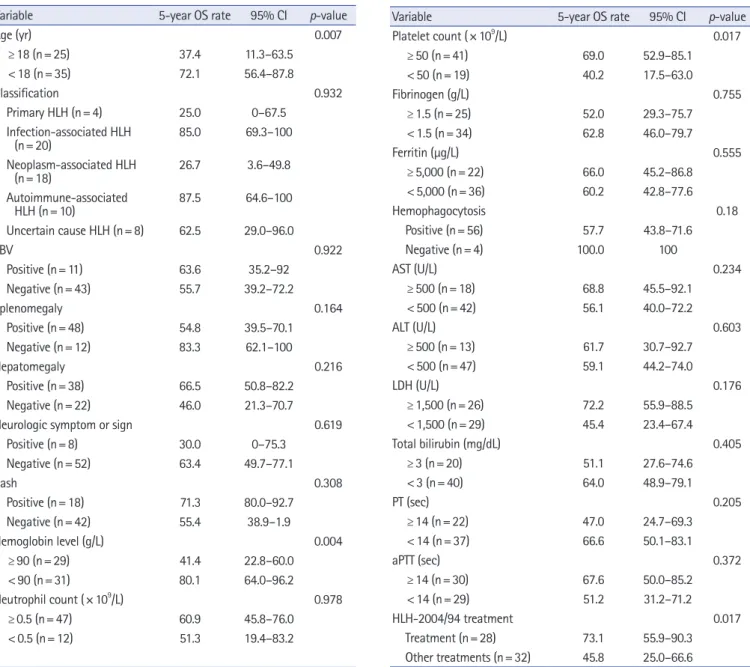
5. Survival of HLH patients
The 5-year OS rate of all patients with HLH (n=60) was 59.9%
(95% CI, 46.6–73.2) (Fig. 3A). The comparison of 5-year OS rates according to the variables by univariate analysis is shown in Table 4. According to age, the difference in 5-year OS between children (72.1%; 95% CI, 56.4–87.8) and adults (37.4%; 95%
CI, 11.3–63.5) was significant (p =0.007). Based on the HLH classification, the 5-year OS rates in patients with primary, infec- tion-associated, neoplasm-associated, autoimmune-associated, and uncertain cause of HLH were 25.0% (95% CI, 0–67.5), 85.0% (95% CI, 69.3–100), 26.7% (95% CI, 3.6–49.8), 87.5%
(95% CI, 64.6–100), and 62.5% (95% CI, 29–96), respectively.
Comparatively, the difference in the 5-year OS between the pri- mary and infection-associated HLHs was significant (p=0.024).
The difference in the 5-year OS between the neoplasm-associat- ed and infection-associated HLHs was significant (p =0.001).
Additionally, the difference in the 5-year OS between the neo- Fig. 2. Underlying causes of hemophagocytic lymphohistiocytosis according to the age groups. (A) Children, aged <18 years (n=35). (B) Adults, aged ≥18 years (n=25).
Uncertain 7 (20.0%)
Primary 3 (9.0%)
Infection 14 (40.0%)
Autoimmune
4 (16.0) Infection
6 (24.0%)
Neoplasm 13 (52.0%)
Primary 1 (4.0%) Uncertain
1 (4.0%)
Neoplasm 5 (14.3%) Autoimmune
6 (17.0%)
B A
■
Primary
■Infection
■Neoplasm
■Autoimmune
■Uncertain
■Primary
■Infection
■Neoplasm
■Autoimmune
■Uncertain
plasm-associated and autoimmune-associated HLHs was signifi- cant (p=0.010). The 5-year OS rate, according to the HLH clas- sification, is also shown as a graph in Fig. 3B. As per laboratory results, hemoglobin level of <90 g/L or platelet count of
<50 ×10
9/L at initial diagnosis of HLH were risk factors for a low 5-year OS rate.
In terms of the treatment regimen, the 5-year OS rate in patients treated with HLH-2004 protocol was 74.5% (95% CI, 54.9– 94.1), in those treated with HLH-94 protocol was 68.6% (95% CI, 32.1–
100), and in patients who received other treatments was 45.8%
(95% CI, 25.0–66.6). The difference in the 5-year OS rate be- tween HLH-2004 and HLH-94 protocol groups was not signifi- cant (p=0.904). The difference in the 5-year OS rate between pa- tients treated with HLH-2004/94 (73.1%; 95% CI, 55.9– 90.3) and other treatments (45.8%; 95% CI, 25.0–66.6) was significant (p=0.017). By multivariate analysis using the Cox proportional hazards model, neoplasm-induced HLH (hazard ratio, 4.446; 95%
CI, 1.876–10.538; p=0.001) and platelet count of <50×10
9/L at
initial diagnosis of HLH (hazard ratio, 3.298; 95% CI, 1.373–7.92;
p=0.008) were identified as independent risk factors of death in patients with HLH.
Discussion
In this study, we retrospectively investigated the causes and surviv- al rates in children and adults with different HLH subtypes. More- over, we analyzed the differences in clinical characteristics between children and adults with HLH. Based on the statistical analyses of this study, HLH was observed primarily in children (median age, 7 years); however, during the period from 2001 to 2019, the number of adult patients diagnosed with HLH had increased. During the review of the medical data for this retrospective study, several pa- tients suspected to have HLH and treated with corticosteroids or immunoglobulins were not included in this study because they had not undergone all tests corresponding to the diagnostic crite- ria of HLH. Therefore, the increase in the number of diagnoses Table 2. Comparison of the variables between children and adults with HLH
Variable Child (n= 35) Adult (n= 25) p-value
Sex (male:female) 21:14 14:11 0.481
EBV 7/34 (20.6) 4/20 (20.0) > 0.050
Fever 35 (100) 25 (100) -
Splenomegaly 24 (68.6) 24 (96.0) 0.010
Hepatomegaly 28 (80.0) 10 (40.0) 0.003
Neurologic symptom or sign 4 (11.4) 4 (16.0) 0.708
Rash 13 (37.1) 5 (20.0) 0.253
Hemoglobin (g/L) 90 (51–121) 89 (64–157) 0.685
Neutrophil count (× 10
9/L) 0.97 (0.06–14.1) 1.00 (0.18–10.2) 0.589
Platelet count (× 10
9/L) 67 (25–384) 52 (15–158) 0.184
Triglyceride (mg/dL) 219 (47–742) 244 (128–274)
a)0.988
Fibrinogen (g/L) 1.32 (0.70–4.22)
b)2.18 (0.45–6.18) 0.051
Ferritin (μg/L) 3,225 (185–204,887) 2,450 (764–19,640)
c)0.765
sCD25 (U/mL) 4,745 (825–27,060)
d)2,817
e)0.714
Hemophagocytosis 32 (91.4) 24 (96.0) 0.634
AST (U/L) 450 (22–5,137) 221 (11–2,237) 0.063
ALT (U/L) 232 (14–1,230) 120 (8–2,019) 0.024
LDH (U/L) 1,565 (266–5,861)
f)1,660 (262–6,369)
c)0.666
Total bilirubin (mg/dL) 1.2 (0.1–11.8) 1.1 (0.3–8.2) 0.584
Direct bilirubin (mg/dL) 0.6 (0.0–8.8) 1.3 (0.2–6.2)
g)0.617
PT (sec) 12.9 (11.7–31.6)
b)13.7 (11.0–19.4) 0.758
INR 1.19 (1.00–2.85)
b)1.18 (0.93–1.85) 0.515
aPTT (sec) 40.1 (25.5–200.0)
b)39.3 (21.6–68.8) 0.921
HLH-2004/94 treatment 28 (80.0) 0 < 0.001
Values are presented as number only, number (%), or median (range).
HLH, hemophagocytic lymphohistiocytosis; EBV, Epstein–Barr virus; sCD25, soluble CD25; AST, aspartate transaminase; ALT, alanine transaminase; LDH, lactate dehydrogenase; PT, prothrombin time; INR, international normalized ratio; aPTT, activated partial thromboplastin time.
a)
n=5,
b)n=34,
c)n=23,
d)n=13,
e)n=1,
f)n=32,
g)n=12.
Table 3. Comparison of the variables between the survival group and death group
Variable Survival group (n= 37) Death group (n= 23) p-value
Sex (male:female) 20:17 15:8 0.432
Age (yr) 5.3 (0.1–76.5) 39.3 (0.1–89) 0.007
Primary HLH 1 (2.7) 3 (13.0) 0.153
Infection-associated HLH 17 (45.9) 3 (13.0) 0.011
Neoplasm-associated HLH 5 (13.5) 13 (56.5) 0.001
Autoimmune-associated HLH 9 (24.3) 1 (4.3) 0.073
Uncertain cause HLH 5 (13.5) 3 (13.0) > 0.050
EBV 6/32 (18.8) 5/22 (22.7) 0.743
Fever 37 (100) 23 (100) -
Splenomegaly 27 (73.0) 21 (91.3) 0.107
Hepatomegaly 25 (67.6) 13 (56.5) 0.421
Neurologic symptom or sign 4 (10.8) 4 (17.4) 0.468
Rash 13 (35.1) 5 (21.7) 0.387
Hemoglobin (g/L) 99 (51–157) 86 (64–116) 0.006
Neutrophil count (× 10
9/L) 0.9 (0.13–14.1) 1 (0.06–10.2) 0.638
Platelet count (× 10
9/L) 67 (25–384) 51 (15–117) 0.030
Triglyceride (mg/dL) 228 (47–742) 206 (48–347) 0.335
Fibrinogen (g/L) 1.48 (0.70–5.00)
a)1.40 (0.45–6.18) 0.893
Ferritin (μg/L) 3,200 (528–204,887) 2,935 (185–57,186) 0.850
sCD25 (U/mL) 4,285 (825–10,013)
b)17,647 (1,139–27,060)
c)0.142
Hemophagocytosis 33 (89.2) 23 (100) 0.288
AST (U/L) 307 (11–5,137) 285 (22–4,829) 0.238
ALT (U/L) 178 (8–2,019) 120 (14–1,043) 0.110
LDH (U/L) 1,749 (262–5.861)
d)1,164 (437–15,062)
e)0.110
Total bilirubin (mg/dL) 1.0 (0.3–11.8) 1.2 (0.1–8.2) 0.171
Direct bilirubin (mg/dL) 0.6 (0.1–8.8) 1.4 (0–6.2)
f)0.710
PT (sec) 12.9 (11.0–22.3)
a)13.8 (12.9–16.5) 0.229
INR 1.17 (0.99–2.07)
a)1.27 (0.93–2.85) 0.368
aPTT (sec) 40.1 (26.0–79.6)
a)38.6 (21.6–200.0) 0.681
HLH-2004/94 treatment 21 (56.8) 7 (30.4) 0.064
Values are presented as number only, number (%), or median (range).
HLH, hemophagocytic lymphohistiocytosis; EBV, Epstein–Barr virus; sCD25, soluble CD25; AST, aspartate transaminase; ALT, alanine transaminase; LDH, lactate dehydrogenase; PT, prothrombin time; INR, international normalized ratio; aPTT, activated partial thromboplastin time.
a)
n=10,
b)n=10,
c)n=4,
d)n=35,
e)n=20,
f)n=16.
1.0 0.8 0.6 0.4 0.2 0.0
Probability of OS (%)
Time (yr)
5-yr OS 59.9%
5.0 10.0 15.0 20.0
1.0 0.8 0.6 0.4 0.2 0.0
Probability of OS (%)
Time (yr)
Uncertain 5-yr OS 62.5%
Primary 5-yr OS 25%
Neoplasm 5-yr OS 26.7%
Infection 5-yr OS 85%
Autoimmune 5-yr OS 87.5%