Address for correspondence: Ju Ock Na, M.D.
Department of Internal Medicine, Soonchunhyang University College of Medicine, 23-20 Bongmyeong-dong, Cheonan 330-721, Korea
Phone: 82-41-570-3666, Fax: 82-41-574-5762 E-mail: [email protected]
Received: Jan. 20, 2008 Accepted: Feb. 22, 2008
Primary Sjögren's Syndrome
Departments of
1Internal Medicine and
2Pathology, Soonchunhyang University College of Medicine, Cheonan, Korea Ji Yon Kim, M.D.
1, Hyun Gyu Hwang, M.D.
1, Jae Sung Choi, M.D.
1, Ki Hyun Seo, M.D.
1, Yong Hoon Kim, M.D.
1, Mee Hye Oh, M.D.
2, Ju Ock Na, M.D.
1다발성 폐 낭종을 보인 쇼그렌 증후군의 폐 침범 1예
김지연1, 황현규1, 최재성1, 서기현1, 김용훈1, 오미혜2, 나주옥1
순천향대학교 의과대학 천안병원
1내과학교실,
2병리학교실
쇼그렌 증후군은 림프구 침윤과 관련된 만성적인 염증성 자가면역 질환으로 아직 정확한 병태생리학적 기전은 밝혀지지 않았다. 45세 여자 환자가 내원 2년 전 전신 쇠약 및 피로감으로 입원하여 혈청 검사에서 anti- Ro/La antibody 양성, 흉부 단순방사선 및 컴퓨터 촬영에서 양 폐야의 다낭성 병변이 관찰되어 비디오 흉강경을 이용한 폐 생검 시행 결과 세기관지 주위에 림프구 침윤 및 다양한 크기의 폐낭종들이 관찰되어 쇼그렌 증후군의 폐 침범 의심하에 추가 검사 시행하려 하였으나 추적 관찰 되지 않았다. 2년 후 폐렴으로 입원하였으며, 다시 시행한 흉부 컴퓨터 단층촬영에서 다발성 낭성 변화는 큰 차이를 보이지 않았다. 쇼그렌 증후군의 폐 침범은 다양한 형태로 나타나는데, 단순히 세기관지 주위에 림프구 침윤에 의한 다낭성 폐 질환에 대한 보고는 극히 드물다. 따라서 본 저자들은 일차성 쇼그렌 증후군 환자에서 비디오 흉강경을 이용한 폐 생검으로 진단된 세기관지 주위에 림프구 침윤을 동반한 다낭성 폐 질환 1예를 경험하였기에 보고하는 바이다.
(Tuberc Respir Dis 2008;64:230-235)Key Words: Sjögren's syndrome, Lung, Cyst, Lymphocyte
Introduction
Sjögren's syndrome, a chronic inflammatory auto- immune exocrinopathy, that is characterized by dry eyes and dry mouth clinically and lymphocytic infiltration of lacrimal and salivary glands pathologically1. Lung in- volvement in Sjögren's syndrome usually consists of lymphocytic infiltration similar to that seen in salivary glands and results in tracheobronchial disease or inter- stitial lung disease2,3. Sjögren's syndrome is associated with various histologic patterns of interstitial lung dis- ease2,3. Although there have been earlier reports on lung involvement in Sjögren's syndrome2,4, there have been few reports on lung involvement with multiple
lung cysts caused by only peribronchiolar lymphocytic infiltration in Sjögren's syndrome. We describe herein the first case of Sjögren's syndrome in Korea which pre- sented as multiple cysts caused by only peribronchiolar lymphocytic infiltration and was confirmed by surgical lung biopsy. A brief review of the literature has been included.
Case Report
Patient: 45-year-old Korean woman
Chief complaints: presented with a 4-week history of dry cough, dyspnea, and dry mouth
Present illness: She was referred to our clinic 2 years ago because of general weakness and fatigue of 2-month duration. Prior to this presentation, she had been admi- tted to a regional hospital and treated under the clinical diagnosis of pneumonia and anemia. However, she did not improved, so she was referred to our clinic. At that time, she had no obvious pulmonary symptoms. Two years later, she was admitted to our hospital via the
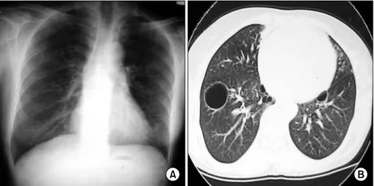
Figure 1. Chest radiographs and computed tomography at initial visit. (A) Chest radiographs showing bilateral multiple cysts, and (B) chest CT showing multiple well-demarcated, variable-sized cystic lesions in both lung fields.
emergency department due to dry cough, dyspnea, and dry mouth of 4-week duration.
Past, family and other history: She had no previous symptoms associated with autoimmune diseases except a history of dry eye for a duration of 6 months. There was neither a family history of autoimmune diseases, nor a history of exposure to drugs or alcohol.
Physical examinations: On physical examination, her blood pressure was 90/60 mmHg, pulse rate 90 beats/min, respiration rate 24/min, and temperature 36.5oC. She had pale conjunctivae and dry tongue, and inspiratory fine crackles were audible in both lower lung fields.
Laboratory findings: Laboratory data on admission showed hemoglobin 8.8 g/dl, hematocrit 29.1%, leuko- cyte count 3,800/mm3, platelet count 126,000/mm3, erythrocyte sedimentation rate (ESR) 60 mm/hr, C-re- active protein (CRP) 194.1 mg/L, serum iron 55μg/dl (normal, 65∼157), total iron binding capacity 217μg/
dl (normal, 250∼437), ferritin 78.9 ng/ml (normal, 30
∼400), aspartate aminotransaminase (AST) 19 IU/L, ala- nine aminotransaminase (ALT) 8 IU/L, sodium 137 mEq/L, potassium 3.1 mEq/L, chloride 104 mEq/L, total protein 7.3 g/dl, albumin 3.2 g/dl, blood urea nitrogen 9.7 mg/dl, creatinine 1.0 mg/dl, and β2 microglobulin was 5154.6 ng/ml (normal, 861∼1533). Arterial blood gas analysis at room air showed pH 7.416, pCO2 33.8 mmHg, pO2 97.2 mmHg, HCO3-
21.2 mmHg, and O2
saturation 97.2%. Serum antinuclear antibody (ANA) positive with a speckled pattern (titer 1:640) and pos- itive for anti- Ro/SS-A (201 IU/ml : 0∼10) and La/SS-B (243 IU/ml : 0∼15) antibody. However, anti-RNP anti- body, anti-smooth muscle antibody, anti-microsomal an- tibody, anti-Scl 70 antibody, ANCA, and anti-ds antibody were negative.
Serum C3 and C4 were 91.4 mg/dl (normal, 90∼180) and 27.0 mg/dl (normal, 10∼40), respectively. The serum immunoglobulin (Ig) showed IgG 1686.9 mg/dl (normal, 700∼1600), IgA 201.7 mg/dl (normal, 70∼400), and IgM 113.1 mg/dl (normal, 40∼230). Serum and urine electrophoresis and immunoelectrophoresis showed un- remarkable findings except that urine electrophoresis demonstrated a polyclonal pattern with increased amount of α1 and γglobulins. The Schirmer test showed 4/4 (right/left) mm/5 min (normal, >5/5 mm/5 min), which was suggestive of decreased lacrimation. Pulmonary function tests (PFT) showed decreased forced vital ca- pacity (FVC) 2.30 L (67% predicted normal), forced ex- piratory volume in 1 second (FEV1) 1.80 L (66% pre- dicted normal), FEV1/FVC 78%, forced expiratory flow (FEF)25∼75 1.64L (59% predicted normal), and diffusing lung capacity of carbon monoxide (DLCO) 10.29 ml/min/
mmHg (46% predicted normal), which indicated mild restrictive impairment combined with small airways ob- structive disease.
Figure 3. Histologic finding of wedge resected lung specimen. (A) Small cyst is noted (H&E stain, ×10). (B) Peribronchiolar lymphocytic infiltration without atypism is revealed (H&E stain, ×100).
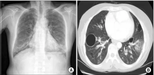
Figure 2. Chest radiographs and computed tomography at second time visit. (A) Chest radiographs showing multiple cystic lesions combined with pneumonic infiltration in both lower lungs, and (B) chest CT showing multiple cystic lesion of variable-size and some consolidations in both lower lungs with bronchovascular bundle thickening.
Radiologic findings: Chest radiographs and computed tomography (CT) showed multiple well-demarcated var- iable-sized cystic lesions in both lung fields (Figure 1).
Two years later, chest radiographs showed multiple cystic lesions in both lung fields combined with pneu- monic infiltration in both lower lungs. When compared with a prior chest CT, chest CT demonstrated multiple cystic lesions in both lung fields without any interval changes except some consolidations in both lower lung fields with bronchovascular bundle thickening (Figure 2).
Histopathologic findings: Bronchoscopy was performed
with transbronchial lung biopsy (TBLB). Bronchofibro- scopy showed no endobronchial lesions, and the speci- men taken from the right posterior basal segment by TBLB exhibited mild interstitial thickening and mild in- filtration of chronic inflammatory cells. Five days after bronchoscopy, lung biopsy was performed using vid- eo-assisted thoracic surgery (VATS). Microscopically, the resected lung specimen revealed multifocal, various- sized cysts and peribronchiolar lymphocytic infiltration without any atypism, but the other parenchymal tissue showed no abnormal findings (Figure 3), which was
consistent with lung involvement in Sjögren's syndrome.
Clinical course and treatment: Two years ago when the patient was suspected to have Sjögren's syndrome with lung, we planned further workup including oph- thalmoloic evaluation, but she was lost to follow-up after being discharged from the hospital. At the second ad- mission, she received 4 boluses of intravenous methy- prednisolone (62.5 mg daily), followed by oral pre- dnisolone (0.5 mg/kg/day) with intravenous antibiotics.
Ten days later, she improved and was discharged from the hospital with oral prednisolone (0.5 mg/kg/day) and oral antibiotics. One month later, dosage of oral prednisolone was tapered to 0.2 mg/kg/day. She has been followed up regularly without any significant inter- val change of multiple lung cysts on chest radiographs.
Discussion
Sjögren's syndrome is a slowly progressive chronic autoimmune disorder of the exocrine glands with asso- ciated lymphocytic infiltrates, which exhibits a wide range of organ-specific and systemic extraglandular manifestations including lung involvement1,3. Despite extensive studies of the underlying causes of Sjögren's syndrome, the pathogenesis remains obscure1. The syn- drome can be seen alone (primary Sjögren's syndrome) with a prevalence of about 0.5% to 3% or in association with other autoimmune diseases (secondary Sjögren's syndrome)3. Primary Sjögren's syndrome mainly affects women with a female-to-male ratio of 9:1, and may occur in patients at all ages, but it typically has its onset in the fourth to the sixth decades1,3.
Patients with Sjögren's syndrome have symptoms re- lated to diminished lacrimal and salivary gland function and frequently present with xerostomia, keratoconjuncti- vitis sicca, and parotid gland enlargement. Although it is commonly observed, pulmonary involvement is seldom clinically significant in patients with Sjögren's syndrome.
Dry cough is often the main respiratory symptom and is usually a manifestation of xerotrachea3. The reported frequency of pulmonary involvement in primary Sjögren's syndrome varies widely, ranging from 9% to 75% de-
pending on the detection method employed. Also, pri- mary Sjögren's syndrome is manifested as various forms of small airway and interstitial lung diseases2,4. Ito et al5 reported that 33 patients with primary Sjögren's syn- drome associated interstitial lung disease (ILD) and that nonspecific interstitial pneumonia (NSIP) was the most common histopathologic pattern, occurring in 61% of patients. Lymphocytic interstitial pneumonia (LIP) is a benign polyclonal proliferation, usually of mature B cells, that is either multifocal or diffusely involves the lungs. Histologically, LIP is characterized by massive in- terstitial lymphoid infiltrates predominately basilar mem- brane and diffusely spreading into the alveolar septa, although there may occasionally be some sparing of the lungs6.
Different kinds of PFT have been reported in many studies. Segal et al7 reported obstructive disease was de- tected in 37% of patients with Sjögren's syndrome, while Constrantopoulos et al8 found small airway disease in 22% of patients with Sjögren's syndrome. In the study of Newball and Brahim9, 46% of patients with Sjögren's syndrome showed obstructive disease. However, most studies reported higher incidences of restrictive dis- ease2,5. Small airway narrowing and obstructive lung disease are thought to be related to peribronchiolar lym- phocytic infiltration10,11. Airway narrowing due to peri- bronchiolar mononuclear cell infiltration causes a check-valve mechanism, which may lead to cyst for- mation11,12. The FEF25∼75 is often considered a more sensitive measurement of early airflow obstruction, par- ticularly in the small airways. However, this measure- ment must be cautiously interpreted because it is less reproducible10.
Although pulmonary involvement associated with Sjögren's syndrome has attracted attention recently, cyst formation has been rarely reported. Cyst formation, a rare pulmonary manifestation in Sjögren's syndrome, has been reported to be mainly associated with LIP.
Cysts with only peribronchiolar lymphocytic infiltration but without LIP features have rarely been reported.
From a review of the literature, only 8 cases were found around the world11-13. Compared with these cases, the
bronchiole were severely involved by dense lympho- cytic infiltrates in our case, but the interstitium of the alveoli were almost intact. Histologic findings of these reported cases showed lymphocytic infiltration through the interstitial space of the alveoli and a widening of the alveolar septa, whereas those of our case favor peri- bronchiolar lymphocytic involvement of Sjögren's syn- drome with multiple cysts than LIP associated with Sjögren's syndrome.
Until recently, there have been several sets of diag- nostic criteria for Primary Sjögren's syndrome1,3. Although minor salivary gland traditionally has been traditionally considered the "gold standard" for the diagnosis of Sjögren's syndrome, newer criteria permit classification of Sjögren's syndrome without necessarily performing this procedure. An American-European consensus com- mittee recently modified and reapproved the criteria.
These criteria encompass the presence of subjective and objective sicca manifestations, antibodies to Ro/SS-A and La/SS-B, and characteristic histopathologic findings in minor salivary glands3. Our case was diagnosed as Sjögren's syndrome based on ocular and oral sicca symp- toms, positive Schirmer test, and positive antibodies to anti- Ro/SS-A and La/SS-B.
Treatment of Sjögren's syndrome is mainly sympto- matic and is directed toward early diagnosis and treat- ment of its complications3. Topical agents have been used for improving moisture and decreaseing inflam- mation. Systemic treatment includes steroidal and non- steroidal anti-inflammatory agents, disease-modifying agents, and cytotoxic agents to address the extraglan- dular manifestations involving the skin, lung, heart, kidney, and nervous systems1.
The 5-year survival rate of Sjögren's syndrome with lung involvement was reported to be 84%. The causes of death were as follows; malignant lymphoma, NSIP, Aspergillus infection and gastrointestinal bleeding dur- ing corticosteroid therapy. Some of the studies showed an association between mortality and baseline PaO25
. Theander et al14 did not find an increase of mortality in patients with lung involvement of Sjögren's syndrome compared with the general population. Kruize et al15 re-
ported that a long-term follow-up (10 to 12 years) of 29 patients with primary Sjögren's syndrome showed a mild and stable course of glandular and extraglandular manifestations and that none of them developed clin- ically significant pulmonary diseases. Although Sjögren's syndrome is a benign and non-life threatening disorder, patients should be managed with appropriate treatment in order to improve quality of life and to avoid compli- cations. However, optimal treatment for patients with Sjögren's syndrome-associated specific disease remains to be defined2.
Summary
We described herein the first case of primary Sjögren's syndrome in Korea which presented with multiple cysts caused by only peribronchiolar lymphocytic infiltration, a rare pulmonary manifestation in Sjögren's syndrome, and was confirmed by surgical lung biopsy. A brief re- view of the literature has been included.
References
1. Fox RI. Sjögren's syndrome. Lancet 2005;366:321-31.
2. Parambil JG, Myers JL, Lindell RM, Matteson EL, Ryu JH. Interstitial lung disease in primary Sjögren syndrome.
Chest 2006;130:1489-95.
3. Kassan SS, Moutsopoulos HM. Clinical manifestations and early diagnosis of Sjögren syndrome. Arch Intern Med 2004;164:1275-84.
4. Jang DY, Shin BC, Jung KY, Kim JO, Yang JT, Joo YC, et al. A case of Sjögren's syndrome with interstitial lung disease and multiple cystic disease. Tuberc Respir Dis 2001;51:597-602.
5. Ito I, Nagai S, Kitaichi M, Nicholson AG, Johkoh T, Noma S, et al. Pulmonary manifestations of primary Sjögren's syndrome: a clinical, radiologic, and patho- logic study. Am J Respir Crit Care Med 2005;171:632-8.
6. Travis WD, Colby TV, Koss MN. Rosado-de-Christenson ML, Müler NL, King Jr TE. Non-neoplastic disorders of the lower respiratory tract. 2nd ed. Washington, DC:
American Registry of Pathology; 2002. p. 265-77.
7. Segal I, Fink G, Machtey I, Gura V, Spitzer SA.
Pulmonary function abnormalities in Sjögren's syn- drome and the sicca complex. Thorax 1981;36:286-9.
8. Constantopoulos SH, Papadimitriou CS, Moutsopoulos
HM. Respiratory manifestations in primary Sjögren's syndrome. A clinical, functional, and histologic study.
Chest 1985;88:226-9.
9. Newball HH, Brahim SA. Chronic obstructive airway disease in patients with Sjögren's syndrome. Am Rev Respir Dis 1977;115:295-304.
10. Papiris SA, Maniati M, Constantopoulos SH, Roussos C, Moutsopoulos HM, Skopouli FN. Lung involvement in primary Sjögren's syndrome is mainly related to the small airway disease. Ann Rheum Dis 1999;58:61-4.
11. Sakamoto O, Saita N, Ando M, Kohrogi H, Suga M, Ando M. Two cases of Sjögren's syndrome with multi- ple bullae. Intern Med 2002;41:124-8.
12. Kobayashi H, Matsuoka R, Kitamura S, Tsunoda N, Saito K. Sjögren's syndrome with multiple bullae and pulmonary nodular amyloidosis. Chest 1988;94:438-40.
13. Gardiner P, Ward C, Allison A, Ashcroft T, Simpson W, Walters H, et al. Pleuropulmonary abnormalities in pri- mary Sjögren's syndrome. J Rheumatol 1993;20:831-7.
14. Theander E, Manthorpe R, Jacobsson LT. Mortality and causes of death in primary Sjögren's syndrome: a pro- spective cohort study. Arthritis Rheum 2004;50:1262-9.
15. Kruize AA, Hene RJ, van der Heide A, Bodeutsch C, de Wilde PC, van Bijsterveld OP, et al. Long-term fol- low up of patients with Sjögren's syndrome. Arthritis Rheum 1996;39:297-303.