• 검색 결과가 없습니다.

Successful High Flow Nasal Oxygen Therapy for Excessive Dynamic Airway Collapse: A Case Report

N/A
N/A
Protected

Academic year: 2021

Share "Successful High Flow Nasal Oxygen Therapy for Excessive Dynamic Airway Collapse: A Case Report"

Copied!
4
0
0

로드 중.... (전체 텍스트 보기)

전체 글

(1)

455

Successful High Flow Nasal Oxygen Therapy for Excessive Dynamic Airway Collapse:

A Case Report

Jisoo Park, M.D.

1,2

, Yeon Joo Lee, M.D.

2

, Se Joong Kim, M.D.

2

, Jong Sun Park, M.D.

2

, Ho Il Yoon, M.D.

2

, Jae Ho Lee, M.D.

2

, Choon-Taek Lee, M.D.

2

and Young-Jae Cho, M.D.

2

1

Division of Pulmonology, Department of Internal Medicine, CHA University, CHA Bundang Medical Center, Seongnam,

2

Division of Pulmonary and Critical Care Medicine, Department of Internal Medicine, Seoul National University College of Medicine, Seoul National University Bundang Hospital, Seongnam, Korea

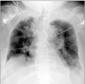
Excessive dynamic airway collapse (EDAC) is a disease entity of excessive reduction of the central airway diameter during exhalation, without cartilage collapse. An 80-year-old female presented with generalized edema and dyspnea at our hospital. The patient was in a state of acute decompensated heart failure due to pneumonia with respiratory failure. We accordingly managed the patient with renal replacement therapy, mechanical ventilation and antibiotics.

Bronchoscopy confirmed the diagnosis of EDAC. We scheduled extubation after the improvement of pneumonia and heart condition. However, extubation failure occurred due to hypercapnic respiratory failure with poor expectoration. Her EDAC was improved in response to high flow nasal oxygen therapy (HFNOT). Subsequently, the patient was stabilized and transferred to the general ward. HFNOT, which generates physiologic positive end expiratory pressure (PEEP) effects, could be an alternative and effective management of EDAC. Further research and clinical trials are needed to demonstrate the therapeutic effect of HFNOT on EDAC.

Keywords: Airway Obstruction; Oxygen Inhalation Therapy; Pressure

chea in forced expiration or during coughing. It is character- ized by an excessive collapse of the posterior membranous trachea towards the lumen without cartilage collapse

1-4

. EDAC presents with a variety of symptoms ranging from mild short- ness of breath or cough to respiratory failure

5,6

. Advances in imaging modalities, including bronchoscopy and dynamic radiographic studies, allow increased recognition of EDAC

7

. Current therapeutic management of EDAC includes conser- vative medical therapy, including bronchodilators and con- tinuous positive airway therapy, and minimally invasive and open surgical interventions

8

.

Recently, there has been growing interest in high flow nasal oxygen therapy (HFNOT). HFNOT has been known to gener- ate physiological positive airway pressure effect. Therefore, it was suggested that HFNOT could be replace to continuous positive airway pressure (CPAP) or non-invasive positive pres- sure ventilation (NIPPV)

9

. However, there was no study about HFNOT was used in management of EDAC.

Herein, we report a case of EDAC that caused inability to Copyright © 2015

The Korean Academy of Tuberculosis and Respiratory Diseases.

All rights reserved.

Introduction

Excessive dynamic airway collapse (EDAC) is defined as more than 50% reduction of the sagittal diameter of the tra-

CASE REPORT

http://dx.doi.org/10.4046/trd.2015.78.4.455

ISSN: 1738-3536(Print)/2005-6184(Online) • Tuberc Respir Dis 2015;78:455-458

Address for correspondence: Young-Jae Cho, M.D.

Division of Pulmonary and Critical Care Medicine, Department of Internal Medicine, Seoul National University Bundang Hospital, Seoul National University College of Medicine, 82 Gumi-ro 173beon-gil, Bundang-gu, Seongnam 13620, Korea

Phone: 82-31-787-7058, Fax: 82-31-787-4052 E-mail: [email protected]

Received: May 22, 2015 Revised: Jun. 19, 2015 Accepted: Jun. 19, 2015

cc

It is identical to the Creative Commons Attribution Non-Commercial

License (http://creativecommons.org/licenses/by-nc/4.0/).

(2)

J Park et al.

456 Tuberc Respir Dis 2015;78:455-458 www.e-trd.org

wean from mechanical ventilation, and which was success- fully managed with HFNOT.

Case Report

An 80-year-old female came to hospital because of general weakness for 1 week. She had diabetes mellitus and diastolic heart failure previously. She had symptoms like cough, in- creased sputum and 14 kg weight gain for 1 month (from 48 to 62 kg). At initial, her saturation (SpO

2

) was 78% in room air. There were wheezing and diffuse crackle sound in both whole lung fields. Multifocal patchy consolidations in right upper lobe and both lower lobe were presented in her chest anteroposterior and chest computed tomography (CT) (Fig- ure 1). White blood cell (WBC) count was 7,900/L, blood urea nitrogen/serum creatinine 48/1.83 mg/dL, FeNa 1.28%,

N-terminal of the prohormone brain natriuretic peptide 6,748 pg/mL, urine WBC ≥100/high-power field (HPF), urine bacte- ria 2+/HPF were checked. We assessed the patients with acute decompensated heart failure due to respiratory infection and acute kidney injury with urinary tract infection. Despite di- uretics, oxygen therapy, empiric antibiotics and bronchodila- tor therapy, her hypoxemia with hypercapnea was aggravated.

Eventually, she was intubated and mechanically ventilated in hospital day (HD) 1. For microbiologic evaluation of pu- rulent sputum, we performed diagnostic bronchoscopy in HD 3, coincidently we found out excessive dynamic airway compression in exhalation and purulent secretion in her bronchoscopy (Figure 2). Oxygen requirement of the patients was reduced in according to pneumonia was improved, so we tried to wean from mechanical ventilation in HD 13. How- ever, carbon dioxide (CO

2

) retention and poor expectoration of secretion cause failure to wean. Follow-up bronchoscopy was done for evaluating airway status, so we found EDAC was sustained (Figure 3). Therefore, we performed percutaneous dilatational tracheostomy and tried HFNOT with FiO

2

0.5 flow 30 L/min in HD 14. We found that the excessive dynamic tra- cheal compression in exhalation was decreased after applying of HFNOT (video file was showed in supplementary material, Video clip 1) (Figure 4), and CO

2

retention and expectoration were improved. After 3 days, she was stable, so she was trans- ferred to general ward with HFNOT.

Discussion

EDAC and tracheobronchomalacia (TBM) are two different disease entities, included in expiratory central airway collapse.

EDAC is a disease characterized by excessive bulging of the posterior membrane into the airway lumen during exhalation.

On the other hand, there is weakness of the tracheobronchial cartilaginous walls in TBM

1-4

. The reported prevalence of TBM and EDAC varies, reporting 4%–23% in patients undergoing bronchoscopy

3,10

, and EDAC was present in 22% in chronic

A B

Figure 2. The initial bronchoscopy shows excessive dynamic airway compression at exhalation (A) and inhalation (B).

Figure 1. The initial chest radiography at our hospital shows patchy

consolidations in left lower lung field and right upper lung field.

(3)

High flow nasal oxygen therapy for excessive dynamic airway collapse

http://dx.doi.org/10.4046/trd.2015.78.4.455 457

www.e-trd.org

obstructive pulmonary disease (COPD) and/or asthma pa- tients

4

.

These diseases are probably underdiagnosed because it shows nonspecific symptoms similar to patients with obstruc- tive lung disease like COPD and asthma

8

. They can cause chronic cough, dyspnea, difficulty clearing secretions, recur- rent bronchitis or pneumonia, syncope during episodes of coughing, pulmonary edema, and respiratory failure

5,6,11

. In the case of respiratory failure, the patient may experience dif- ficulties to wean or unexplained extubation failure

12

. Physical examination may reveal wheezing, rhonchi, and decreased breath sounds

6

.

EDAC can be diagnosed using dynamic bronchoscopy or dynamic radiologic imaging such as dynamic CT. Suggestive symptoms and pulmonary function test help to diagnosis of EDAC, but they have no diagnostic value and is not predictive of disease progress. Otherwise, we should know that EDAC di- agnosis is difficult because of bronchoscopy or CT performed under mechanical ventilation could give negative results

2

.

Treatment for EDAC depends on the severity of symptoms,

degree, and extent of airway collapse. Many patients with EDAC are asymptomatic, therefore they should not be treat- ed

8

. Proposed management strategies include conservative therapy (bronchodilators, CPAP, and NIPPV), minimally inva- sive therapy (endoluminal airway stents and laser therapy), surgical therapy (tracheostomy, airway splinting, and tracheal resection)

8

. Among them, CPAP and NIPPV act as pneumatic stents. Positive pressure application was known to decrease pulmonary resistance and improve airflow in patients with expiratory airflow obstruction

13

.

In this case, we diagnosed EDAC in patients by bronchos- copy. We could not perform pulmonary function test because of the patient was bed-ridden state in hospital. We could guess that EDAC of the patients contributed to retention of CO

2

and poor expectoration, therefore it caused failure to wean from mechanical ventilation. We tried HFNOT for applying intermittent positive end expiratory pressure (PEEP) in this case, and it worked well. To our knowledge, this case is the first case about HFNOT applying in management of EDAC.

HFNOT is known to have a number of physiological effects.

A B

Figure 3. The follow-up bronchoscopy in hospital day 13 showed that excessive dynamic tracheal compression was sus- tained. (A) At exhalation. (B) At inhala- tion.

A B

Figure 4. After applying of high flow na-

sal oxygen therapy, the bronchoscopy

shows excessive dynamic tracheal com-

pression was decreased at exhalation (A)

and inhalation (B).

(4)

J Park et al.

458 Tuberc Respir Dis 2015;78:455-458 www.e-trd.org

In previous several studies, HFNOT generates a low level of PEEP

14

, improves oxygenation, increased the end-inspiratory lung volume, reduces airway resistance, increased functional residual capacity

14

, and makes better mucociliary clearance

15

. Therefore, we recommend that HFNOT can be an effective management method in EDAC.

There are no randomized studies about HFNOT applying in EDAC. Therefore, further research and clinical trials are needed to demonstrate the therapeutic effect of HFNOT on EDAC.

Supplementary Material

Supplementary material can be found in the journal homepage (http://www.e-trd.org).

Video clip 1. The bronchoscopic video of case. While the sagittal diameter of the trachea collapses about 90% in initial 15 seconds, after high flow nasal oxygen therapy (HFNOT) applying, the diameter of the trachea was recovered. Then, the diameter of the trachea re-collapses after taking off the HF- NOT in the last 15 seconds.

Conflicts of Interest

No potential conflict of interest relevant to this article was reported.

References

1. Murgu SD, Colt HG. Description of a multidimensional clas- sification system for patients with expiratory central airway collapse. Respirology 2007;12:543-50.

2. Murgu SD, Colt HG. Tracheobronchomalacia and excessive dynamic airway collapse. Respirology 2006;11:388-406.

3. Kalra A, Abouzgheib W, Gajera M, Palaniswamy C, Puri N,

Dellinger RP. Excessive dynamic airway collapse for the in- ternist: new nomenclature or different entity? Postgrad Med J 2011;87:482-6.

4. Park JG, Edell ES. Dynamic airway collapse: different from tracheomalacia. Rev Port Pneumol 2005;11:600-2.

5. Murgu SD, Cherrison LJ, Colt HG. Respiratory failure due to expiratory central airway collapse. Respir Care 2007;52:752-4.

6. Nuutinen J. Acquired tracheobronchomalacia: a clinical study with bronchological correlations. Ann Clin Res 1977;9:350-5.

7. Murgu SD, Colt HG. Tracheobronchomalacia and excessive dynamic airway collapse: novel diagnostic tools clarify the is- sues. Pulm Perspect 2005;22:7-10.

8. Murgu SD, Colt HG. Treatment of adult tracheobronchoma- lacia and excessive dynamic airway collapse: an update. Treat Respir Med 2006;5:103-15.

9. Gotera C, Diaz Lobato S, Pinto T, Winck JC. Clinical evidence on high flow oxygen therapy and active humidification in adults. Rev Port Pneumol 2013;19:217-27.

10. Ikeda S, Hanawa T, Konishi T, Adachi M, Sawai S, Chiba W, et al. Diagnosis, incidence, clinicopathology and surgical treat- ment of acquired tracheobronchomalacia. Nihon Kyobu Shi- kkan Gakkai Zasshi 1992;30:1028-35.

11. Firdose R, Elamin E. Pulmonary edema secondary to dynam- ic tracheal collapse. J Bronchol 2004;11:118-21.

12. Carden KA, Boiselle PM, Waltz DA, Ernst A. Tracheomalacia and tracheobronchomalacia in children and adults: an in- depth review. Chest 2005;127:984-1005.

13. Martin JG, Shore S, Engel LA. Effect of continuous positive air- way pressure on respiratory mechanics and pattern of breath- ing in induced asthma. Am Rev Respir Dis 1982;126:812-7.

14. Sztrymf B, Messika J, Bertrand F, Hurel D, Leon R, Dreyfuss D, et al. Beneficial effects of humidified high flow nasal oxygen in critical care patients: a prospective pilot study. Intensive Care Med 2011;37:1780-6.

15. Parke RL, Eccleston ML, McGuinness SP. The effects of flow on airway pressure during nasal high-flow oxygen therapy.

Respir Care 2011;56:1151-5.

수치

Figure 2. The initial bronchoscopy shows  excessive dynamic airway compression  at exhalation (A) and inhalation (B)
Figure 3. The follow-up bronchoscopy  in hospital day 13 showed that excessive  dynamic tracheal compression was  sus-tained

참조

관련 문서

this study analysed cast speed and solidification in thermal and flow perspectives and based on the results, conducted a confidence test on the high-speed general purpose

Effect of Soru Lower-leg Massage Therapy on Heart Rate Variability and Reactive Oxygen Species Concentration in

Applying Design Thinking as a Method for Developing Multi Competencies of Technical High School Student..

Martonosi, "Dynamic thermal management for high- performance microprocessors," Proceedings of International Symposium on High-Performance Computer

In addition, if the patients had a high MPV level (cut-off value of 7.95 fL) without low-dose aspirin therapy, they were at risk for ischemic stroke, especially in

The change in active oxygen was decreased after exercise in the exercise group than in the control group, and there was a statistically significant

7.2 Turbulent Flow and Eddy Viscosity 7.3 Fluid Flow Past Solid Boundaries 7.4 Characteristics of Boundary Layers 7.5 The Laminar Boundary Layer*.. 7.6 The

- Turbulence is generated primarily by friction effects at solid boundaries or by the interaction of fluid streams that are moving past each other with different velocities