Res. Plant Dis. 15(3) : 170-174 (2009)
©The Korean Society of Plant Pathology
국내 복숭아에서 분리한 Prunus necrotic ringspot virus의 특성
김현란*·이신호1·신일섭·김정희·조강희·허 성·김정수2·최용문3 농촌진흥청 국립원예특작과학원 과수과
,
1한국과수농협연합회,
2농촌진흥청 국립농업과학원
,
3농촌진흥청Characterization of Prunus necrotic ringspot virus Isolate from Peach in Korea
Hyun Ran Kim
*, Sin-Ho Lee
1, Il-Sheob Shin, Jeong Hee Kim, Kang-Hee Cho, Seong Heo, Jeong Soo Kim
2and Yong Mun Choi
3Fruit Research Division, National Institute of Horticultural & Herbal Science, Rural Development Administration, Suwon 441-440, Korea
1
Korea Fruit Agricultural Co. Federation, Seoul 137-133, Korea
2
National Academy of Agricultural Science, R.D.A. Suwon 441-707, Korea
3
Rural Development Administration, Suwon 150, Korea (Received on July 23, 2009; Accepted on October 28, 2009)
In this paper, we report a characterization of Prunus necrotic ringspot virus (PNRSV) isolate. The virus was identified from ‘Yumyeong’ peach showing mild mosaic on leaves in commercial orchard of ‘Umsung’, Chungbuk province in Korea. The virus isolate produced ringspot symptom on the inoculated cotyledons and systemic mosaic and malformation on the upper leaves of Cucumis sativus . Systemic mottles were appeared in Chenopodium quinoa . When the buds of the virus infected stem were grafted on the healthy young Prunus persica GF305 seedlings, line pattern with mosaic appeared within 3 months. Isometric virus-like particles were found in parenchyma cells and plasmodesmata of C. sativus leaves inoculated mechanically with the virus. The cDNA fragments of PNRSV coat protein (CP) region, approximately 675bp, were synthesized from genomic RNA extracted from virus-infected leaves by RT-PCR using specific primer pairs. Partial nucleotide sequences of the CP regions were determined and analyzed with the known PNRSV. The CP gene of PNRSV- Korea isolates showed 93.9~94.7% similarity to the 4 known PNRSV isolates.
Keywords : Ilarvirus, Indicator assay, RT-PCR detection, Peach, PNRSV
복숭아
,
살구,
자두 등핵과류에는 전세계적으로 심각한문제를 일으키는
Plum pox virus (PPV)
를포함하여 약18
여종의 바이러스와3
종의바이로이드가 보고되어있다.
국내에서는 복숭아나무에서의
Prunus necrotic ringspot virus (PNRSV)
발생이김 등(2001)
에 의해발표된 보고가있을뿐 복숭아 바이러스병에관한논문은 거의없는 실 정이다
.
한편
, PNRSV
는 전 세계적으로 복숭아,
양앵두 등 핵과류에서 경제적인 피해를 주는 주요 바이러스 중의 하 나로 보고되어 있으며
(Smith
등, 1972; Wood
등, 1979)
분류학적으로는
Family Bromoviridae , Genus Ilarvirus subgroup 3
에 속하는 바이러스이다(Alan
등, 1995).
바이러스 입자는 직경
23 nm
정도의 크기로 구형과 간상형의 입자로존재한다
.
접목,
화분에 의해전염되고 종자로도 전염이가능한 것으로 알려져 있으며 초본식물에서는 즙액접종으로감염되는것으로보고되어있다
(Fulton, 1970;
1983).
현재까지국내에서
Ilarvirus
에관한연구는Apple mosaic virus (ApMV)
의 진단기술에 대한 보고가 있지만(
이 등, 2002;
최등, 2003)
생물검정을통한바이러스 분리특성및 유전자 분석에 관한국내 보고는 없다
.
따라서
,
본연구는Ilarvirus
그룹에속하는 바이러스중에 우리나라 복숭아 과원에서 검출된
PNRSV isolate
의특성을 조사하고자 초본 및 목본 지표식물을 이용한 생
*Corresponding author
Phone) +82-31-240-3673, Fax) +82-31-240-3709
Email) [email protected]
물검정과 세포 내 바이러스입자 존재양식을 관찰하였으 며
,
특이프라이머를 디자인하여RT-PCR
에 의해증폭된국내 분리주 외피단백질 유전자 특성을 확인하여 기 보 고된분리주와의 유전적 상동성을비교함으로써국내 분 리주를 동정하였다
.
재료 및 방법
바이러스 생물검정. 충북 음성지역 등 복숭아 주산단 지의 과수원에 재식되어 있는 ‘유명’ 등 다양한 품종의
성목을 대상으로스위스
BIOREBA
사의 항혈청을 이용하여
ELISA
진단법으로 바이러스 감염여부를 스크리닝하여
PNRSV
감염주를선발하였다. PNRSV
단독검출주로부터 잎과가지 부위를 채취하여초본지표식물 및 목 본지표식물을이용한생물검정에활용하였다
.
초본지표식물 검정에서는
Ilarvirus
의 분리기주로 많이 활용되는C. sativus
를포함하여5
종의지표식물을이용하였으며 모자이크증상을 보이는 어린 복숭아 잎을
C. sativus
의 자엽조직에 즙액 접종한 후 접종엽과 상엽의 병징 발현여 부를관찰하였다
.
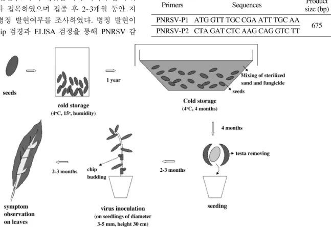
목본지표식물검정은핵과류바이러스에대해감수성품종으로보고되어있는
P. persica GF305
품종의 종자를이용하여
Fig. 1
과 같이 실생묘를 양성한다음
PNRSV
감염주로부터채취한 녹색가지의 눈 부위를 메스로 잘라 접목하였으며 접종 후
2~3
개월 동안 지표식물 잎의 병징 발현여부를 조사하였다
.
병징 발현이관찰된 잎은
dip
검경과ELISA
검정을 통해PNRSV
감염여부를 확인하였다
.
전자현미경검경. 인위접종한
C. sativus cv. Beakbong
에서 상엽의 모틀
(mottle)
과 기형증상을 보이는 잎을 전자현미경검경용시료로 이용하였다
.
잎절편을1~2 mm
2크기로 잘라서
karnovsky
고정액으로 고정하고Spurr's
resin
으로블록을만든다음다이아몬드나이프로자른초박절편을
2% uranyl acetate
와lead citrate
로 이중염색하는방법
(Hayat, 1972)
을이용하였으며Carl Zeiss LEO 906 TEM
으로 세포내 바이러스 입자를 관찰하였다.
RT-PCR 및염기서열분석. 병징을나타내는
C. sativus
상엽을 절취하여
Qiagen RNeasy Plant kit
를 이용, total
RNA
를 추출하여PCR
진단에 사용하였다. GenBank
database
에 공개되어 있는 염기서열정보와RT-PCR
기술(Spiegel
등, 1996, Hammond
등, 1998)
을 참고하여PNRSV
외피단백질 유전자부분을 기준으로 특이프라이머를
Table 1
과 같이 디자인하여RT-PCR
에이용하였다. RT-PCR
증폭조건은45
oC 45
분(1cycle)
→94
oC 2
분(1 cycle)
처리 후
94
oC 30
초→60
oC 1
분→68
oC 2
분간40 cycle
처리하였으며 최종적으로
68
oC
에서7
분1 cycle
처리한 후1% Agarose gel
상에서 전기영동하여ethidium bromide
Fig. 1. Bioassay protocols using P. persica ‘GF305’ woody indicator.
Table 1. Oligonucleotide primers used to amplify the PNRSV coat protein gene
Primers Sequences Product size (bp) PNRSV-P1 ATG GTT TGC CGA ATT TGC AA
PNRSV-P2 CTA GAT CTC AAG CAG GTC TT 675
로염색하여특이밴드를확인하였다
.
증폭된 유전자의염기서열 분석은
ABI Prism 377 Genetic Analyzer
를 이용,
Bioneer
사에 의뢰하여 결과를 획득하였으며분석된 염기서열 결과는
DNASTR
프로그램을 이용하여Genbank
에기 보고된 분리주와의 상동성을 비교하였다
.
결과 및 고찰
충북 음성 등 복숭아 주요 재배과원의 성목을 대상으
로
ELISA
법으로PNRSV
감염여부를 스크리닝 한 결과음성지역 농가의 ‘유명’ 품종의 약한 모자이크증상의 잎
에서
PNRSV
양성반응이 확인되었다.
모자이크 증상을나타내는 잎을
Table 1
에서와 같이Chnopodium quinoa
등
5
종의초본지표식물에 접종하여반응을조사한 결과, C. quinoa
와C. amaranticolor
의상엽에mottle
병징이 각각 관찰되었으며
C. sativus
의 접종자엽에ringspot
와 상엽에
mosaic
병징이 나타나다가 잎이기형화되고선단부의 생육이 불량해지면서 고사되는 것을 관찰할 수 있었 다
(Table 2).
이러한 지표식물의반응은Salem
등(2004)
이보고한 결과와 일치하였다
.
P. persica GF305
품종을 이용한목본지표식물 검정에서는 접종
3
개월후부터 신엽에vein banding
증상이 관찰되었다
(Fig. 2). C. sativus
의 접종엽에 나타난 국부 병반을 채취하여 재 접종한 다음
2
차 병징이 발현된 잎의혈청학적 반응을 조사하기 위해 잎을 절취하여
PNRSV,
Prune dwarf virus (PDV)
및Cherry leafroll virus (CLRV)
등 핵과류에 감염 가능한 구형 바이러스
3
종(Alan
등,
1995)
의항혈청에 대해ELISA
진단한 결과, C. sativus
와P. persica GF305
품종모두PDV
와CLRV
항혈청에 대해서는반응이 나타나지않은 반면
PNRSV
에 대해서만 양성반응을 보여
PNRSV
감염에 의한 병징인 것으로 확인할 수 있었다
.
기보고된생물검정에서도
C. sativus
에서자엽에yellow chlorotic local lesion
이 나타나고point stunt
가 나타난다고보고되어 있으며
, C. quinoa
에서systemic mottle
증상이 나타나는 것으로 보고되어 있어
(Fulton, 1970, 1983),
본연구에서 얻은결과와 유사하였다
.
보통PNRSV
의 유지 증식 기주로서
C. sativus
를 사용하는 것으로 보고되어 있으며
(ICTV DB 00.010.0.02.015)
본 연구에서도 시험용 바이러스원을
C. sativus
접종주를 활용하였다. C. sativus
이병세포에 대한TEM
검경에서 엽육조직세포내에 구형의 바이러스입자가 존재하는 것을관찰할
수 있었으며
Plasmodesmata
내 바이러스 입자가 존재하Table 2. Reaction of indicator plants inoculated with PNRSV-Ko Indicator plants Response on leaves
Inoculated Upper Chenopodium quinoa Moa Mo Chenopodium amranticolor NS NS Nicotiana glutinosa NS NS Nicotiana tabacum cv. Xanthi NS NS Cucumis sativus RS M, Mf, TN
a
M; mosaic, Mf; malformation, Mo; mottle, NS; no symptoms, RS;
ringspot, TN; Top necrosis.
Fig. 2. Symptoms of PNRSV on peach and indicator plants. (A) mild mosaic on peach, (B) ringspots on C. sativus cotyledons, (C) mosaic and malformation on C. sativus, (D) mosaic with line pattern on P. persica GF305.
Fig. 3. Electron micrographs of C. sativus leaves inoculated mechanically with PNRSV-Ko. Arrows indicate virus particles.
는형태도 확인할 수있었다
(Fig. 3).
이는바이러스 입자형태가 구형인 바이러스 종인
Nepovirus , Comovirus , Fabavirus , Ilarvirus
등이 공통적으로 가지는 세포병리학 적 특성으로 보고되어 있다(Martelli, 1980).
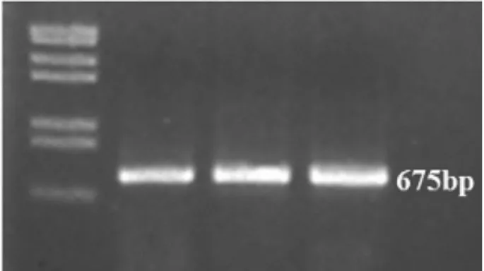
PNRSV
외피단백질유전자를 특이적으로 증폭하도록디자인한
Table 1
의프라이머를이용하여C. sativus
접종엽을
RT-PCR
한 결과약675bp
크기의특이적인 증폭산물을 얻을수 있었다
(Fig. 4).
증폭된 특이산물을 회수하여 염기서열 분석을 수행하
고
NCBI
에 공개된 외국의 기보고 분리주4
개와 유전적상동성을 비교한결과약
93.9~94.7%
의상동성을나타내는것으로확인되었고
(
자료생략),
이에대한phylogenetic tree
를 작성하였다(Fig. 5).
본 논문은
PNRSV
에 대한국내 첫보고로서 생물검정결과와
RT-PCR
진단 기술은금후전국적인 발생모니터링과 피해구명에 관한 연구의 기반기술로 활용될 수 있 을 것으로 판단된다
.
요 약
국내의 복숭아 과수원 중에 충북 음성지역의 유명 품 종에서 약한 모자이크증상 잎으로부터
Prunus necrotic
ringspot virus (PNRSV)
를 분리 동정하였다.
지표식물을이용한 생물검정에서
Cucumis sativus
의 접종 자엽에 원 형모양의 반점이 관찰되었으며 상엽으로 전이되어 모자 이크와 잎기형 증상을 나타내다가고사하는 것으로 나타 났다.
접종한Chenopodium quinoa
접종엽과상엽에도 모 틀증상을나타내었다.
목본지표식물Prunus persica GF305
를 활용한 검정에서는 바이러스 감염된 줄기의 눈을 인 위적으로 접종한 지
3
개월 후에 잎에 라인 패턴의 모자이크 증상을 관찰할 수 있었다
.
인위접종한
Cucumis sativus
잎조직의 세포 검경에서parenchyma cells
과plasmodesmata
내에존재하는구형바이러스 입자가 관찰되었다
.
PNRSV
의 외피단백질 유전자 분석을 위해RT-PCR
진단프라이머와조건을 설정하고분석한 결과
675bp
위치에서특이적으로증폭된것을확인할 수있었다
.
또한증폭산물을 회수하여염기서열 분석한 결과기보고된
4
개의
PNRSV isolate
와93.9~94.7%
의유전적상동성을보였다.
결론적으로복숭아과수원에서분리한바이러스주의생 물적 특성과 유전자분석결과를 종합하여
Ilarvirus
에 속하는
PNRSV
인 것으로 최종동정하였다.
참고문헌
Alan, B., Karen, C., Michael, D., Adrian, G. and Leslie, W. 1995.
Viruses of Plants ; Descriptions and lists from the VIDE Database. pp.100-102. CAB International.
Choi, S. H. and Ryu, K. H. 2003. Rapid screening of Apple mosaic virus in cultivated apples by RT-PCR.
Plant Pathology J.19: 159-161.
Fulton, R. W. 1970. Prunus necrotic ringspot virus. CMI/AAB Description of plant viruses. No. 5.
Fulton, R. W. 1983. Ilarvirus group. CMI/AAB Description of plant viruses. No. 275.
Hammond, R., Howell, W. E., Mink, G. I. and Crosslin, J. M.
1998. Strain-specific polymerase chain reaction assays for discrimination of Prunus necrotic ringspot virus isolates. Acta Hort. 472.
Hayat, M. A. 1972. Principles and Techniques of Electron Microscopy. New York : Van Nostrand Reinhold.
http://www.ncbi.nlm.nih.gov/ICTVdb/ICTVdB/00.010.0.02.015.
Prunus necrotic ringspot virus
Kim, H. R., Chung, B. N., Choi, G. S., Choi, Y. M. and Kim, J. S.
2001. Identification and characterization of Prunus necrotic ringspot virus, the family Ilarvirus, from peach.
Plant Pathol.J.
17: 386-387.
Lee, G. P., Ryu, K. H., Kim, H. R., Kim, C. S., Lee, D.W., Kim, J.
S., Park, M. H., Noh, Y. M., Choi, S. H., Han, D. H. and Lee, C. H. 2002. Cloning and phylogenetic characterization of coat
Fig. 4. Agarose gel electrophoretic analysis of cDNA products from RT-PCR of PNRSV-infected C. sativus leaves using PNRSV-CP gene-specific primers.
Fig. 5. Phylogenic tree analysis of the CP gene nucleotide sequences of PNRSV-Ko isolate. PNRSV-ku, Genbank accession No. AY037791; PNRSV-gg, AY037788 ; PNRSV-nt, AY037790 ; PNRSV-yug, AY037789 ; PNRSV-Ko.
protein genes of two isolates of Apple mosaic virus from Fuji apple.
Plant Pathol. J.18: 259-265.
Martelli, G. P. 1980. Ultrastructural aspects of possible defence reactions in virus-infected plant cells.
Microbiologica3: 369- Salem, N., Mansour, A., Al-Musa, A., Al-Nsour, A. and 391.
Hammond, R. 2004. Identification and partial characterization of Prunus necrotic ringspot virus on stone fruits in Jordan.
J.Plant Pathology.