․교신저자: 설인찬 대전광역시 중구 대흥동 22-5번지 대전대학교 부속 대전한방병원 심계내과 TEL: 042-229-6805 FAX: 042-254-3403 E-mail: [email protected]
循環器加減方이 고지혈증 유발 白鼠에 미치는 영향
김동현, 임승민, 안정조, 조현경, 유호룡, 김윤식, 설인찬 대전대학교 한의과대학 심계내과학교실
Effect of Sunhwangigagambang (SHG) on Hyperlipidemia in SD Rats Induced by High Cholesterol Diet
Dong-hyun Kim, Seung-min Lim, Jung-jo Ahn, Hyun-kyung Jo, Ho-ryong Yoo, Yoon-sik Kim, In-chan Seol Dept. of Internal Medicine, College of Oriental Medicine, Dae-jeon University
ABSTRACT
Objective :
This study was performed to investigate the effect of Sunhwangigagambang(SHG) on hyperlipidemia in SD rats induced by high cholesterol diet.
Method :
After treatment with SHG, cytotoxicity, body weight, liver weight, AST, ALT, ALP, total cholesterol, LDL-cholesterol, HDL-cholesterol, triglyceride, glucose, albumin, total protein in serum, malondialdehyde, and gene expression for ACAT and HMG-CoA reductase in hepatic tissue were analyzed.
Result :
1. SHG didn't show any cytotoxicity in both human fibroblast cell line and SD rats.
2. SHG significantly inhibited the increase of liver weight by high cholesterol diet compared to the control group.
3. SHG significantly ameliorated the increase of total cholesterol, LDL-cholesterol, and triglyceride and reduction of HDL-cholesterol compared to the control group.
4. SHG significantly reduced glucose level in serum compared to the control group.
5. SHG significantly reduced malondialdehyde in hepatic tissue compared to the control group.
6. SHG significantly down-regulated gene expression of ACAT and HMG-CoA reductase compared to the control group.
Conclusion :
These results suggest that SHG might be effective in treatment and prevention of hyperlipidemia.
Key words : Sunhwangigagambang(SHG), Hyperlipidemia, HMG-CoA reductase, Cholesterol
Ⅰ. 서 론
뇌혈관 질환 및 관상동맥 질환의 공통되는 위험 인자에는 고지혈증, 고혈압, 당뇨, 흡연 등이 있다
1,2
. 이들 중 동맥경화의 위험인자로 잘 알려진 고 지혈증을 관리하는 것이 순환기 질환의 예방에 중
요하다
3.
고지혈증은 한의학적으로 痰濁, 血瘀의 범주에 속하고, 嗜食肥甘厚味의 外因과, 肝脾腎機能失調의 內因에 의해 痰濕, 瘀血이 生하여 발병한다고 보았 다. 따라서 滋補肝腎, 調理脾胃로 扶正하고, 利濕化 痰, 活血化瘀로 祛邪하는 補瀉兼施의 治法을 응용 할 수 있다
4.
최근 降脂通脈飮
5, 雙降湯
6, 加味地黃湯
7등의 고
지혈증 관련 인자에 대한 유효성이 실험적으로 밝
혀지고 있다. 이에 저자는 기존 논문들에서 고지혈
증에 대한 유효성이 확인 된 豨簽
8, 芍藥
9, 當歸
10등의 약재로 구성된 循環器加減方을 이용하여 항 고지혈증 효과를 실험적으로 규명하고자 고콜레스테 롤 식이로 유발된 고지혈증 Sprague-Dawley(SD) rat 에 循環器加減方을 투여 후 aspartate aminotransferase (AST), alanine aminotransferase (ALT), alkaline phosphatase (ALP), 몸무게, 간무게, TC, LDL-C, HDL-C, TG, glucose, albumin, total protein, 간조 직의 과산화지질 함량, 간조직의 acetocoenzyme A acetyltransferase (ACAT) 및 3-hydroxy-3-methylglutaryl -coenzyme A (HMG-CoA) reductase의 mRNA 유 전자 발현 등을 측정하여 유의성 있는 결과를 얻 었기에 보고하는 바이다.
Ⅱ. 실 험
1. 재 료
1) 동물 및 사육조건
본 실험에 사용된 실험용 쥐는 체중 180∼220g 의 웅성 SD rat으로 실험 당일까지 일반 사료와 고콜레스테롤 사료 (Bio-serv, USA)를 자유식이하 면서 식수를 충분히 공급하였다. 실온 22 ± 2℃, 상 대습도 50 ± 10%, 조명시간 12시간 (07:00∼19:00), 조도 150∼300Lux로 설정하여 2주일간 실험실 환 경에 적응시킨 후 체중 변화가 일정하고 건강한 쥐만을 선별하여 실험에 사용하였다.
2) 약 재
본 실험에 사용한 循環器加減方( Sunhwangigagambang , 이하 SHG)의 구성 약물은 대전대학교 부속한방병 원에서 구입하여 정선한 후 사용하였다(Table 1).
Name Herb General Name Amount
豨 簽 Siegesbeckiae Herba 12g
白芍藥 Paeoniae Radix 8g
當 歸 Angelicae Gigantis Radix 6g
牛膽南星 Arisaematis Rhizoma 3g
半 夏 Pinelliae Rhizoma 3g
白 朮 Atractylodes Rhizome Alba 3g
白茯笭 Hoelen Alba 3g
天 麻 Gastrodiae Rhizoma 3g
川 芎 Cnidii Rhizoma 3g
酸棗仁 Zizyphi Spinosi Semen 3g 陳 皮 Aurantii Nobilis Pericarpium 3g
黃 芩 Scutellariae Radix 3g
熟地黃 Rehmanniae Radix Preparata 3g
防 風 Peucedani Radix 3g
桃 仁 Persicae Semen 3g
薄 荷 Menthae Herba 3g
桂 枝 Cinnamomi Ramulus 3g
羌 活 Angelicae Koreanae Radix 3g
牛 膝 Achyranthis Radix 3.2g
生地黃 Rehmanniae Radix Crudus 3.2g
紅 花 Carthami Flos 1.8g
炙甘草 Glycyrrhizae Radix 1.8g
黃 柏 Phellodendri Cortex 1.2g 竹 瀝 Bambusae Caulis in Liquamen (10㎖) 10g
Total Amount 92.2g Table 1. Composition of Sunhwangigagambang (SHG)
3) 시약 및 기기 (1) 시 약
Dulbecco's phosphate buffered saline (DPBS-A;
Sigma Co., U.S.A.), RPMI 1640 (Sigma Co., U.S.A), Collagenase A (BM, U.S.A), DNase type Ⅰ (Sigma.
Co., U.S.A.), Penicillin (Sigma. Co., U.S.A.), Streptomycin
(Sigma. Co., U.S.A.), Deoxycorticosterone Acetate
(DOCA; Sigma., U.S.A), Trypsin (invitrogen., U.S.A),
Ethylenediaminetetraacetic acid (EDTA; Sigma., U.S.A),
3-(4,5-dimethylthiazol-2-yl)-2,5-diphenyltetrazolium
bromide (MTT; Sigma., U.S.A), Dimethyl sulfoxide
(DMSO; Sowa chemical., Japan), 3.8% Sodium citrate (Sigma Co., U.S.A), Angiotensin converting enzyme (ACE, Sigma Co., U.S.A), Hippuryl-his-leu acetate (Sigma Co., U.S.A), Ethylacrtate(Junsei., Japan), Potassium Phosphate Monobasic (KH2PO4; Yakuri., Japan), Bovine serum albumin (BSA; Sigma, U.S.A) 등을 사용하였고, 이 밖에 일반 시약은 특급 시약 을 사용하였다.
(2) 기 기
본 연구에 사용된 기기는 Accoutered GC (Roche, Germany), Ice-maker (Vision, Korea), Serum separator (녹십자, Korea), Minos-ST (Cobas Co., France), Centrifuge (Beckman Co., U.S.A.), Rotary vaccum evaporator (Büchi 461, Switzerland), Deep freezer (Sanyo Co., Japan), Freeze dryer (Eyela Co., Japan), Autoclave (Hirayama, Japan), Ultrasonic cleaner (Branson Ultrasonics Co., U.S.A.), ELISA reader (Molecular Divice., U.S.A), Roller Mixer (Gowon scientific technology Co., Korea), 한약유출기 (DWP -1800T, 웅진, Korea), Spectrophotometer (UV-2450, Shimazu, Japan), Balance (Cass, korea), 생화학기 기 (AU400, Olimpus, U.S.A) 등을 사용하였다.
2. 방 법 1) 약물 추출
시료 추출 방법은 SHG 2첩을 한약유출기에 넣 고, 증류수 1500㎖와 같이 혼합하여 3시간 열탕하여 추출한 후 흡입 여과하였다. 이를 rotary vacuum evaporator에서 감압 농축하여 분리한 후, 다시 freeze dryer에서 24시간 동결 건조하여 분말 34.5g 을 얻었으며, 얻어진 분말은 초저온냉동고 (-80℃) 에 보관하면서, 실험에 따라 필요한 농도로 증류수 에 희석하여 사용하였다.
2) In vitro (1) 독성 검사
① Human fibroblast cells (hFCs) 배양 피부 조직을 cool DPBS-A로 3회 세척한 후 작
은 조각으로 절단한 다음, conical tube (15㎖)에 넣어 1,400rpm에서 5분간 원심분리 하였다. 이 tube 에 RPMI 1640 {containing collagenase A (5㎎/㎖) 와 DNase type Ⅰ(0.15㎎/㎖), antibiotics (penicillinm 10
4U/㎖, streptomycin 10㎎/㎖, amphote-ricin B 25㎍/㎖)}을 넣고 37℃ CO
2배양기에서 hFCs를 2 시간 동안 배양하였다. 여기에 0.5% trypsin-0.2%
EDTA를 첨가하여 30분간 배양하고, phosphate buffered saline (PBS)로 약 2회 1,500rpm에서 원심 분리한 후 RPMI 1640-10% FBS로 1주일 동안 배 양하였다. 이를 다시 0.5% trypsin-0.2% EDTA로 분리하여 연속으로 1주일씩 3회 반복하여 살아있 는 부착세포를 RPMI 1640-10% FBS 배양액에서 배양하였다.
② 세포독성 측정
세포독성 측정은 MTT assay로 하였다. 배양한 hFCs를 96 well plate에 2×10
4cell씩 분주한 후 배양 하고, 24시간 후 SHG를 500, 250, 125, 62.5, 31.25 (㎍/㎖)의 농도로 투여하였다. 다시 48시간 동안 배양 후 부유액을 제거하고, 각 well에 MTT solution 100㎕씩 첨가하여 4시간 동안 배양 후 부유액을 제 거하고 각 well에 100㎕의 DMSO를 첨가하여 37℃
CO
2배양기에서 30분간 반응 시킨 후 ELISA reader 를 사용하여 wave length 540㎚에서 흡광도를 측정 하였다.
③ 간독성 측정
실험 종료 후 혈액을 채취해 혈청을 분리하여 생화학분석기를 이용하여 AST, ALT, ALP를 측정 하였다.
3) In vivo
(1) 고콜레스테롤 사료에 의한 고지혈증 유발 과 약물투여
각 실험군은 정상군, 대조군 그리고 SHG 투여
군으로 나누어 정상군은 일반 사료와 식수를 충분
히 공급하였고, 대조군과 SHG 투여군은 고콜레스
테롤 사료와 식수를 충분히 공급하였다. SHG 투여
군은 고콜레스테롤 사료 투여 2주 후부터 SHG를
115㎎/㎏/0.5㎖의 양으로 존대를 사용하여 4주간 정해진 시간에 매일 1회 투여하였고 대조군은 동 일한 방법으로 동량의 증류수를 투여하였다.
(2) 몸무게 및 간무게 측정
몸무게는 SHG 투여가 끝나고 sacrifice 직전에 측정하였고, 간무게는 Sacrifice 후 몸의 혈액을 제 거한 후 간을 적출하여 측정하였다.
(3) 채혈 및 혈청 분리
SHG 투여 종료 후 12시간 절식 후 ether로 마취 시킨 다음 심장에서 12㎖ 이상의 혈액을 취하여 5
㎖를 15㎖ conical tube에 넣어 6,500rpm에 15분간 원심분리시켜 혈청을 분리하였다.
(4) 혈청 및 혈장 성분 측정
분리한 혈청은 생화학분석기를 이용하여 TC, LDL-C, HDL-C, TG, glucose 그리고 albumin을 측정하였다.
(5) 간조직의 효소활성 측정
① 효소원 제조
적출한 간은 PBS로 세척하여 수분을 제거하고 1g의 간조직에 10㎖의 0.25M sucrose / 0.5mM EDTA / 5mM N-2-hydroxylethyl-piperazine-N-2-ethane sulfonic acid (HEPES) 용액을 가하여 ultraturax homogenizer를 이용하여 빙냉 하에서 마쇄하였다.
마쇄한 용액을 4℃, 600xg로 15분간 원심분리하여 핵 및 미마쇄 부분을 제거한 후 4℃, 8000xg로 30분 간 원심분리하여 1㎖의 상등액을 thiobarbituric acid reactive substance(TBARS) 함량 측정에 사 용하였다.
② 과산화지질(TBARS) 함량 측정
간조직의 과산화지질 정량은 Sato법을 이용하여 측 정하였다. 제조한 간조직 효소원 0.5㎖에 trichloroacetic acid (TCA) 용액 2.5㎖를 가하여 잘 섞은 다음 실 온에서 10분간 방치한 후 1500xg에서 10분간 원심 분리하여 상등액을 버리고 침전물은 0.05M 황산으 로 1회 세척 후 침전물에 0.05M 황산 2.5㎖와 0.6%
TBA 3.0㎖를 가하여 잘 섞은 후 95℃의 항온 수조에 서 30분간 가열하였다. 가열 후 n-butanol:pyridine
(15:1) 혼합액 3.0㎖를 가하여 잘 섞은 후 1500xg에 서 10분간 원심분리하고 상등액을 취하여 530㎚에 서 흡광도를 측정하였다.
(6) Real Time Quantitative RT-PCR
① RNA 추출
미량의 간조직에 RNAzolB 500㎕를 넣고 homogeniger 로 마쇄한 후 chloroform (CHCl
3) 50㎕를 첨가한 후 15초간 다시 혼합하였다. 이를 얼음에 15분간 방치한 후 13,000rpm에서 원심분리한 후 약 200㎕
의 상층액을 회수하여 2-propanol 200㎕와 동량 혼 합 후 천천히 흔들고 얼음에서 15분간 방치하였다.
이를 다시 13,000rpm에서 원심분리한 후 80% EtOH 로 수세하고 3분간 vaccum pump에서 건조하여 RNA 를 추출하였다. 추출한 RNA는 diethyl pyrocarbonate (DEPC)를 처리한 20㎕의 증류수에 녹여 heating block 75℃에서 불활성화 시킨 후 first strand cDNA 합성에 사용하였다.
② 역전사-중합효소 연쇄반응
역전사 (reverse transcription) 반응은 준비된 total RNA 3㎍을 DNase I (10U/㎕) 2U/tube를 37℃
heating block에서 30분간 반응한 후 75℃에서 10분 간 변성시키고, 여기에 2.5㎕ 10mM dNTPs mix, 1
㎕ random sequence hexanucleotides (25pmole/25㎕), RNA inhibitor로서 1㎕ RNase inhibitor (20U/㎕), 1㎕
100mM DTT, 4.5㎕ 5×RT buffer (250mM Tris-HCl, pH 8.3, 375mM KCl, 15mM MHCl
2)를 가한 후, 1
㎕의 M-MLV RT (200U/㎕)를 다시 가하고 DEPC 처리된 증류수로서 최종 부피가 20㎕가 되도록 하 였다.
이 20㎕의 반응 혼합액을 잘 섞은 뒤 2,000rpm 에서 5초간 원심침강하여 37℃ heating block에서 60분간 반응시켜 first-strand cDNA를 합성하였다.
이를 다시 95℃에서 5분간 방치하여 M-MLV RT를 불활성화 시킨 후 합성이 완료된 cDNA를 polymerase chain reaction (PCR)에 사용하였다.
③ Real Time Quantitative RT-PCR
간조직으로부터의 총 RNA는 TRI 시약으로 분
리하고, DNase I (Life Technologies, Grand Island, NY)로 염색체의 DNA를 제거하기 위해 digested 하였다. 75℃에서 20분 동안 DNase와 5㎍을 넣어 total RAN은 First Strand cDNA Synthesis kit (Amersham Pharmacia, Piscataway, NJ)로 cDNA 로 transcription하였다.
기술된 것에 따라 Real-Time PCR은 Applied (Applied Biosystems, USA)를 사용하면서 수행되
었다. Probes는 6-carboxy-fluorescein으로 라벨을 붙이고, beta-actin cDNA는 모든 cDNA와 같은 양 을 포함한 각 cDAN 표본을 AmpliTaq Gold DNA Polymerase를 포함시켜 TaqMan Universal PCR로 증폭시켰다. PCR 조건은 40cycles를 위한 50℃에서 2분, 95℃에서 10분, 60℃에서 15초 수행하였다. 사 용된 probe는 아래와 같다(Table 2).
ACAT Sense CCTCCCGGTTCATTCTGATA
Antisense ACACCTGGCAAGATGGAGTT HMG-CoA reductase Sense TGCTGCTTTGGCTGTATGT
Antisense TGAGCGTGAACAAGAACCA
y = x(1+e)n, x = starting quantity, y = yield, n = number of cycles,e = efficiency로 계산하여 RQ (relative quantitative)를 측정하였다.
Table 2. Primer Sequence
4) 통계 처리
본 실험에서 얻은 결과를 ANOVA multi t-test (JAVA, Bonferroni Ver 1.1)로 분석하여 p값을 구 하였다. 각 대조군을 정상군과, 실험군을 대조군과 비교하여 p<0.05일 때 유의성이 있는 것으로 판정 하였다.
Ⅲ. 실험결과
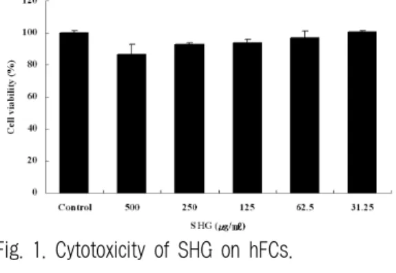
1. 세포독성 평가
hFCs는 대조군 세포생존율이 100.2 ± 1.5 (%), SHG의 500, 250, 125, 62.5, 31.25 (㎍/㎖) 농도에서 세포생존율이 각각 86.7 ± 5.9, 92.6 ± 1.5, 93.7 ± 3.1, 97.2 ± 3.9, 100.4 ± 1.2 (%) 였다(Fig. 1).
Fig. 1. Cytotoxicity of SHG on hFCs.
hFCs were treated with various concentration (500, 250, 125, 62.5, 31.25 ㎍/㎖) of the SHG extract.
2. 간독성 평가 1) AST
AST는 정상군이 118.0 ± 7.0 (I.U/L), 대조군이
206.0 ± 33.1 (I.U/L), SHG 투여군이 145.0 ± 10.8
(I.U/L)로 대조군에 비하여 유의성 있게 (*p<0.05)
감소되었다(Fig. 2).
Fig. 2. Effect of SHG on the AST in rats fed high cholesterol diets.
Normal : Basal diet group. Control : High cholesterol diet and normal saline (0.5㎖/day) treated group.
SHG : High cholesterol diet and SHG (115㎎
/0.5㎖/day) treated group. The result represents the mean ± S.D (n=6). Statistically significant value compared with normal group (++p<0.01).
Statistically significant value compared with control group (*p<0.05).
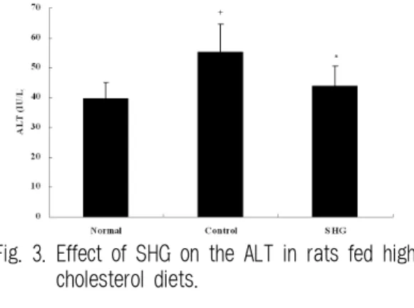
2) ALT
ALT는 정상군이 39.7 ± 5.5 (I.U/L), 대조군이 55.3 ± 9.5 (I.U/L), SHG 투여군이 44.0 ± 6.6 (I.U/L) 로 대조군에 비하여 유의성 있게 (*p<0.05) 감소되 었다(Fig. 3).
Fig. 3. Effect of SHG on the ALT in rats fed high cholesterol diets.
Normal : Basal diet group. Control : High cholesterol diet and normal saline (0.5㎖/day) treated group.
SHG : High cholesterol diet and SHG (115㎎/0.5
㎖/day) treated group. The result represents the mean ± S.D (n=6). Statistically significant value compared with normal group (+p<0.05). Statistically significant value compared with control group (*p<0.05).
3) ALP
ALP는 정상군이 178.0 ± 10.6 (I.U/L), 대조군이 235.7 ± 35.7 (I.U/L), SHG 투여군이 175.7 ± 8.5 (I.U/L)로 대조군에 비하여 유의성 있게 (*p<0.05) 감소되었다(Fig. 4).
Fig. 4. Effect of SHG on the ALP in rats fed high cholesterol diets.
Normal : Basal diet group. Control : High cholesterol diet and normal saline (0.5㎖/day) treated group.
SHG : High cholesterol diet and SHG (115㎎/0.5
㎖/day) treated group. The result represents the mean ± S.D (n=6).. Statistically significant value compared with normal group (+p<0.05). Statistically significant value compared with control group (*p<0.05).
3. 몸무게 및 간무게에 미치는 영향 1) 몸무게에 미치는 영향
몸무게는 정상군이 406.0 ± 36.2 (g), 대조군이 452.0
± 20.5 (g), SHG 투여군이 436.1 ± 32.7 (g)로 대조 군에 비하여 감소하였으나 유의성은 없었다.
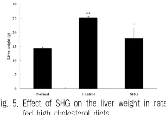
2) 간무게에 미치는 영향
간무게는 정상군이 14.3 ± 0.4 (g), 대조군이 25.2
± 0.4 (g), SHG 투여군이 17.8 ± 3.7 (g)로 대조군 에 비하여 유의성 있게 (*p<0.05) 감소되었다(Fig.
5).
Fig. 5. Effect of SHG on the liver weight in rats fed high cholesterol diets.
Normal : Basal diet group. Control : High cholesterol diet and normal saline (0.5㎖/day) treated group.
SHG : High cholesterol diet and SHG (115㎎
/0.5㎖/day) treated group. The result represents the mean ± S.D (n=6). Statistically significant value compared with normal group (++p<0.01).
Statistically significant value compared with control group (*p<0.05).
4. Total cholesterol에 미치는 영향
TC는 정상군이 55.0 ± 4.4 (㎎/㎗), 대조군이 271.0 ± 28.0 (㎎/㎗), SHG 투여군이 120.3 ± 22.8 (㎎/㎗)로 대조군에 비하여 유의성 있게 (**p<0.01) 감소되었다(Fig. 6).
Fig. 6. Effect of SHG on the TC level in rats fed high cholesterol diets.
Normal : Basal diet group. Control : High cholesterol diet and normal saline (0.5㎖/day) treated group.
SHG : High cholesterol diet and SHG (115㎎/0.5
㎖/day) treated group. The result represents the mean ± S.D (n=6). Statistically significant value compared with normal group (+++p<0.001). Statistically significant value compared with control group (**p<0.01).
5. LDL cholesterol에 미치는 영향
LDL-C는 정상군이 6.7 ± 1.5 (㎎/㎗), 대조군이 43.0 ± 7.5 (㎎/㎗), SHG 투여군이 20.7 ± 3.1 (㎎/㎗) 로 대조군에 비하여 유의성 있게 (**p<0.01) 감소 되었다(Fig. 7).
Fig. 7. Effect of SHG on the LDL-C level in rats fed high cholesterol diets.
Normal : Basal diet group. Control : High cholesterol diet and normal saline (0.5㎖/day) treated group.
SHG : High cholesterol diet and SHG (115㎎/0.5
㎖/day) treated group. The result represents the mean ± S.D (n=6). Statistically significant value compared with normal group (++p<0.01). Statistically significant value compared with control group (**p<0.01).
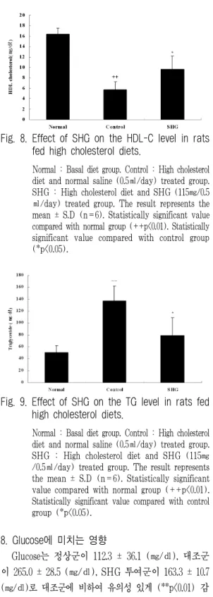
6. HDL cholesterol에 미치는 영향
HDL-C는 정상군이 16.3 ± 1.2 (㎎/㎗), 대조군 이 5.7 ± 1.5 (㎎/㎗), SHG 투여군이 9.7 ± 2.5 (㎎/㎗) 로 대조군에 비하여 유의성 있게 (*p<0.05) 증가되 었다(Fig. 8).
7. Triglyceride에 미치는 영향
TG는 정상군이 50.7 ± 11.5 (㎎/㎗), 대조군이
137.0 ± 25.2 (㎎/㎗), SHG 투여군이 79.3 ± 30.0
(㎎/㎗)로 대조군에 비하여 유의성 있게 (*p<0.05)
감소되었다(Fig. 9).
Fig. 8. Effect of SHG on the HDL-C level in rats fed high cholesterol diets.
Normal : Basal diet group. Control : High cholesterol diet and normal saline (0.5㎖/day) treated group.
SHG : High cholesterol diet and SHG (115㎎/0.5
㎖/day) treated group. The result represents the mean ± S.D (n=6). Statistically significant value compared with normal group (++p<0.01). Statistically significant value compared with control group (*p<0.05).
Fig. 9. Effect of SHG on the TG level in rats fed high cholesterol diets.
Normal : Basal diet group. Control : High cholesterol diet and normal saline (0.5㎖/day) treated group.
SHG : High cholesterol diet and SHG (115㎎
/0.5㎖/day) treated group. The result represents the mean ± S.D (n=6). Statistically significant value compared with normal group (++p<0.01).
Statistically significant value compared with control group (*p<0.05).
8. Glucose에 미치는 영향
Glucose는 정상군이 112.3 ± 36.1 (㎎/㎗), 대조군 이 265.0 ± 28.5 (㎎/㎗), SHG 투여군이 163.3 ± 10.7 (㎎/㎗)로 대조군에 비하여 유의성 있게 (**p<0.01) 감
소되었다(Fig. 10).
Fig. 10. Effect of SHG on the glucose level in rats fed high cholesterol diets.
Normal : Basal diet group. Control : High cholesterol diet and normal saline (0.5㎖/day) treated group.
SHG : High cholesterol diet and SHG (115㎎/0.5
㎖/day) treated group. The result represents the mean ± S.D (n=6). Statistically significant value compared with normal group (++p<0.01).
Statistically significant value compared with control group (**p<0.01).
9. Albumin에 미치는 영향
Albumin은 정상군이 3.9 ± 0.1 (g/㎗), 대조군이 4.6 ± 0.2 (g/㎗), SHG 투여군이 4.5 ± 0.3 (g/㎗) 로 대조군에 비하여 감소하였으나 유의성은 없었 다.
10. Total protein에 미치는 영향
Total protein은 정상군이 6.2 ± 0.1 (g/㎗), 대조 군이 7.6 ± 0.3 (g/㎗), SHG 투여군이 7.5 ± 0.6 (g/㎗)로 대조군에 비하여 감소하였으나 유의성은 없었다.
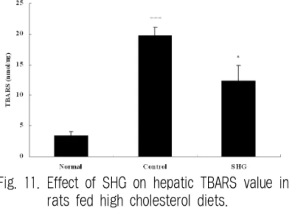
11. 간조직의 과산화지질에 미치는 영향
간조직의 과산화지질 함량은 정상군이 3.4 ± 0.6
(nmol/㎎), 대조군이 19.8 ± 1.4 (nmol/㎎), SHG
투여군이 12.4 ± 2.5 (nmol/㎎)로 대조군에 비하여
유의성 있게 (*p<0.05) 감소되었다(Fig. 11).
Fig. 11. Effect of SHG on hepatic TBARS value in rats fed high cholesterol diets.
Normal : Basal diet group. Control : High cholesterol diet and normal saline (0.5㎖/day) treated group.
SHG : High cholesterol diet and SHG (115㎎/0.5
㎖/day) treated group. The result represents the mean ± S.D (n=6). Statistically significant value compared with normal group (+++p<0.001).
Statistically significant value compared with control group (*p<0.05).
12. 간조직의 콜레스테롤 인자에 미치는 영향 1) ACAT에 미치는 영향
간조직의 ACAT mRNA 유전자 발현은 대조군 의 유전자 발현을 1 (RQ)로 했을 때 정상군이 0.18
± 0.1 (RQ), SHG 투여군이 0.6 ± 0.1 (RQ)로 대 조군에 비하여 유의성 있게 (**p<0.01) 감소되었다 (Fig. 12).
Fig. 12. Effects of SHG on the gene expression level of ACAT in hepatic tissue of rats fed high cholesterol diets.
Normal : Basal diet group. Control : High cholesterol diet and normal saline (0.5㎖/day) treated group.
SHG : High cholesterol diet and SHG (115㎎/0.5
㎖/day) treated group. The result represents the mean ± S.D (n=6). Statistically significant value compared with normal group (+++p<0.001).
Statistically significant value compared with control group (**p<0.01).
2) HMG-CoA reductase에 미치는 영향 간조직의 HMG-CoA reductase mRNA 유전자 발현은 대조군의 유전자 발현을 1 (RQ)로 했을 때 정상군이 0.36 ± 0.03 (RQ), SHG 투여군이 0.55 ± 0.08 (RQ)로 대조군에 비하여 유의성 있게 (**p<0.01) 감소되었다(Fig. 13).
Fig. 13. Effects of SHG on the gene expression level of HMG-CoA reductase in hepatic tissue of rats fed high cholesterol diets.
Normal : Basal diet group. Control : High cholesterol diet and normal saline (0.5㎖/day) treated group.
SHG : High cholesterol diet and SHG (115㎎/0.5
㎖/day) treated group. The result represents the mean ± S.D (n=6). Statistically significant value compared with normal group (+++p<0.001).
Statistically significant value compared with control group (**p<0.01).