黃藥子 메탄올 추출물의 염증억제 효과
정지윤1,2․이종록1․변성희1․정지욱3․김용한3․김상찬1,2*
1 : 대구한의대학교 한의과대학, 2 : 한방신약개발팀(BK21 Team), 3 : 한약재약리학과
Inhibitory Effect of Dioscorea Bulbifera MeOH Extract on Pro-inflammatory Mediator In Vitro and In Vivo
Ji Yun Jung1,2, Jong Rok Lee1, Sung Hui Byun1, Ji Wook Jung3, Yong Han Kim3, Sang Chan Kim1,2*
1 : College of Oriental Medicine, 2 : BK21 Team, 3 : Department of Herbal Medicinal Pharmacology, Daegu Haany University
Dioscorea bulbifera is one of the traditional medicinal herb. It commonly used in the treatment of hematemesis, epistaxis, tuberculous cervical lymphadenitis, laryngitis, acute infectious disease in East Asia. In the present study, we have demonstrated the anti-inflammatory effects of Dioscorea bulbifera MeOH extract (DBME) in macrophage cell line.
To investigate mechanism of the anti-inflammatory activity, we examined the effects of the lipopolysaccaride (LPS)-induced production of nitric oxide (NO), prostaglandin E 2 (PGE 2 ), pro-inflammatory cytokines and expression of inducible NO synthase (iNOS), cyclooxygenase-2 (COX-2), p-inhibitory κBα (p-IκBα), and nuclear factor-κB (NF-κB) in a murine macrophage cell line RAW 264.7. The RAW 264.7 cells were cultured in DMEM + serum medium for 24 hrs.
After serum starvation for 24 hrs, the cells were treated with DBME 0.03, 0.10, 0.30 ㎎/㎖ for 1 h, followed by stimulation with LPS (1 ㎍/㎖) for activation of immune response. After treatment, cell viability was measured by MTT assay, and NO production was monitored by measuring the nitrite content in culture medium. The protein band of iNOS, COX-2, p-Iκ Bα, and NF-κB was determined by immunoblot analysis and levels of cytokine were analyzed by sandwich immunoassays. There were three experimental groups: carrageenan, DBME 0.3, 1.0 g/kg. Rats were administrated either carrageenan (40% PEG) or carrageenan + DBME (0.3, 1.0 g/kg body weight) for 4 days (p.o.). To induce acute paw edema, rats were injected 1% carrageenan (100 ㎕/rat, dissolved in sterilized saline). The effect of DBME in the carrageenan-induced rat paw edema. As results, DBME has an inhibitory effect on the production of NO, PGE 2 , TNF-α, IL-1β and IL-6 and on the expression of iNOS, COX-2, p-IκBα and translocation of NF-κB to nuclear from cytosol. In addition, DBME effectively inhibited the increases of paw edema induced by carrageenan treatment in vivo. These results suggest that DBME can inhibit production of pro-inflammatory mediators and might be a useful source for treatment of acute inflammatory disease.
Key words : dioscorea bulbifera, LPS, cytokines, carrageenan, paw edema
* 교신저자 : 김상찬, 대구시 수성구 상동 165 대구한의대학교 한의과대학
․E-mail : [email protected], ․Tel : 053-770-2247
․접수 : 2010/02/08 ․수정 : 2010/03/09 ․채택 : 2010/03/15
서 론
염증반응은 생체나 조직에서 물리적 작용이나 세균감염, 화 학적 물질 등의 기질적 변화를 가져오는 침습이 가해질 때 손상 부위를 재생하려는 기전이다. 활성화된 macrophage는 염증반응 으로 질소 화합물 및 다양한 cytokine을 생성한다. 그러나, 과도 하고 지속적인 염증매개물질의 발현은 만성염증질환 및 종양 등
의 유발 원인이 되기도 한다 1,2) .
Macrophage는 외부자극 중 하나인 lipopolysaccharide (LPS) 로 인해 활성이 증대되며, 활성화된 macrophage cell은 자연 면역 뿐 아니라 획득면역 등 숙주 반응에 관여하여 숙주 방어와 항상 성 유지에 관여하는 것으로 알려져 있고 3) , 염증반응시 cytokine, nitric oxide (NO), prostaglandin E 2 (PGE 2 )등을 생산한다.
NO 생성은 NOS중에서 iNOS에 의한 생성이 많으며 이렇게
생성된 NO는 박테리아를 제거하는 역할 4) 을 하지만 과도한 NO
형성은 염증을 유발시켜 조직의 손상, 유전자 변이, 신경 손상을
유발 한다 5) . iNOS는 외부자극에 반응하여 생체를 방어하려는 목
적으로 단시간에 과량의 NO를 생성하게 된다 6-8) .
Cyclooxygenase (COX)는 arachidonic acid를 prostaglandin (PG)로 변화시키며 COX-1과 COX-2의 isoform 형태로 존재한다
9) . 그중 COX-2는 유도성의 형태로서 염증부위에서 많은 양의 전 염증성 PG의 생산에 반응한다 10) .
Tumor necrosis factor-α (TNF-α)는 활성화된 대식세포, 섬 유모세포 등 다른 여러 세포에서 생성되고, 이것은 종양세포에 영향을 미치는 숙주 방어인자 및 염증매개물질로 알려져 있다.
Interleukin-6 (IL-6)의 생성은 IL-1β나 TNF-α와 같은 요인 외에 도 LPS에 의해 유도되고 11) , 염증유도 cytokine으로서 내인성 발 현인자로 작용하고 면역 체계와 조혈에 영향을 미친다 12) .
NF-κB는 면역과 염증반응 조절의 중요한 역할을 하는 전사인 자이며 21) , 활성화된 NF-κB는 핵 내로 translocation되면 target 유 전자의 promoter regions에 있는 κB 결합 자리에 결합하여 iNOS, COX-2, cytokine 등 염증 매개물질의 전사를 촉진 시킨다 13) .
黃藥子 (Dioscorea bulbifera L.)는 덩굴성 초본인 薯蕷科의 둥근 마의 괴경으로 경엽과 협잡물을 제거하고 수염뿌리를 잘라 내고 깨끗이 씻은 다음에 햇볕에 말린 한약재로써 味苦性平無毒 하며, 肝心經에 入한다. 凉血, 降火, 消癭, 解毒 효과가 있어 주로 토혈증, 비출혈, 결핵성 경부림프선염, 후두염 등의 치료에 사용 되었다 14) . 黃藥子의 연구동향은 항암작용에 대한 연구 15) , 덩이뿌 리에서 추출된 Lectin 단백질의 항균작용 16) 에 대한 연구 등이 이 루어져 있으며, 黃藥子의 항염증효과에 대한 연구는 전무하여 본 연구를 시행하였다.
재료 및 방법
1. 시약
LPS (Escherichia coli 026:B6), carrageenan과 3-(4,5-dimethylthiazol-2-yl)-2,5-diphenyltetrazoleum bromide (MTT)는 Sigma (St. Louis, MO, USA)에서 구입하였고, Dulbecco's modified Eagle's medium (DMEM), penicillin, fetal bovine serum (FBS)는 Gibco/BRL (Eggenstein, Germany)에서 구입하였다. Griess reagent는 Fluka (Seelze, Germany)에서 구입 하였고, Antibody는 BD Bioscience (San Jose, CA, USA), Cayman (Ann Arbor, Mi, USA)과 Cell Signaling (Danvers, MA, USA), Santa Cruz (Santacruz, CA, USA)로부터 구입하였으며 NC paper는 Schleicher & Schuell (Dassel, Germany)에서 구입 하였다. TNF-α, IL-1β, IL-6의 ELISA Kits는 Pierce endogen (Rockford, IL, USA)으로 부터 구입하였고, PGE 2 는 R&D Systems (Minneapolis, MN, USA)를 사용하였다.
2. 추출물 제조
黃藥子 추출물 (Dioscorea bulbifera MeOH extract; DBME) 은 黃藥子 265 g에 2 L의 100% 메탄올을 가하고 상온에서 72시 간 동안 침지하고, No.2 filter paper (Nalgene, New York, NY, USA)로 여과하여, 여액을 진공회전농축기 (EYELA, Tokyo, Japan)를 사용하여 농축하고, 그 농축액을 Ultra-low
temperature freezer (Operon, Korea)에 12시간 동안 동결하고 동결건조기 (LABCONCO, USA)로 동결건조 하였다. 黃藥子의 최종 수율은 2.52%이며, in vitro에서는 DMSO, in vivo에서는 40% PEG에 녹여 사용하였다.
3. 세포배양
Murine macrophage cell line인 RAW 264.7 cell (KCLRF, Korean Cell Line Research Foundation, Seoul, Korea)을 DMEM 에 10% FBS, 100 U/㎖ penicillin 및 100 ㎍/㎖ streptomycin을 혼합한 배지를 사용하였고, 37℃, 5% CO 2 조건의 incubator (Sanyo, Japna)에서 배양하였다. 실험과정의 모든 cell은 80~90%
의 confluence에서 실험하였고, 20 passage를 넘기지 않은 cell만 사용하였다.
4. NO의 생성량 측정
RAW 264.7 cell을 24 well plate에 1×10 5 cells/well로 분주 한 다음 24시간 후에 serum을 포함하지 않는 배지로 교환하여 24시간 동안 배양하였다. DBME 0.03, 0.10, 0.30 ㎎/㎖로 처리한 다음 1시간 후에 LPS (1 ㎍/㎖)를 처리하고 37℃, 5% CO 2 조건의 incubator에서 배양하였다. LPS 처리 후 12, 18, 24시간에 배지를 수거하여 세포배양 상등액 50 ㎕와 Griess시약 50 ㎕를 혼합하여 96 well plate에서 10분 동안 반응시킨 후 540 nm에서 Titertek Multiskan Automatic ELISA microplate reader (Model MCC/340, Huntsville, AL)로 흡광도를 측정하였다.
5. 세포생존율 측정
RAW 264.7 cell을 24 well plate에 1×10 5 cells/well로 분주 한 다음 serum을 포함하지 않는 배지로 교환하여 24시간 동안 배양하였다. DBME 0.03, 0.10, 0.30 ㎎/㎖처리 한 다음 1시간 후 에 LPS (1 ㎍/㎖)를 처리하고 37℃, 5% CO 2 조건의 incubator에 서 24시간 동안 배양하였다. 배양 후 생존세포에 MTT solution 을 넣고 4시간동안 배양한 다음 MTT solution을 제거하고 생성 된 formazan crystal을 DMSO에 녹여 Titertek Multiskan Automatic ELISA microplate reader (Model MCC/340, Huntsville, AL)로 570 nm에서 흡광도를 측정하였다. 세포생존 율은 control cell에 대한 백분율로 나타내었다. [i.e. viability (%
control) = 100×/(absorbance of treated sample)/(absorbance of control)].
6. Immunoblot analysis
20 mM Tris Cl (pH 7.5), 1% Triton X-100, 137 mM sodium chloride, 10% glycerol, 2 mM EDTA, 1 mM sodium orthovanadate, 25 mM b-glycerophosphate, 2 mM sodium pyrophosphate, 1 mM phenylmethylsulfonylfluoride와 1 ㎎/㎖
leupeptin을 함유하는 buffer를 사용하여 단백질을 추출한 후,
15,000 × g로 10분간 원심 분리하여 상등액을 얻어 BCA법으로
정량하여 protein을 확인하였고, SDS-PAGE를 이용하여 분리하
였다. 전기영동이 끝난 후 gel로부터 단백질을 NC membrane으
로 transfer하였다. 각각의 primary antibody를 사용하여 상온에 서 2시간동안 처리 하고 secondary antibody는 alkaline phosphatase conjugated anti-rabbit을 사용하였으며, ECL western blotting detection reagents (Amersham, Berkshire, UK) 를 사용하여 발색한 후 image analyzing system (Ultra-Violet Products, Upland, CA, USA)을 사용하여 관찰하였다.
7. 핵분획의 제조
배양된 세포를 PBS로 세척한 후 cell을 수거하여 microcentrifuge tube에 담고, 4℃에서 3,000 × g로 3분간 원심 분리하여 PBS를 제거하였다. 세포에 10 mM Hepes (pH 7.9), 10 mM KCl, 0.1 mM EDTA, 0.5% Noridet P-40, 1 mM dithiothreitol, 0.5 mM phenylmethylsulfonyl fluoride를 함유한 완충액을 100 ㎕ 넣어 vortex하고 10분간 ice에서 배양한 후 4℃
에서 7,800 × g로 5분간 원심분리 하였다. 원심분리 후 상층액은 버리고 핵이 포함된 침전물에 20 mM Hepes (pH 7.9), 400 mM NaCl, 1 mM EDTA, 10 mM dithiothreitol, 1 mM phenylmethylsulfonyl fluoride를 함유한 완충액을 40 ㎕ 넣은 후 현탁시켜 ice에서 1시간 배양하였다. 배양 후 4℃에서 15,000 × g 로 10분간 원심분리 하여 핵분획인 상층액을 얻었다. 핵분획물은 사용시까지 -70℃에 보관하였다.
8. Cytokine의 측정
Cytokine을 측정하기 위하여 24 well plate에 세포를 1×10 5 cells/well을 분주 하여 배양하고 DBME를 0.03, 0.10, 0.30 ㎎/㎖
처리한 다음 1시간 후에 LPS (1 ㎍/㎖)를 처리하였다. LPS 처리 후 24시간 경과 후 배지를 수거하여 cytokine을 측정하였다. 수 거된 배지는 측정 전 까지 -70℃에서 보관하였고, ELISA Kit를 사용하여 측정하였다. 실험방법은 manufacturer's instruction에 따랐다. Cytokine 생성률은 control cell에 대한 백분율로 나타내 었다.
9. PGE 2 의 측정
PGE 2 를 측정하기 위하여 24 well plate에 세포를 1×10 5 cells/well을 분주 하여 배양하고 DBME를 0.03, 0.10, 0.30 ㎎/㎖
처리한 다음, 1시간 후 LPS (1 ㎍/㎖)를 처리하고 24시간 후에 배지를 수거하여 PGE 2 를 측정하였다. Coating된 96 well plate에 배지를 100 ㎕씩 첨가하고 primary antibody solution과 PGE 2
conjugate를 50 ㎕씩 각각의 well에 넣은 후 상온에서 2시간 동 안 배양하였다. Washing buffer로 4회 세척하고 substrate solution 200 ㎕를 처리하여 30분간 반응시키고 stop solution 50
㎕를 처리한 후 Titertek Multiskan Automatic ELISA microplate reader로 450 nm에서 흡광도를 측정하였다.
10. 실험동물
실험동물은 4주령 (80~100 g)된 Sprague-Dawley계 수컷 흰 쥐 (효창 Science, Daegu, Korea)를 1주일 동안 환경에 적응시킨 후 실험에 사용하였으며, 사육실 환경은 온도 20~23℃, 습도
60%, 12시간 light/dark cycle을 유지하고, 사료 (Nestle Purina Petcare Korea, Seoul, Korea)와 물은 자유롭게 섭취하도록 하였 고, 측정 24시간 전에 절식시켜 실험을 실시하였다.
11. 약물 투여
기염제인 carrageenan만 투여한 군을 Control군으로 하였으 며, 0.30 g/kg의 DBME과 carrageenan을 투여한 군을 0.30 g/kg 의 DBME군으로 하고, 1.0 g/kg의 DBME과 carrageenan을 투여 한 군을 1.0 g/kg의 DBME군으로 하였으며, 각 군당 n수는 5마 리로 하였다. 40% PEG와 DBME는 4일 동안 매일 1회 p.o.투여 하였으며, 마지막 약물 투여 1시간 후 0.9% 식염수에 녹인 1%
carrageenan을 100 ㎕/rat으로 rat의 오른쪽 발바닥에 주입하였다.
12. Paw edema의 유도 및 측정
4일째 약물 투여 1시간 후 carrageenan을 주입하여 paw edema를 유발시켰다. Paw edema측정은 carrageenan을 주입한 다음 주입 직후와 4시간 후에 부종측정기 (UGO BASILE ; Comerio, VA, ITALY)를 이용하여 측정하였다.
13. 조직 처리
Paw edema의 유발과 회복여부를 살펴보기 위해, carrageenan으로 염증이 유발된 rat의 오른쪽 발 발목아래를 절 단하여 발등 및 발바닥의 피부 실질조직을 분리하여 10% 중성 포르말린에 6시간 이상 고정시킨 후 3~4 ㎛의 longitudinal 절편 을 만들어 Hematoxylin-eosin (HE)으로 염색하여 slide glass에 부착시키고 광학현미경 (Nikon, Japan)으로 관찰하였다.
14. Histomorphometry
발등 및 발바닥 피부 (상피에서 진피)의 두께를 mm 단위로 40배 현미경 시야에서 자동영상 분석 장치 (DMI-300 Image Processing; DMI Korea)를 이용하여 각각 측정하였으며, 각 1 mm 2 의 발등 및 발바닥 피부에 침윤된 염증세포의 수 역시 자동 영상 분석 장치를 이용하여, 200배 현미경 시야에서 측정하였다.
15. 통계적 검증
실험결과는 SPSS를 사용하여 mean ± S.D로 나타내었으며, ANOVA Tukey LSD의 통계처리방법으로 통계적 유의성 검정을 조사하였다. 유의수준은 P < 0.05로 하였다.
결 과
1. DBME가 LPS로 유도된 RAW 264.7 cell의 NO production에 미치는 영향
RAW 264.7 cell에서 DBME의 nitric oxide 생성억제정도를
관찰하기 위해서 DBME 0.03, 0.10, 0.30 ㎎/㎖를 세포에 전 처리
하여 12, 18, 24시간에서 생성되는 NO양을 측정하였다. 그 결과
12~24시간에서 LPS 단독처리군의 NO생성량이 유의하게 증가하
였고, DBME 0.10, 0.30 ㎎/㎖ 전 처리군 또한, 12~24시간에서 유
의하게 NO 생성을 억제하였다(Fig. 1).
Fig. 1. Effects of DBME on the production of NO.
RAW 264.7 cells were treated with (0.03, 0.10 and 0.30 ㎎/㎖) of DBME for 1 h prior to the addition of LPS (1 ㎍/㎖). The NO concentration in the culture medium was measured for 12, 18 and 24 h. The date represent the mean ± S.D of three reparate experiments. (**, significant as compared to control.**P < 0.01;##, significant as compared to LPS alone,##P < 0.01).2. DBME가 RAW 264.7 cell의 생존율에 미치는 영향
DBME의 RAW 264.7 cell에서 LPS로 유도된 NO 생성의 감 소가 DBME의 독성으로 인한 RAW 264.7 cell의 수적 감소에 기 인한 것인지를 관찰하기 위하여 MTT assay를 시행하여 세포생 존율을 확인하였다. LPS 단독처리군은 control군과 비교하여 cell viability가 60%정도로 나타났다. DBME 0.03, 0.10, 0.30 ㎎/㎖ 전 처리군에서는 LPS 단독처리군과 비교하여 세포독성을 나타내지 않았다(Fig. 2).
Fig. 2. Effects of DBME on the cell viability in LPS stimulated RAW 264.7 cells.
RAW 264.7 cells were treated with (0.03, 0.10 and 0.30 ㎎/㎖) of DBME for 1 h prior to the addition of LPS (1 ㎍/㎖), and the cells were further incubated for 24 h. Data represent the mean ± S.D. with three separate experiments. (**, significant as compared to control.**P < 0.01).3. DBME가 LPS로 유도된 RAW 264.7 cell의 PGE 2 에 미치는 영향
Arachidonic acid로부터 생성되는 PGE 2 는 부종, 통증, 염증 등의 증상을 일으키며, 염증반응이 일어날 경우 PGE 2 는 COX-2 에 의해 생성되며 혈관을 확장시키고 혈관투과성을 증가시켜 염 증을 유발한다 8) . 본 실험에서는 DBME의 RAW 264.7 cell에서 LPS에 의한 PGE 2 억제 효과를 확인하기 위해 Kit를 사용하여 관 찰하였다. PGE 2 의 경우 LPS 단독처리군은 control군에 비하여 유의한 증가가 나타났으며, 이러한 증가는 DBME 0.10, 0.30 ㎎/
㎖의 전 처리에 의하여 유의성 있게 억제되었다(Fig. 3).
Fig. 3. Inhibition of LPS-activated PGE 2 production by DBME in RAW 264.7 cells.
RAW 264.7 cells were cultured with LPS (1 ㎍/㎖) in the presence or absence of DBME for 24 h to determine the level of PGE2. The cultured medium was collected and directly assayed for PGE2. The data represent the mean ± SD of three separate experiments. (**: significant as compared with the control,**P < 0.01,##: significant as compared with the LPS alone, ##P < 0.01). PGE2, prostaglandin E2.4. DBME가 LPS로 유도된 RAW 264.7 cell의 iNOS 및 COX-2의 발현에 미치는 영향
산화질소는 L-arginine으로부터 nitric oxide synthase (NOSs)를 경유하여 생산되므로 세포질 내에서의 iNOS 단백질 발현량을 확인하였다. 그 결과 LPS 단독처리군은 iNOS 단백질 의 발현이 유의성 있게 증가되었으나, DBME 0.10, 0.30 ㎎/㎖의 전처리에 의하여 iNOS의 발현량이 유의성 있게 감소하였다 (Fig. 4A, B). 염증반응에 중요한 역할을 하는 COX-2의 발현량을 확인하였다. LPS처리로 인해 COX-2 단백질의 발현이 유의성 있 게 증가하였으며, 증가한 COX-2는 DBME 0.10, 0.30 ㎎/㎖ 전 처 리에 의해 발현량이 유의성 있게 감소하였다(Fig. 4A, C).
A.
B.
C.
Fig. 4. Effect of DBME on the expression of in LPS stimulated RAW 264.7 cells iNOS and COX-2.
The levels of iNOS and COX-2 protein were monitored 24 h after treatment of cells with LPS (1 ㎍/㎖) with or without 0.03, 0.10 and 0.30 ㎎/㎖ of DBME pretreatment (i.e. 1 h before LPS). Equal amounts of total protein were resolved by SDS-PAGE. Expressions of iNOS and COX-2 protein were determined by immunoblot analysis using specific antibodies (A). The relative levels of iNOS and COX-2 were measured by scanning densitometry (B, C). The actin was used as a loading control (**: significant as compared with the control,**P < 0.01,##: significant as compared with the LPS alone,#P < 0.05,##P< 0.01).
5. DBME가 LPS로 유도된 RAW 264.7 cell의 NF-κB 발현에 미치 는 영향
DBME가 LPS로 인해 발현이 증가된 iNOS 및 COX-2 protein을 유의하게 억제하였기 때문에, iNOS 및 COX-2 의 발현 에 핵심적 역할을 하는 NF-κB의 발현을 확인하였다. 핵 분획에 서의 NF-κB는 LPS처리에 의해 증가하였으며, DBME 0.03, 0.10, 0.30 ㎎/㎖ 전처리에 의하여 NF-κB량이 감소하였다(Fig. 5).
A.
B.
Fig. 5. Effect of DBME on the nuclear translocation of NF-κB.
The level of NF-κB protein was monitored 60 min after treatment of cells with LPS (1㎍/㎖) with or without DBME pretreatment, respectively (i.e. 1 h before LPS) (A).
The relative levels of NF-κB was measured by scanning densitometry (B). The actin was used as a loading control.
6. DBME가 LPS로 유도된 RAW 264.7 cell의 IκBα, p-IκBα 발현
에 미치는 영향
RAW 264.7 cell은 LPS자극에 의해 NF-κB signaling cascade 가 활성화 되면서 IκB가 인산화되고 NF-κB가 핵으로 전위하여 COX-2, iNOS등의 전사를 유도하게 된다. 세포질에서 IκBα는 LPS 처리에 의하여 뚜렷하게 감소하였고, DBME 0.10, 0.30 ㎎/㎖
로 인해 감소된 IκBα가 유의성있게 증가하였다(Fig. 6A, B). p-IκB α 는 LPS처리에 의해 증가하였고, 이 증가는 DBME 0.30 ㎎/㎖ 전 처리에 의해 p-IκBα가 감소한 것을 확인하였다(Fig. 6C, D).
A.
B.
C.
D.
Fig. 6. Effects of DBME on the expression of IκBα and p-IκBα.
The levels of IκBα and p-IκBα protein were monitored 15 min after treatment of cells with LPS (1 ㎍/㎖) with or without DBME pretreatment, respectively (i.e. 1h before LPS) (A, C). The relative levels of IκBα and p-IκBα were measured by scanning densitometry (B, D). The actin was used as a loading control (**, significant as compared to control.**P < 0.01;#, significant as compared to LPS alone,#P < 0.05,##P < 0.01).
7. DBME가 LPS로 유도된 RAW 264.7 cell의 cytokines에 미치는 영향
염증반응에 있어서 중요한 역할을 하는 TNF-α는
macrophage와 mast cell등에서 분비되며 LPS반응의 주요 매개 체로서 내재면역에서 중요한 역할을 한다 17) . IL-6는 B cell에서 plasma로 분화되도록 촉진시키고 항체의 분비를 자극하는 cytokine으로 B-cell 분화단계의 후기에 주로 작용하며, T-cell의 분화 및 증식에도 관련되어 있다고 알려져 있다 18) . IL-1β는 pro-inflammatory cytokine으로서 여러 면역학적 작용들과 연관 되어 있다. 본 연구에서는 TNF-α, IL-6, IL-1β가 LPS 단독처리에 의해 유의성 있게 증가 하였으며, 이러한 증가는 TNF-α의 경우 DBME 0.30 ㎎/㎖ 전 처리에 의해 유의성 있게 억제 되었고(Fig.
7A), IL-6, IL-1β에서는 DBME 0.10, 0.30 ㎎/㎖ 전 처리에 의해 유의성 있게 억제 되었다(Fig. 7B, C).
A.
B.
C.
Fig. 7. The effect of DBME on LPS-inducible cytokines production.
Production of TNF-α, IL-6 and IL-1β were measured in the medium of RAW 264.7 cells cultured with LPS (1 ㎍/㎖) in the presence or absence of DBME for 24 h.
The levels of TNF-α, IL-6 and IL-1β in culture medium were monitored as described in the methods section. Data represent the mean ± SD of three separate experiments. (**: significant as compared with the control,**P < 0.01,##: significant as compared with the LPS alone, ##P < 0.01).
8. DBME가 carrageenan으로 유도된 rat의 급성염증에 미치는 효과
DBME이 carrageenan으로 유도된 rat의 급성염증에 미치는 효과를 알아보기 위해 rat의 오른쪽 발에 carrageenan주입 직후 와 4시간 후 paw volume를 측정하였다. 측정 결과, carrageenan 처리 후 4시간이 경과한 실험군에서는 paw volume이 유의성 있 게 증가하였고, DBEM 0.3, 1.0 g/kg 투여군에서는 paw volume 이 유의성 있게 감소한 것을 확인할 수 있었다(Fig. 8).
Fig. 8. The effect of DBME on the formation of carrageenan-induced paw edema.
Rats were administrated with either carrageenan or carrageenan + DBME (0.3, 1.0 g/kg body weight) for 4 days (p.o.). To induce acute paw edema, rats were injected 1% carrageenan (100 ㎕/rat, dissolved in sterilized saline). Date were measured at 0 and 4 hours after injection of 1% carrageenan.Data represent the mean ± SD. (**: significant as compared with the 0h carrageenan,**P < 0.01,##: significant as compared with the 4h carrageenan,##P
< 0.01).
Fig. 9. Change on histological profiles of the dorsum pedis skin in carrageenan (a,b), DBME 0.3 g/kg (c,d) and DBME 1.0 g/kg (e,f) groups.
Rats were administrated with either carrageenan or carrageenan + DBME (0.3, 1.0 g/kg body weight) for 4 days (p.o.). To induce acute paw edema, rats were injected 1% carrageenan (100 ㎕/rat, dissolved in sterilized saline).Note that marked increases of skin thicknesses due to edematous changes were detected by carrageenan treatment with increases of inflammatory cell infiltrations.
However, these increases of skin thicknesses and inflammatory cell infiltrations were effectively inhibited by treatment of both two different dosages of DBME 0.3 and 1.0 g/kg respectively. Arrow indicated total thicknesses measured; All HE stain; Scale bars = 160 ㎛.
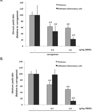
9. DBME가 carrageenan으로 유도된 급성 염증조직에 미치는 효과
carrageeana으로 유도된 paw edema의 발등 및 발바닥두께
를 조직 병리학적으로 분석한 결과, carrageenan주입으로 인해
발등 및 발바닥 두께의 증가가 일어났으며, DBME 0.3, 1.0 g/kg
투여로 인해 유의성 있게 억제 되었다. carrageenan에 의해 유의 성 있게 증가한 발등 피부조직내 염증세포가 DBME 0.3, 1.0 g/kg 투여로 인해 억제되었으나, 발바닥 피부 염증 세포의 침윤 억제는 DBME 1.0 g/kg에서만 일어났다(Fig. 9, 10, 11).
Fig. 10. Change on histological profiles of the ventrum pedis skin in carrageenan (a,b), DBME 0.3 g/kg (c,d) and DBME 1.0 g/kg (e,f) groups.
Rats were administrated either carrageenan or carrageenan + DBME (0.3, 1.0 g/kg body weight) for 4 days (p.o.). To induce acute paw edema, rats were injected 1% carrageenan (100 ㎕/rat, dissolved in sterilized saline). Note that marked increases of skin thicknesses due to edematous changes were detected by carrageenan treatment with increases of inflammatory cell infiltrations.However, these increases of skin thicknesses and inflammatory cell infiltrations were effectively inhibited by treatment of both two different dosages of DBME 0.3 and 1.0 g/kg except for inflammatory cell numbers of ventrum pedis skin in DBME 1.0 g/kg, in which quite similar numbers of infiltrated inflammatory cells were observed as compared with carrageenan control. Arrow indicated total thicknesses measured; All HE stain; Scale bars = 160 ㎛.
A.
B.
Fig. 11. Change on the histomorphometrical analysis of Dorsum pedis skin and Ventrum pedis skin by DBME.
Rats were administrated either carrageenan or carrageenan + DBME (0.3, 1.0 g/kg body weight) for 4 days (p.o.). To induce acute paw edema, rats were injected 1% carrageenan (100㎕/rat, dissolved in sterilized saline). Values are expressed as mean ± SD of 5 histological fields; ##P < 0.01 compared with carrageenan control.
고 찰
본 연구에서는 黃藥子 메탄올 추출물이 LPS로 활성화된 RAW 264.7 cell에 미치는 iNOS 억제 기전 및 염증관련 cytokine 의 발현과 rat에서 carrageenan으로 유도된 paw edema의 염증 억제를 관찰하고자 하였다. 黃藥子 (Dioscorea bulbifera L.)는 토 혈증, 코피, 결핵성 경부림프선염, 후두염 등의 치료제로 사용되 었다 14) . 현재까지 黃藥子는 항암에 대한 효과가 연구 되었고 15) , 덩이뿌리에서 추출된 Lectin 단백질이 장운동을 증가시키고, 항 균 작용 16) 등의 약리효과가 알려져 있다.
nitric oxide는 향균작용과 종양제거에 중요한 역할을 하지 만 19) , 염증시 iNOS에 의해 생성된 NO는 부종, 혈관투과성 등의 염증반응을 촉진하고 염증매개물질 생합성이 촉진되어 염증을 심화시킨다고 알려져 있다 20,21) .
RAW 264.7 cell에서 DBME로 인한 NO 생장억제를 LPS 처 리하여 생성되는 NO양을 시간별로(12, 18, 24 h)측정하였다. 실 험결과 LPS 처리에 의하여 12~24시간에서 NO의 생성량이 증가 하였으며, DBME 0.10, 0.30 ㎎/㎖ 전 처리에 의해 NO의 생성량 이 유의성 있게 감소하였다. DBEM 0.10, 0.30 ㎎/㎖이 LPS로 유 도된 NO의 생성 감소가 DBME의 세포 독성으로 인해 세포에 영 향을 미칠 수 있기 때문에 세포 독성의 여부를 확인하기 위하여 MTT assay로 cell viability를 측정한 결과 LPS 처리로 인해 cell viability가 60% 정도 나타났으며, DBME 0.03, 0.10, 0.30 ㎎/㎖
전 처리에 의해서는 독성이 나타나지 않았다.
L-arginine으로부터 NOS를 경유하여 NO가 생성되기 때문 에 22) DBME의 NO생성억제와 iNOS의 상관성을 확인하기 위해 세포질 내에서의 iNOS 단백질 발현량을 확인해 보았다. LPS 처 리로 인해 iNOS 단백질 발현량이 유의성 있게 증가하였으며, 이 러한 증가는 DBME 0.10, 0.30 ㎎/㎖ 전 처리에 의해 iNOS의 발 현량이 유의성 있게 감소한 것을 확인할 수 있었다. 이결과 DBME로 인해 iNOS의 발현량이 억제됨으로써 NO 생성이 억제 함을 의미한다.
Arachidonic acid로 부터 prostaglandin 합성과정에서 중요 한 역할을 하는 COX-2는 호르몬, 성장인자, cAMP, 염증인자나 cytokine, 종양유전자 등과 같은 자극에 의해 분비가 유도되는 효소이다 23) . 염증 반응에 중요한 역할을 하는 COX-2의 발현량은 LPS 처리로 인해 유의성있게 증가 하였고, DBME 0.10, 0.30 ㎎/
㎖ 전 처리에 의해 유의성 있게 감소하였다. PGE 2 의 경우 LPS 처리로 인하여 유의성 있게 증가하였으며, 이와 같은 증가는 DBME 0.10, 0.30 ㎎/㎖ 전 처리에 의하여 유의성 있게 억제 되 었다. 이러한 결과 DBME가 prostaglandin 생합성에 관여하는 COX-2를 억제함으로써 염증유발 물질인 PGE 2 의 활성 역시 유의 성 있게 감소함을 확인 할 수 있었다.
대식세포는 염증반응시 IL-1β, TNF-α, IL-6 같은 cytokine을
생산하여 감염초기에 생체 방어에 중요한 역할을 하는 세포로
알려져 있다 24) . 면역반응과 염증반응을 유도하는 염증매개 물질
cytokine인 TNF-α는 대식세포나 단핵세포에서 세포의 분화와 성
장 등의 기능에 관여하고 혈관투과성을 증가 시킨다 25) . 단핵구와
대식세포에서 분비되어 T cell과 B cell을 활성화시키는 IL-1β는 다양한 lymphokine을 분비시켜 세포증식과 세포외 기질 축적을 자극한다 26) .
IL-6는 단핵구를 포함한 여러세포에서 분비가 되고, 숙주방 어, 면역반응, 신경세포의 기능 등에서 중요한 기능을 하는 물질 이지만, 과도한 분비는 여러 가지 질환에서 분비가 증가된 것을 볼 수 있다 27) . 본 연구에서는 TNF-α, IL-1β, IL-6에서 모두 LPS에 의해 유의성 있게 증가하였고, TNF-α는 DBME 0.30 ㎎/㎖ 전 처 리에 의해 유의성 있게 감소하였고, IL-1β, IL-6는 DBME 0.10, 0.30 ㎎/㎖ 전 처리에 의해 유의성 있게 감소한 것을 확인할 수 있었다.
NF-κB는 저해단백질인 IκBα에 의해 불활성화 상태로 세포 질에서 존재하는데 28) 외부자극을 가하면 IκB kinase에 의해 IκBα 가 분해되는데, IκBα가 분해되면 세포질의 NF-κB가 핵으로 이동 한다. 본 연구에서는 세포질에서 IκBα는 LPS의 자극에 의하여 유의성 있게 감소하였고, DBME 0.10, 0.30 ㎎/㎖ 전 처리에 의해 감소된 IκBα가 유의성 있게 증가하였고, p-IκBα는 LPS처리로 인 해 증가하였으며, DBME 0.30 ㎎/㎖ 전 처리에 의해 p-IκBα가 감 소한 것을 확인 하였다. 핵분획에서의 NF-κB는 LPS의 처리에 의 하여 증가하였으나, DBME 0.10 ,0.30 ㎎/㎖ 전 처리에 의하여 NF-κB가 감소하였다. 이러한 결과는 DBME가 세포질에서 IκBα 의 인산화 억제로 인해 IκBα가 세포내에서 다량 존재하게 됨으 로 NF-κB가 핵으로 전위되는 것을 억제할 수 있는 가능성을 시 사한다.
기염제인 carrageenan으로 급성 부종을 유발하였으며, DBME 0.3, 1.0 g/kg 투여로 인해 paw volume이 유의성 있게 감소하였다. 또한 발바닥 피부 염증 세포의 침윤억제는 DBME 1.0 g/kg 투여군에서만 억제 되었다. 이러한 carrageenan 유발 급성 부종성 염증소견은 DBME의 투여에 의해 유의성 있게 억제 되었다.
이러한 결과로 보아 DBME는 항염증에 효과적인 물질로 확 인되어 염증성질환의 예방 및 면역 치료의 이용과 약물의 개발 에 활용 될 수 있을 것으로 판단된다.
결 론
黃藥子 메탄올 추출물의 염증억제 효과를 평가한 결과 다음 과 같은 결론을 얻었다.
LPS에 의해 증가한 NO의 생성을 DBME 전 처리에 의해 유 의성 있게 억제함으로써, iNOS 발현 역시 유의성 있게 억제하였 다. 또한 DBME 처리군은 LPS단독처리군에 비교하여 유의한 세 포독성을 나타내지 않았다. PGE 2 생성이 감소하여 COX-2 발현 역시 DBME 전 처리에 의해 유의하게 억제 되었다. LPS로 인해 유의성 있게 감소한 IκBα는 DBME 전 처리에 의해 유의성 있게 증가하였고, LPS로 인해 증가한 p-IκBα를 DBME 전 처리에 의해 억제됨으로써, 핵 분획에서 LPS로 증가한 NF-κB가 DBME 전 처 리에 의해 발현을 감소 시켰다. 또, TNF-α, IL-1β, IL-6등의 cytokine은 LPS로 인해 생성량이 유의성 있게 증가 하였고
DBME에 의해 유의성 있게 감소하였다.
Carrageenan으로 유도된 급성 부종의 paw volume이 유의 성 있게 증가 하였으며, DBME 투여로 인하여 paw volume이 감 소하여 발등 및 발바닥 피부조직을 조직병리학적으로 검사한 결 과 발등 및 발바닥 피부 부종에 의한 두께 증가 및 발등 염증세 포 침윤을 억제하였다.
감사의 글
본 연구는 지식경제부 지역혁신센터사업(대구한의대학교 한 방생명자원연구센터)의 지원에 의하여 이루어진 것입니다.
참고문헌