J. Fish Pathol., 34(1) : 047~053 http://dx.doi.org/10.7847/jfp.2021.34.1.047
Introduction
Scuticociliates are one of the serious parasitic threats in aquaculture industry worldwide. They pro- voke severe systemic infection through rapid invasion of internal organs and finally induce mass mortality in host populations (Cheung et al. 1980; Bassleer 1983). To date, several scuticociliates have been re- ported as the culprits of scuticociliatosis in marine fish including Uronema nigricans in southern bluefin tuna (Munday et al. 1997; Crosbie & Munday 1999), Philasterides dicentrarchi in sea bass, turbot and olive flounder (Dragesco et al. 1995; Iglesias et al.
2001; Parama et al. 2003; Kim et al. 2004a), Uronema marinum in olive flounder (Jee et al. 2001), Pseudo-
cohnilembus persalinus in olive flounder (Kim et al.
2004b) and Miamiensis avidus in olive flounder (Jung et al. 2005; Jung et al. 2007; Song et al. 2009). In addition, an unidentified scuticociliate was also re- ported as the cause of mortality in olive flounder (Yoshinaga & Nakazoe 1993).
The farming of olive flounder Paralichthys oliva- ceus, which is one of major marine fish species raised by fish farmers, is widespread in Korea. Chun (2000) recorded that serious outbreaks of scuticociliatosis from cultured olive flounder in Korea were first no- ticed in the early 1990s. Until now, Uronema mar- inum (Jee et al. 2001), Philasterides dicentrarchi (Kim et al. 2004a), Pseudocohnilembus persalinus (Kim et al. 2004b), or Miamiensis avidus (Jung et al. 2005; Jung et al. 2007; Song et al. 2009) have been shown to be the causative agents of scuticocilia- tosis in olive flounder in Korea.
First report of Paranophrys marina (Protozoa, Ciliophora, Scuticociliatia) isolated from olive flounder Paralichthys olivaceus in Korea:
morphological and phylogenetic analysis
Hyun-Sil Kang1†, Ilson Whang2† and Jae-Kwon Cho1
1Southeast Sea Fisheries Research Institute, National Institute of Fisheries Science (NIFS) of Korea, Tongyeong 53085, Republic of Korea
2National Marine Biodiversity Institute of Korea (MABIK), 75, Jangsan-ro 101beon-gil, Janghang-eup, Seochun-gun, Chungchungnam-do 33662, Republic of Korea
Scuticociliates are one of the serious parasitic threats faced by the marine aquaculturists worldwide.
To date, Uronema nigricans, Philasterides dicentrarchi, Miamiensis avidus, Uronema marinum, and Pseudocohnilembus persalinus have been reported as the important culprit species causing scuticocilia- tosis in fish. The present paper reports the finding of an additional scuticociliate isolate from the gill of diseased olive flounder Paralichthys olivaceus in Korea. Based on the morphological character- istics, a scuticociliate in this study was identified as Paranophrys marina. Phylogenetic analysis placed P. marina as a sister lineage to three species of Pseudocohnilembus and Mesanophrys carcini within the order Philasterida.
Key words: Paranophrys marina, scuticociliatosis, olive flounder, morphology, SSU rRNA, phylogeny
†Corresponding author: Hyun-Sil Kang Tel: +82-55-640-4731; Fax: +82-55-641-2036 E-mail: kanghs97@korea.kr
The present study reports the finding of another scuticociliate species screened during an outbreak of scuticociliatosis from farmed olive flounder in Korea.
Based on morphological characteristics, we identified this scuticociliate as Paranophrys marina. Moreover, the complete small subunit ribosomal RNA (SSU rRNA) gene of P. marina was sequenced for classi- fication and identification of its phylogenetic position.
Materials and Methods
Ciliate isolation and in vitro culture
Scuticociliates were isolated from the gill tissue of diseased olive flounder Paralichtys olivaceus ob- tained from a local fish farm in Jeju, Korea in 2004.
When live ciliates were observed under the light mi- croscope in wet preparation of gill tissue, the gill tis- sue had nearly been destroyed by the ciliates’ feeding action. Live ciliates were cultured in sterilized sea- water containing 1% yeast extract and 1% proteose peptone as primary culture medium at 15oC for 5 days.
After 5 days in culture, ciliates were cloned several times using a series of dilutions from the original cul- ture until one ciliate remained in each well of a 96- well tissue-culture plate containing primary culture medium. The cloned ciliate was sub-cultured and maintained in the same medium on a petri-dish at 15
oC.
Staining and microscopic characteristics Cultured ciliates were wet-mounted and observed under a differential-interference-contrast (DIC) mi- croscope. For the study of the somatic and oral in- fraciliature, the ciliates were stained by the wet Chatton-Lwoff silver nitrate impregnation method de- scribed by Foissner (1991), and the silver carbonate impregnation method described by Ma et al. (2003).
Stained ciliates were examined by light microscopy and measured using an ocular micrometer and im- age-analyzing software (Image-Pro Plus 3.0, USA).
Nuclear DNA extraction, PCR amplification of SSU rRNA and sequence analysis
Cultured ciliates were collected by centrifugation at 1000 × g for 10 min and washed with sterilized artificial seawater. Genomic DNA was extracted us- ing the QIAmp DNA Mini kit (Qiagen, Germany), and the concentration of total genomic DNA was measured on a SmartSpecTM Plus Spectrophotometer (Bio-Rad, USA). Based on SSU rRNA sequences of scuticociliates in GenBank (Accession No. Z22881, AY103190, U83128), primer SSUF 5'-AACCTGGTT GATCCTGCCAG-3' and primer SSUR 5'-GATCYW TCTGCAGGTTCACCTAC-3' were designed to am- plify the SSU rRNA sequences of the ciliate. PCR reactions were performed in a 50 μl PCR reaction mixture containing 20 pmol of each primer, 2.5 U of Ex Taq polymerase (Takara, Japan), and 50 ng of genomic DNA. The reaction was processed for 30 cy- cles using a Takara PCR Thermal cycler (Takara, Japan) at 95oC for 30 s, 55oC for 35 s, and 72oC for 2 min with pre-denaturation at 95oC for 5 min. The amplified product was ligated into pBluescript II SK(-) and used to transform Escherichia coli DH10b (Stratagene, USA). Recombinant plasmid was prepared by the alkaline lysis method using the AccuprepTM Plasmid Extraction kit (Bioneer, Korea). Sequencing reactions were carried out using the ABI PRISM Big Dye Terminator Cycle Sequencing Ready Reaction Kit and an ABI 377 DNA sequencer (Applied Biosystems, USA) according to the manufacturer's instructions. To determine the complete sequences of SSU rRNA, we used the SSU-IF 5'-CGGTAATTCCAGCTCCAAT AG-3' with the universal primers SK and T7.
Phylogenetic analyses
The sequences were aligned with other SSU rRNA gene sequences using ClustalW 1.80 (Thompson et al. 1994). The sequence similarity and evolutionary distances were calculated between pairs of nucleotide sequences using the Kimura two-parameter model. A distance matrix tree was then constructed using the
neighbor-joining (NJ) methods (Saitou & Nei 1987) with the MEGA 4.0 program. The confidence esti- mate was obtained based on bootstrap generation of 10,000 replicates. The nucleotide sequences used in this paper are available from the GenBank/EMBL da- tabases under the following accession numbers:
Apofrontonia dohrni, AM072621; Anophryoides hae- mophila, U51554; Cardiostomatella vermiforme, AY 881632; Cohnilembus verminus, Z22878; Cyclidium plouneouri, U27816; Cyclidium porcatum, Z29517;
Dexitrichides pangi, AY212805; Entodiscus borealis, AY541687; Entorhipidium tenue, AY541688; Ento- rhipidium triangularis, AY541690; Frontonia leucas, AM072622; Ichthyophthirius multifiliis, U17354;
Lembadion bullinum, AF255358; Mesanophrys carci- ni, AY103189; Metanophrys similis, AY314803;
Miamiensis avidus, AY550080; Ophryoglena cat- enula, U17355; Paranophrys magna, AY103191;
Paramecium caudatum, AF217655; Paramecium mul- timicronucleatum, AF255361; Paramecium putrinum,
AF255360; Paramecium woodruffi, AF255362; Par- auronema longum, AY212807; Parauronema virgin- ianum, AY392128; Philasterides dicentrachi, AY 642280; Plagiopyliella pacifica, AY541685; Pleur- onema coronatum, AY103188; Pseudocohnilembus hargisi, AY833087; Pseudocohnilemus marinus, Z22880; Pseudocohnilemus persalinus, AY835669;
Tetrahymena corlissi, U17356; Thyrophylax vorax, AY541686; Uronema elegans, AY103190; Uronema marinum, AY551905.
Results and Discussion
Morphological characteristics of Paranophrys marina
Morphological characteristics of the scuticociliate under study are shown in Fig. 1 and Table 1. The body shape was generally slim and slender type with a sharply pointed anterior and narrowly rounded pos- terior end. Cell size was approximately 40 (range,
Fig. 1. Live observation and silver impregnated specimens of Paranophrys marina. (A) live P. marina ciliate in vivo. (B) live ciliate in vivo in fast growth phase. (C) buccal field of infraciliature. (D) caudal view of silverline system. (E) ventral view of silverline system. BF: buccal field; CC: caudal cilium; CCo: caudal cilium complex;
Cs: cytostome; CV: contractile vacuole; CVP: contractile vacuole pore; CYP: cytopyge; M1, 2, 3: membranelles 1, 2, 3; PM: paroral membrane; Sc: scutica. Scale bar = 20 µm.
33-46) × 11 (range, 8-15) µm in vivo. Cytoplasm looked transparent, which was filled with many trans- parent food vacuoles in the growth phase (Fig. 1B).
A number of small light reflecting granules and sev- eral bar- shaped crystals were also observed (Fig. 1B).
A contractile vacuole (CV) was located in posterior end and contained one caudal cilium (CC) about 11.5 (9-14) µm in length (Fig. 1A). The morphometric characteristics of scuticociliates stained using silver impregnation are given in Table 1. The ciliate was variable in size, ranging from 39.1 to 48.7 µm in length, and from 17.1 to 24.6 µm in width (n = 40).
The Buccal field (BF) consisted of tripartite mem- branes on the left side and paroral membrane (PM) on the right side (Fig. 1C and Table 1). Membranelle 1 (M1) was slightly apart from anterior pole and lo- cated in the anterior portion of buccal cavity. It was well-developed with 2 long rows of kinetids and 7.9 µm in length. Membranelle 2 (M2) was just posteri- orly located on M1, and was 3.2 µm in length.
Membranelle 3 (M3) was close to M2 with 3 short rows and 1.0 µm in length. The paroral membrane began near the anterior end of M2, and terminated near the posterior end of the buccal cavity. The ante- rior portion of PM was straight and the posterior por- tion was curved around the cytostome (Cs) (Fig. 1C
& Table 1). Ten somatic kineties were longitudinally
arranged on both ends of the body. Continuing on to the caudal cilium complex (CCo), silverline of ki- nety 10 crossed over the caudal area and had a linkage between kinety 5 and 6 (Fig. 1D). The contractile va- cuole pore (CVP) was positioned on the posterior end of kinety 2. A cytopyge (CYP), located between ki- nety 1 and 10, showed a wavy line at the posterior end in the ventral area (Fig. 1E). One macronucleus appeared in the middle of the body with one ante- riorly-positioned micronucleus.
Based on the body shape of ciliate, organization of oral apparatus, the number of somatic kineties and position of contractile vacuole pore, we identified the scuticociliate screened in this study as Paranophrys marina. It was originally described by Thompson and Berger (1965) from hydroids (Plumularia sp.) and was re-evaluated by Song et al. (2002) after isolating from farmed scallop (Argopecten irradians). The only dissimilarity between the two descriptions was the body shape of this ciliate: Song et al. (2002) de- scribed the body shape as slim, spindle-shaped with a pointed anterior end, while Thompson and Berger (1965) depicted their form with a rounded anterior end. In the present study, a pointed shape was ob- served in the anterior end with a sharp apical pole in live and fixed specimens, which is consistent with Song et al. (Table 2).
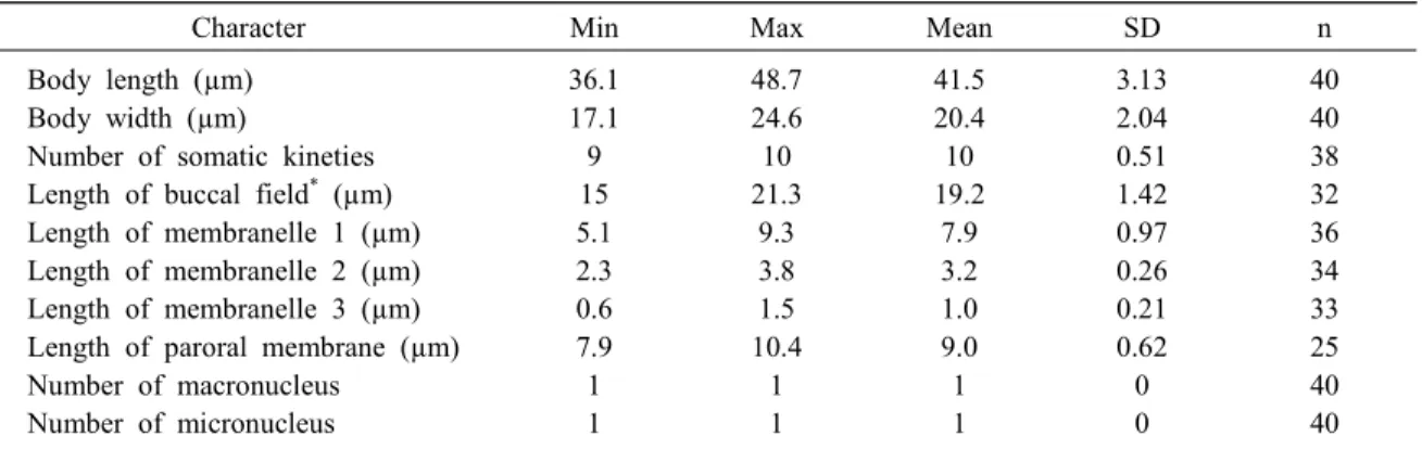
Table 1. Morphometric characterization of Paranophrys marina isolated from flounder.
Character Min Max Mean SD n
Body length (µm) Body width (µm)
Number of somatic kineties Length of buccal field* (µm) Length of membranelle 1 (µm) Length of membranelle 2 (µm) Length of membranelle 3 (µm) Length of paroral membrane (µm) Number of macronucleus
Number of micronucleus
36.1 17.1 9 15 5.1 2.3 0.6 7.9 1 1
48.7 24.6 10 21.3
9.3 3.8 1.5 10.4
1 1
41.5 20.4 10 19.2
7.9 3.2 1.0 9.0 1 1
3.13 2.04 0.51 1.42 0.97 0.26 0.21 0.62 0 0
40 40 38 32 36 34 33 25 40 40
* - distance from apex to posterior end of paroral membrane
Max-maximum; Mean-arithmetic mean; Min-minimum; n-number of samples; SD-standard deviation.
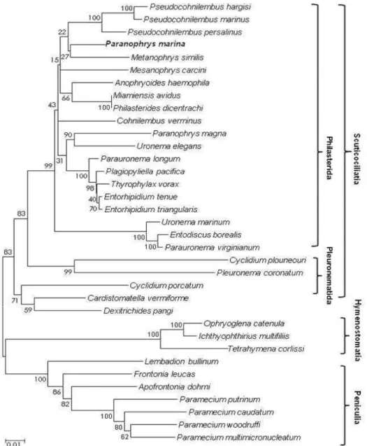
Phylogenetic analysis
The P. marina SSU rRNA sequence showed 96.3%
identity with the Parauronema longum SSU rRNA gene sequence (GenBank accession no. AY212807).
The SSU rRNA sequence of P. marina was aligned with SSU rRNA sequences of 34 other ciliates from three subclasses (Scuticociliatia, Hymenostomatia and Peniculia) in the GenBank database. After the re- moval of ambiguous sequences in the alignment site, a total of 1620 nucleotides remained for phylogenetic analysis. Phylogenetic analysis demonstrated that the subclass Scuticociliatia is supported as a monophyly in which it forms a polyphyletic clade (Order Philas- terida and Pleuronematida). Within the Philasterida, P. marina and Metanophrys similis form as a mono- phyletic clade which is a sister lineage to three species of Pseudocohnilembus and Mesanophrys carcini (Fig. 2).
To date, molecular information has not been eluci- dated for Paranophrys marina. According to the anal- ysis of SSU rRNA sequence data, P. marina SSU rRNA sequence was 96.3% homologous to that of Parauronema longum, indicating that these two spe- cies are closely related. Phylogenetic analysis dis- played that P. marina is included in the order philater- ida, within the subclass Scuticociliatia, the phylum
Ciliophora, as a monophyletic clade which is a sister lineage to three species of Pseudocohnilembus and Mesanophrys carcini.
P. marina is reported as an ectocommensal within the mantle cavity of farmed scallop Argopecten irra- dians in China (Song et al. 2002). It is also known that Paranophrys sp. are opportunistic secondary par- asites of cultured prawn Penaeus chinensis, in which they colonize pre-existing wounds, and subsequently invade the hemolymph and damage various organs including the gills (available at: http://www.pac.d- fo-mpo.gc.ca/sci/shelldis/pages/cildsp_e.htm).
Although the present study reports that Paranophrys marina is another scuticociliate species screened dur- ing an outbreak of scuticociliatosis from farmed olive flounder in Korea, it cannot be ruled out that scutico- ciliatosis may have occurred in conjunction with other scuticociliate species. Further investigations are need- ed to prove the pathogenicity of P. marina.
Acknowledgements
This research was supported by National Institute of Fisheries Science under a grant (R2021018).
Table 2. Morphological comparison of Paranophrys marina in literatures
Character P. marina*
(Thomson & Berger, 1965) Present study* P. marina**
(Song et al. 2002) Body length × width (µm) in vivo
Body length × width (µm) fixed No. of somatic kineties
Length of buccal field (µm) Length of membranelle 1 (µm) Length of membranelle 2 (µm) Body shape
Position of contractile vacuole pore Host
Sample location
- 39.3 × 19.2
10 19.6
8.4 2.8
Rounded both anterior and posterior
end of SK2 Hydroid Washington, USA
33-46 × 8-15 41.5 × 20.4
10 19.2
7.9 3.2 Slim, slender with
pointed anterior end of SK2
Flounder Jeju, Korea
30-45 × 10-15 - 10 about 2/5 very long
short Slim, spindle shaped with pointed anterior
end of SK2 Scallop Qingdao, China
*- Data from silver nitrate impregnation
**- Data from protargol impregnation
References
Bassleer G. (1983) Uronema marinum, a new and com- mon parasite on tropical saltwater fishes. Freshwater and Marine Aquarium 6, 78-81.
Chun S.K. (2000) Scuticociliatosis, disease of cultured and marine fish. Hanguk Susan Sinbo Press, Seoul (in Korean).
Cheung P.J., Nigrelli R.F. & Ruggieri G.D. (1980) Studies on the morphology of Uronema marinum Dujardin (Ciliatea: Uronematidae) with description of histopathology of the infection in marine fishes.
Journal of Fish Diseases 3, 295-303.
Crosbie P.B.B. & Munday B.L. (1999) Environmental factors and chemical agents affecting the growth of the pathogenic marine ciliate Uronema nigricans.
Fig. 2. A phylogenetic tree of SSU rDNA sequences of P. marina and scuticociliates constructed by the NJ method.
Numbers at the nodes are bootstrap values representing their robustness (10,000 replicates). The new sequence is shown in boldface.
Diseases of Aquatic Organisms 36, 213-219.
Dragesco A., Dragesco J., Coste F., Gasc C., Romestand B., Raymond J.C. & Bouix G. (1995) Philasterides dicentrarchi, n. sp., (Ciliophora, Scuticociliatida), a histophagous opportunistic parasite of Dicentrachus labrax (Linnaeus, 1758) a reared marine fish. Eur- opean Journal of Protistology 31, 327-340.
Foissner W. (1991) Basic light and scanning electron microscopic methods for taxonomic studies of cili- ated protozoa. European Journal of Protistology 27, 313-330.
Iglesias R., Paramá A., Álvarez M.F., Leiro J., Fernán- dez J. & Sanmartin M.L. (2001) Philasterides dicen- trarchi (Ciliophora, Scuticociliatida) as the causative agent of scuticociliatosis in farmed turbot Scophthal- mus maximus in Galicia (NW Spain). Diseases of Aquatic Organisms 46, 47-55.
Jee B.Y., Kim Y.C. & Park M.S. (2001) Morphology and biology of parasite responsible for scuticocilia- tosis of cultured olive flounder Paralichthys olivaceus.
Diseases of Aquatic Organisms 47, 49-55.
Jung S.J., Kitamura S.I., Song J.Y., Jeong I.Y. & Oh M.J. (2005) Complete small subunit rRNA gene se- quence of the scuticociliate Miamiensis avidus pa- thogenic to olive flounder Paralichthys olivaceus.
Diseases of Aquatic Organisms 64, 159-162.
Jung S.J., Kitamura S.I., Song J.Y. & Oh M.J. (2007) Miamiensis avidus (Ciliophora:Scuticociliatida) cau- ses systemic infection of flounder Paralichthys oli- vaceus and is a senior synonym of Phiasterides dicentrarchi. Diseases of Aquatic Organisms 73, 227- 234.
Kim S.M., Cho J.B., Kim S.K., Nam Y.K. & Kim K.H.
(2004a) Occurrence of scuticociliatosis in olive flounder Paralichthys olivaceus by Philasterides di- centrarchi (Ciliophora: Scuticociliatida). Diseases of Aquatic Organisms 62, 233-238.
Kim S.M., Cho J.B., Lee E.H., Kwon S.R., Kim S.K., Nam Y.K. & Kim K.H. (2004b) Pseudocohnilembus persalinus (Ciliophora:Scuticociliatida) is an addi- tional species causing scuticociliatosis in olive floun- der Paralichthys olivaceus. Diseases of Aquatic Organisms 62, 239-244.
Ma H., Choi J.K. & Song W. (2003) An improved silver
carbonate impregnation for marine ciliated protozoa.
Acta Protozoologica 42, 161-164.
Munday B.L., O'Donoghue P.J., Watts M., Rough K.
& Hawkesford K. (1997) Fatal encephalitis due to the scuticociliate Uronema nigricans in sea-caged, southern blue tuna Thunnus maccoyi. Diseases of Aquatic Organisms. 30, 17-25.
Parama A., Iglesias R., Alvarez M.F., Leiro J., Aja C.
& Sanmartn M.L. (2003) Philasterides dicentrarchi (Ciliophora, Scuticociliatida): experimental infection and possible routes of entry in farmed turbot (Sco- phalmus maximus). Aquaculture 217, 73-80.
Saitou N. & Nei M. (1987) The neighbor-joining meth- od: a new method for reconstructing phylogenetic trees. Molecular Biology and Evolution 4, 406-425.
Song J.Y., Kitamura S.I., Oh M.J., Kang H.S., Lee J.H., Tanaka S.J. & Jung S.J. (2009) Pathogenicity of Miamiensis avidus (syn. Philasterides dicentrarchi), Pseudocohnilembus persalinus, Pseudocohnilembus hargisi and Uronema marinum (Ciliophora: Scutico- ciliatida). Diseases of Aquatic Organisms 83, 133- 143.
Song W., Ma H., Wang M. & Zhu M. (2002) Compar- ative studies on two closely related species Urone- mella filificum (Kahl, 1931) and Uronema elegans Maupas, 1883 with redescription of Paranophrys marina Thompson et Berger, 1965 (Ciliophora: Scu- ticociliatida) from China Seas. Acta Protozoologica 41, 263-278.
Thompson J.C. & Berger J. (1965) Paranophrys marina n. g., n. sp., a new ciliate associated with a hydroid from the Northeast Pacific (Ciliata: Hymenostomatida).
Journal of protozoology 12, 527-531.
Thompson J.C., Higgins D.G. & Gibson T.J. (1994) CLUSTAL W: Improving the sensitivity of pro- gressive multiple sequence alignment through se- quence weighting, positions-specific gap penalties and weight matrix choice. Nucleic Acids Research 22, 4673-4680.
Yoshinaga T. & Nakazoe J. (1993) Isolation and in vitro cultivation of an unidentified ciliate causing scutico- ciliatosis in Japanese flounder (Paralichthys oliva- ceus). Fish Pathology. 28, 131-134.
Manuscript Received : Apr 29, 2021 Revised : May 31, 2021 Accepted : Jun 03, 2021