논문 2016-53-12-22
VR, AR 시뮬레이션 및 3D Printing을 활용한 어깨와 팔꿈치 수술실습
( VR, AR Simulation and 3D Printing for Shoulder and Elbow Practice )
임 원 봉*, 문 영 래**
( Wonbong Lim and Young Lae Moon
ⓒ)
요 약
최근 의료 영상 기술의 발전은 진단, 수술계획, 또는 교육에 도움이 되는 수술 시뮬레이션을 만들어 왔다. 개선된 고화질 영 상과 3차원 시각화는 의료 영상 가용성을 향상시키고 수술, 교육 분야에서 더 잘 이용할 수 있게 되었다. 실제 인간의 시각은 입체이다. 따라서, 외과의사의 판단을 통해 2차원 영상을 스테레오로 재구성하여 처리하는 것이 함께 필요하다. 이러한 과정을 줄이기 위해, 3차원 (3D) 이미지가 사용되어 왔다. 3D 영상은 복잡한 상황에서 외과 의사가 매우 짧은 시간에 판단할 수 있도 록 3D 시각화를 강화하여 제공한다. 3D 화상 데이터 세트에 기초하여, 가상 내시경 수술 계획, 실시간 상호 작용 가상 의료 시뮬레이션이 가능하게 되었다. 본 논문은 새로운 이미징 기술의 최근 응용 프로그램을 설명하고 이의 기본과 특별히 주목할 만한 의료 3D 복원 기술에 관한 것이다. 최근 CT, MR 및 기타 영상 양식의 기술발전은 흥미로운 새로운 솔루션과 어깨 영상 의 활용 가능성을 넓혀왔다. 특히, 의료 기기에서 파생 된 3차원 (3D) 이미지는 고급 정보를 제공한다. 이 프레젠테이션은 어 깨와 팔꿈치의 수술실습에서 원리, 3D 영상기술의 잠재적 응용가능성, 시뮬레이션, 3D프린팅을 설명한다.
Abstract
Recent advances in technology of medical image have made surgical simulation that is helpful to diagnosis, operation plan, or education. Improving and enhancing the medical imaging have led to the availability of high definition images and three-dimensional (3D) visualization, it allows a better understanding in the surgical and educational field. The Real human field of view is stereoscopic. Therefore, with just 2D images, stereoscopic reconstruction process through the surgeon’s head, is necessary. To reduce these process, 3D images have been used. 3D images enhanced 3D visualization, it provides significantly shorter time for surgeon for judgment in complex situations. Based on 3D image data set, virtual medical simulations, such as virtual endoscopy, surgical planning, and real-time interaction, have become possible. This article describes principles and recent applications of newer imaging techniques and special attention is directed towards medical 3D reconstruction techniques. Recent advances in technology of CT, MR and other imaging modalities has resulted in exciting new solutions and possibilities of shoulder imaging. Especially, three-dimensional (3D) images derived from medical devices provides advanced information. This presentation describes the principles and potential applications of 3D imaging techniques, simulation and printing in shoulder and elbow practice.
Keywords
:VR, AR, Simulation, 3D Printing, Orthopedic
*
정회원, 조선대학교 의과대학 (School of Medicine, Chosun University, Gwangju, Korea)
**
정회원, 조선대학교 의과대학 (School of Medicine, Chosun University, Gwangju, Korea)
ⓒ
Corresponding Author (E-mail: [email protected])
※ 이 논문은 2014년도 조선대학교 학술연구비의 지원을 받아 연구되었음.
※ This study was supported by research fund from Chosun University, 2014.
Received ; October 27, 2016 Accepted ; November 30, 2016
Ⅰ. 서 론
Diagnosis and surgical planning in orthopedic
surgery and other surgical disciplines has evolved rapidly in the past decade. Modern minimally invasive surgery with arthroscopic surgical techniques demand
Fig. 1. Procedure of simulation and 3D printing from medical images.
그림 1. 의료영상 기반 시뮬레이션 및 3D프린팅 절차
high level of skills especially with regard to triangulation in space as well as working within a particular shoulder and elbow while observing the X-ray, CT or MRI images on a separate screen. The learning curve to obtain these skills is very long and steep. It may take several years of training to learn and accomplish proficiency in these techniques.Current teaching techniques involve traditional apprenticeship with many years of assisting senior surgeons, learning on plastic models (rarely mimicking the real life), participating in courses with workshops on human cadavers and eventually learning on real patients. There are obviously difficulties with these methods of training and practice resources are limited.
Using the Virtual Reality (VR), Augmented Reality (AR) and 3D printing of models with tactile feedback within an arthroscopic simulator may overcome these difficulties and improve the ability to make a plan for surgery and train surgeons in performing these complex arthroscopic procedures without risk. The VR and AR arthroscopic simulator provides an
unlimited resource for training and surgery simulation readily available to all. In addition, surgeons can practice on 3D printing simulators for unlimited times until they achieve good enough skills to enable them to perform these procedures safely on patients. These arthroscopic simulators with tactile feedback provides an excellent teaching tool and quantitative skill assessment tool. Several factors are driving the push toward VR and AR simulators in orthopaedics[1]. Surgery is an apprenticeship technique that requires additional time for training in the operating room. In one study, orthopaedic residents took over 40 minutes longer to repair an anterior cruciate ligament tear than did the faculty surgeon[2]. Training residents is an expensive endeavor for the supporting hospital with some estimates reaching $48,000 per resident per year in operating room time alone[3]. Therefore, training simulators could specifically allow practice of this key skill. Simulating these key procedures could improve the skill of novice shoulder and
Fig. 2. Human 3D Volume model from CT.
그림 2. CT에서 추출한 인체 3D 볼륨 모델
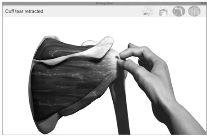
Fig. 3. Shoulder Practice in Leap Motion VR device.
그림 3. 립모션 VR장치를 활용한 어깨 수술실습
elbow surgeons and reduce the risk of injuryto patients.
Ⅱ. 본 론
1. Application of AR and VR simulation for medical image
Medical imaging using by X-ray, MRI, and ultrasounds is used to create visual representations of the interior of the body for clinical analysis and medical intervention of complex diseases in a
very rapidly. The medical imaging market is poised to grow significantly over the next five years as medical providers continue to seek innovativeways to enhance patient care — it was worth as much as $24.39 billion in 2012 and is predicted to grow to $35.35 billion by 2019.
Traditional medical
imaging systems provide 2D visual representations of human organs while more advanced digital medical imaging systems can create both 2D and in many cases 3D images of human organs[4]. Systems capable of 3D digital medical imaging are currently only a small part of the overall medical imaging market — projected to hit $2.9 billion by 2020—but it has effectively doubled in size over thelast two years and is already rapidly expanding into practice areas such as oncology, orthopedics, obstetrics/gynecology, cardiology, and dentistry.
Displays are an integral part of these digital medical imaging systems. Current systems with displays that can only visually represent the imaging data collected in 2D or at best in simulated 3D on 2D. As advances in 3D displays, including glasses-free 3D volumetric displays, the potential applications are endless.
A typical CT scan or MRI produces hundreds of images at one time that then need to be analysis. It is time consuming to ensure all angles and images are accounted not only for the patient but also for radiologists and referring doctors who need to look at every single cross-section image before deciding on a course of treatment. By utilizing 3D technology within
medical imaging, surgeons able to take those cross-section slices and combined them into a concise 3D visual of the area scanned. The 3D data is viewable without any additional viewing aids, in true 3D. This reduces eye fatigue of the viewers and increases cognitive awareness. Certain 3D display technologies currently under development can display these images with resolutions as high as 80,000 voxels (volumetric pixels), a factor of 10x higher resolution than commercially available 2D displays. The Fig. 1 shows procedure of simulation from medical image.
An example would be a collaborative evaluation of an
MRI, CT, or ultrasonic image to determine the presence
of an
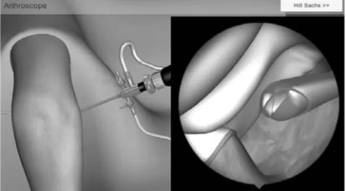
anomaly that otherwise be interrupted when viewed on conventional 2D or simulated 3D on 2D display — like looking for a tumor in dense breast tissue. Benefits include improved diagnostic confidence from patients, the replacement of more invasive diagnostic procedures, and an easy-to-read solution forFig. 4. Elbow Practice in Virtual Reality Arthroscope.
그림 4. 가상현실 관절경의 팔꿈치 수술
Fig. 5. Establishment of preoperative planning using 3D model.
그림 5. 3D모델을 이용한 수술계획 수립
Fig. 6. 3D Printing and Simulation from CT.
그림 6. CT영상기반 3D프린팅과 시뮬레이션 과정
patient education.2. Application for shoulder and elbow surgery
It would be said easy to assume that “everything is same” on the inside, but it is difficult to guarantee what you’ll find until you open someone up. Therefore, it could be considered if a surgeon had a complete understanding of a patient’s actual anatomy before stepping into the operating room. Recently, surgeons have to consider a patient’s problem based on what they know about their anatomy from 2D images. With 3D imaging, a surgeon can see real pictures of the anatomy and interact with it in any way they choose, allowing them to solve the issue at hand before picking up a scalpel. In Fig. 5, establishment of preoperative planning using 3D image was presented.
Utilizing this process minimizes exploratory surgeries and procedures, and decreases damage to surround healthy tissues by more exactly pinpointing
the treatment area. Being able to view a comprehensive 3D image of the area before surgery limits surprises in the operating room and increases efficiency of treatment.
3. Application for 3D printing
3D printing is an ideal partner for 3D medical imaging because of the uniqueness of every patient and the challenges this creates in sustainable business models that require selling large volumes of similar products. One example is prosthetics, while another is neonatal-specific devices for short-term care. One near-term application is the printing of physical models from 3D data that would be used for planning critical
surgeries or general training. In both
cases, the primary challenge with 3D printing
continues to be speed. It will take time to create a
printed model from actual 3D medical imaging data,
and if 3D medical imaging data is not readily
available, additional time to create the image through
various modeling tools to be need. Any additional
iterations of the printed part just continue to extend
this time. In the case of planning or training, a 3D
display would displace or compliment the need for a
physical model because the image created maintains
many of the benefits of the physical model, but it
creates in seconds versus hours, with additional
iterations at a similar speed. The benefit of speed is
significant, as the medical user may require
information much faster than a printer might allow,
even when the 3D imaging data is available in real
time.
저 자 소 개 임 원 봉(정회원)
1999년 전남대학교 생물화학공학 학사 졸업
2002년 전남대학교 물질생물화학 공학 석사 졸업
2007년 전남대학교 의공학 박사 졸업.
<주관심분야 : 의료기기, 물질생물화학, 의공학>
문 영 래(정회원)
1990년 조선대학교 의학 학사 졸업.
1994년 조선대학교 의학 석사 졸업.
2004년 조선대학교 의학 박사 졸업.
<주관심분야 : 의료영상, IOT, AR, VR, 3D프린팅>
Ⅳ. 결 론
Development of CT, MR and other imaging modalities has ensued in new applications and possibilities of imaging. Among them, 3D images are enthusiastic issue on clinical and research field. It can help the clinician to more effectively and shortly understand and decision. In addition, 3D images provide advanced medical related applications, which is considered as a powerful instrument for clinician, students, as well as public health.
REFERENCES
[1] Mabrey JD, Reinig KD, Cannon WD. Virtual reality in orthopaedics: is it a reality? Clin Orthop Relat Res. 2010;468: 2586-2591.
[2] Farnworth LR, Lemay DE, Wooldridge T, et al.
A comparison of operative times in arthroscopic ACL reconstruction between orthopaedic faculty and residents: the financial impact of orthopaedic surgical training in the operating room. Iowa Orthop J. 2001;21: 31-35.
[3] Bridges M, Diamond DL. The financial impact of teaching surgical residents in the operating room. Am J Surg. 1999;177: 28-32.
[4] Fenster A, Downey DB, Cardinal HN. Three- dimensional ultrasound imaging. Phys Med Biol.
2001;46: R67-99.