www.issisglobal.org 9 Journal of International Society for Simulation Surgery 2017;4(1):9-12
Introduction
Craniopagus is rare and the degree of attachment is diverse. Since there may be many other anatomical abnormalities, in-cluding shared venous system or scalp and skull defects, pre-cise inspection is necessary (1). 2D imaging such as magnetic resonance imaging (MRI) or computerized tomography (CT) have a limited viewing angle and may obscure spatial relation-ships. However, 2D images can be easily transformed to 3D
vol-ume images on a computer screen. The limitations of an angled or curved surface can be partly overcome by lighting, perspec-tive, and shading effects. These can be helpful in visualizing re-alistic volume displays; however, the perception of structures is still limited. For presenting the fine details and for an in-depth understanding of the structures, a variety of stereoscopic displays are widely used in medical applications such as diagnosing, pre-operative-planning, minimally invasive surgery (MIS), teach-ing, and training (2).
O
riginal ArticleVirtual Reality and 3D Printing for Craniopagus Surgery
Gayoung Kim1, Eungjune Shim1, Hussein Mohammed2, Youngjun Kim1*, Yong Oock Kim2*
1Center for Bionics, Korea Institute of Science and Technology, Seoul, Korea
2Department of Plastic Surgery, Institute of Human Tissue Restoration, Yonsei University Medical School, Seoul, Korea
PurposeZZSurgery for separating craniopagus twins involves many critical issues owing to complex anatomical features. We demonstrate a 3D printed model and virtual reality (VR) technologies that could provide valuable benefits for surgical planning and simulation, which would improve the visualization and perception during craniopagus surgery.
Material & MethodsZZWe printed a 3D model extracted from CT images of craniopagus patients using segmentation software developed in-house. Then, we imported the 3D model to create the VR environment using 3D simulation software (Unity, Unity Technologies, CA). We utilized the HTC Vive (HTC & Valve Corp) head-mount-display for the VR simulation.
ResultsZZWe obtained the 3D printed model of craniopagus patients and imported the model to a VR environment. Manipu-lating the model in VR was possible, and the 3D model in the VR environment enhanced the application of user-friendly 3D modeling in surgery for craniopagus twins.
ConclusionZZThe use of the 3D printed model and VR has helped understand complicated anatomical structures of craniopagus patients and has made communicating with other medical surgeons in the field much easier. Further, interacting with the 3D model is possible in VR, which enhances the understanding of the craniopagus surgery as well as the success rate of separation surgery while providing useful information on diagnosing and surgery planning.
Key WordsZZVirtual reality ㆍ3D printing ㆍCraniopagus ㆍSurgical simulation.
Received: April 18, 2017 / Revised: April 27, 2017 / Accepted: May 3, 2017 Address for correspondence: Youngjun Kim
Center for Bionics, Korea Institute of Science and Technology, 5 Hwarang-ro 14-gil, Seongbuk-gu, Seoul 02792, Korea
Tel: 82-2-958-5606, Fax: 82-2-958-5649, E-mail: [email protected] Address for correspondence: Yong Oock Kim
Plastic and Reconstructive Surgery, Institute of Human Tissue Restoration, Yonsei University, 50-1 Yonsei-ro, Seodaemun-gu, Seoul 03722, Korea
Tel: 82-2-2228-2218, Fax: 82-2-362-5680, E-mail: [email protected]
pISSN 2383-5389 / eISSN 2383-8116 https://doi.org/10.18204/JISSiS.2017.4.1.009
This is an Open Access article distributed under the terms of the Creative Commons Attribution Non-Commercial License (http://creativecommons.org/licenses/ by-nc/4.0/) which permits unrestricted non-commercial use, distribution, and reproduction in any medium, provided the original work is properly cited.
ORCID
Gayoung Kim: orcid.org/0000-0002-4748-0027 Eungjune Shim : orcid.org/0000-0003-0325-7517 Hussein Mohammed: orcid.org/0000-0002-5807-6628 Youngjun Kim: orcid.org/0000-0002-7832-3200 Yong Oock Kim: orcid.org/0000-0002-3756-4809
10
Journal of International Society for Simulation Surgery█ 2017;4(1):9-12
Some of today’s surgical separations utilize a solid 3D model for diagnosing and pre-operative planning and communica-tion with medical experts (3, 4, 5, 6). A physical model provides actual distance and scale, which increases the understanding of complex anatomical structures.
Virtual reality (VR), a 3D visualization system in a virtual environment, has proven to be useful in medical applications (8). This technology facilitates the education of medical proce-dures, diagnoses, and attainment of knowledge of the human body by interacting with a 3D model, which can be resized and reused (9). In fact, surgery for separating craniopagus twins is an ideal field in which VR can be employed because the shape of conjoined cranium varies, and the internal struc-ture requires detailed observation.
This study constructs a 3D printed model and VR environ-ment using CT images of craniopagus patients to overcome the issues associated with the traditional visualization systems of medical data.
Materials and Methods
The CT images of craniopagus patients were exported in DI-COM format, and a 3D model was developed using segmenta-tion software (Mimics, Materialise, Belgium). This model was saved as an STL file to print the 3D model and was simulated in virtual reality software (Unity, Unity Technologies, CA).
3D Printing
The craniopagus model was printed at a 1:1 scale using an Object260 Connex3 (Stratasys, Eden Prairie, MN) with mate-rial VeroWhite to resemble a realistic skull.
Virtual Reality Simulation
The 3D printed model is imported to Unity to create a virtual environment. C# programming language was used in Unity to build interface functions. The device used for the simulation was HTC Vive (HTC, Valve Corp.).
Similar to the 3D printed model, the novel user interface was built to allow certain interaction functionality using a control-ler, which included the freedom to manipulate and separate parts of the geometry. The “Grip button” of the controller con-nects the object and the controller, so practitioners may ascer-tain when they grab the model. The collider event is utilized to detect collision between the structure and the controller. Fur-ther, to control the model’s position through a physics simula-tion, a Rigidbody component should be included. “Use Gravity” was checked and “Is Kinetic” was unchecked for this simulation.
Mesh splitting is implemented by pressing the “Trigger”
but-ton of the controller. The start point of the cutting line is set when the “Trigger” button is pushed down, and it ends when the “Trig-ger” button is released. The cutting plane includes the cutting line and is parallel to the viewing direction of the camera. Con-tinuous slicing is available and the sliced pieces contain the same attributes as the original.
Results
Separation of craniopagus twins is a tremendous challenge largely because of their complex and unusual anatomy. Sur-geons using the virtual reality simulation make use of improved 3D visualization and can interact with the structure.
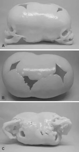
Fig. 1. A-D: 3D printed model of a craniopagus patient. The print-ed model can provide rich information compare to the traditional medical data visualization system.
A
B
Virtual Reality and 3D Printing for Craniopagus Surgery █Kim G, et al
www.issisglobal.org 11 3D printing illustrates how physical modeling can help enhance
the perception of the anatomical structure to medical specialists. Seeing the exact size and shape of the tactile structure makes com-municating with others involved much easier (Fig. 1).
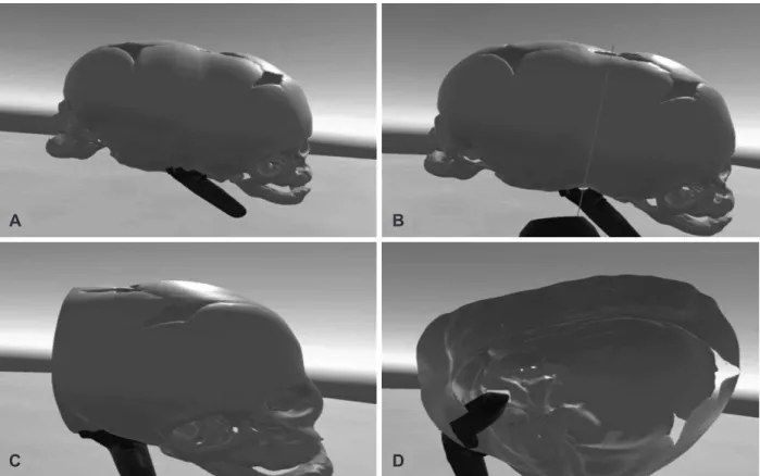
The developed VR system has the capacity to represent the 3D anatomical structure of a craniopagus skull in an interac-tive VR environment. Functionalities were added to enhance interaction with the model such as grabbing and cutting. View-ers can navigate the structure from multiple viewpoints, and take a closer look when the “Grip” button is activated. The “Trig-ger” button generates the cutting line, and the object is split down the line. The sliced parts can also be explored using the “Grip” button. Subsequent additional cutting is also possible (Fig. 2).
This system strives to provide customizable and accessible functionalities. The virtual reality anatomy system can be cus-tomized based on the viewer’s preference, as the model is not limited to a specific size, viewpoints, or cut parts. Through these characteristics, the following benefits are obtained: help in un-derstanding of the complex craniopagus structure; increase in the details of the craniopagus skull; ability to cut the structures through a cutting plane. Moreover, each function is easy to han-dle because only a single button needs to be pressed for manip-ulation.
Discussion
The brains of conjoined twins within a single cranial cavity are unusual and its shape is diverse. There are some points to consider during the separation procedure. With regard to scalp and dural cover, the position and shape of the skin incision is important (3, 5). Without proper closure, fluid leakage or infec-tion may occur. Visualizainfec-tion of the anatomy in 3D clearly shows the surgeons the shared bone and brain, which allows surgeons to better understand how it is twisted or attached to each other.
Another critical factor in the surgery is the shared venous sys-tem. Anomalous venous drainage is a critical factor that deter-mines fetal survival (1, 3, 5, 6).
Virtual reality is widely used in the medical field. It can pro-vide more than just a realistic and modifiable visualized model. Surgeons can implement their operation plan and predict the result. Scalp Incision and closure can be performed virtually. A guided incision line may suggest the optimized coverage area. Further, regarding a shared venous system, venous system di-vision while observing the resulting blood drainage, blood flow, and junction may be possible.
Fig. 2. A-D: 3D model in VR environment. A: The model is grabbed by the controller. B: Cutting plane is created. C: External surface of the sliced model. D: Internal surface of the sliced model.
A B
12
Journal of International Society for Simulation Surgery█ 2017;4(1):9-12
Conclusion
The work presented in this paper shows how a 3D printed model and virtual reality system can be helpful in the surgery for separating of craniopagus twins. The 3D techniques discussed for anatomical analysis provide improved visualization and in-teraction with complex anatomy, surgical rehearsal, customiza-tion, and precise communication between medical specialists.
Developing the VR anatomy system will improve model ma-nipulation and provide anatomical information for each stage of the surgery procedure.
Acknowledgements
This research was supported by the KIST institutional pro-gram (2E26880, 2V05430).
References
1. WALKER, Marion; BROWD, Samuel R. Craniopagus twins:
embry-ology, classification, surgical anatomy, and separation. Child’s Ner-vous System 2004;20(8-9):554-566.
2. VAN BEURDEN, Maurice HPH, et al. Stereoscopic displays in med-ical domains: a review of perception and performance effects. In: IS&T/SPIE Electronic Imaging. International Society for Optics and Photonics;2009:72400A-72400A-15.
3. SWIFT, Dale M., et al. Total vertex craniopagus with crossed venous drainage: case report of successful surgical separation. Child’s Ner-vous System 2004;20(8-9):607-617.
4. CAMPBELL, Scott. Separation of craniopagus twins: the Brisbane experience. Child’s Nervous System 2004;20(8-9):601-606. 5. GOH, Keith YC. Separation surgery for total vertical craniopagus
twins. Child’s Nervous System 2004;20(8-9):567-575.
6. DUNAWAY, David; JEELANI, NU Owase. Staged separation of cra-niopagus twins. In: Seminars in pediatric surgery. WB Saunders, 2015:241-248.
7. CHRISTENSEN, Andrew M, et al. Advanced “tactile” medical im-aging for separation surgeries of conjoined twins. Child’s Nervous System, 2004;20(8-9):547-553.
8. AL-KHALIFAH, Ali, et al. Using virtual reality for medical diag-nosis, training and education. International Journal on Disability and Human Development 2006;5.2:187.
9. FALAH, Jannat, et al. Virtual Reality medical training system for anatomy education. In: Science and Information Conference (SAI), 2014. IEEE, 2014:752-758.