신경재활치료과학 제6권 제2호
Therapeutic Science for Neurorehabilitation Vol. 6. No. 2. 2017.
The Effect of Prism Adaptation Following Traumatic Brain Injury: A case report
Jeong, Eun-Hwa*, Min, Yoo-Seon**
*Dept. of Occupational Therapy, College of Health Science, Far East University
**Dept. of Occupational Therapy, College of Health Science, Yonsei University
Abstract
Background: The presence of visuospatial impairment can make patients slow functional recovery and impede the re- habilitation process in TBI patients.
Objective: The aim of this study is to investigate effects of prism adaptation treatment for functional outcomes in patients following traumatic brain injury.
Methods: The subject received prism adaptation treatment for 2 weeks additionally during traditional rehabilitation for 4 weeks. The Patient has prism adaptation treatment while wearing wedge prisms that shift the external environ- ment about 12° leftward. The patient received 10 sessions, 15-20min each session. Outcome measures were vi- suospatial deficit(line bisection, latter cancellation), Visual and spatial perception(LOTCA-visual perception and spa- tial perception), motor function of upper extremity(FMA U/E; Fugl-Meyer motor assessment upper extremity, ARAT;
Action research arm test), balance(BBS; Berg Balance Scale), mobility(FAC; Functional ambulation classification) and functional level(FIM; Functional independent measure). All Assessments took place on study entry and post-treat- ment assessments were performed at discharge from the hospital.
Results: After prism adaptation, the visuospatial impairment scores improved as indicated in the line bisection(-15.2 to -6.02), latter cancellation(2 to 0) and LOTCA- spatial perception scores(7 to 9). The upper motor function im- proved as indicated in the scores of affected FMA U/E(21 to 40) and ARAT(4 to 22). Ambulation and balance im- proved as indicated in the BBS scores(25 to 38) and FAC scores(0 to 4). ADL function improved as indicated in the FIM total scores 54 to 70(motor 34 to 61, cognition 20 to 29).
Conclusion: Prism adaptation did improve functional level such as motor functions and ADL abilities in TBI patient.
Further research is recommended.
Key words: Prism adaptation, TBI, Visuospatial impairment
교신저자 : 정은화([email protected]) I I 접수일: 2017. 7. 12 I I 심사일: 2017. 8. 11
Ⅰ. Introduction
Traumatic brain injury occurs when a head injury has by an external force. The most common causes of TBI include vehicle accidents, safety accident, falls, and sports(Kusgber, 1988; Comper et al., 2005). The in- cidence of TBI is frequently stated to be 200 per 100,000 population at risk per year in develop coun- tries(Bruns & Hauser, 2013).
Brain trauma causes of confused level of conscious- ness, seizure, coma, or focal sensory or motor neuro- logic deficit. TBI can cause a host of physical, cognitive, social, emotional, and behavioral effects. The outcome can range from complete recovery to permanent dis- ability or death(Hall, Hall, & Chapman, 2005).
Cognitive deficits following TBI include impaired at- tention, memory loss, deficits in executive functions and reduced processing skills(Hall et al, 2005; Arlinghaus, Shoaib, & Price, 2005). Visuospatial impairment has been linked with right and left hemispheric lesions in the acute stage following brain injury(Golisz & Toglia, 2003; Toglia, 1991; McKenna, Cooke, Fleming, Jefferson,
& Ogden, 2006).
Visuospatial function is process of receiving and or- ganizing visuospatial information. Visuospatial impair- ments occur loss of knowledge of the spatial relations, such as right-left discrimination or distinction of body parts. They may occur motor planning and attentional problems toward contralesional side(Grieve, 2000;
Kerkhoff, 1998). The presence of visuospatial impair- ment can make patients slow functional recovery and impede the rehabilitation process(Suter, 1995).
Visuospatial treatments for improvement of visuo- spatial functions are such as visual search, spatial repre- sentation, reading and prism adaptation(Frassinetti, Angeli, Meneghello, Avanzi, & Làdavas, 2002). Prism adaptation is a simple way of producing visuomotor
correspondences, demonstrated by after-effects of prism exposure(Frassinetti et al, 2002; Pisella, Rode, Farne, Tilikete, & Rossetti, 2006). Prism adaptation requires the patient to perform visuomotor tasks while wearing pris- matic goggles. The goggle induces a deviation of the visual field toward the contralesional side(Serino, Barbiani, Rinaldesi, & Làdavas, 2009). After-effects, the patients have to orient the pointing movement toward contralesion side, resulting in a sensorimotor coordinates.
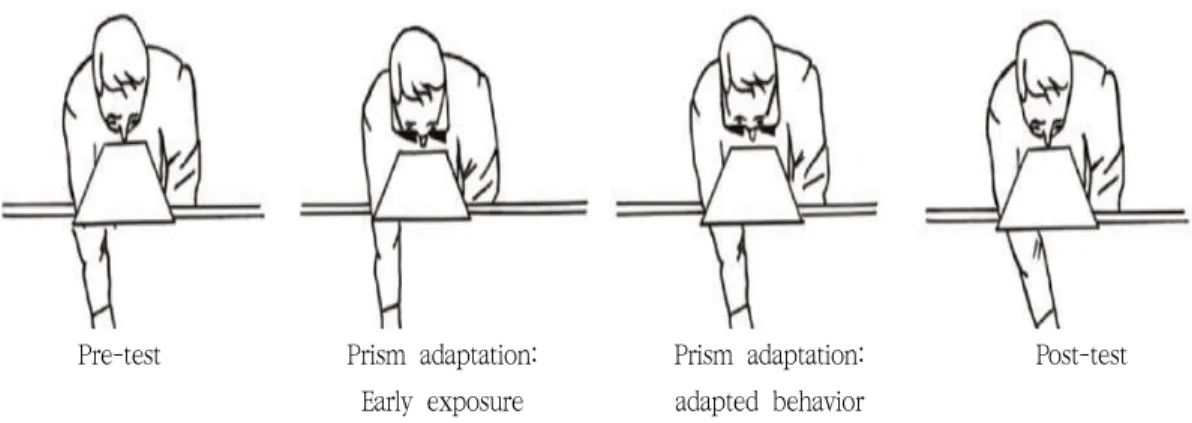
Prism adaptation can improve higher visuospatial neglect both for a short term effect and long term, up to 6 months after prism treatment(figure 1)(Frassinetti et al, 2002; Pisella et al, 2006; Serino et al, 2009).
In later studies, it was reported that the effects of prism adaptation could generalize across various clinical aspect of unilateral spatial neglect in stroke patients, in- cluding wheelchair navigation(Jacquin-Courtois, Rod, Pisella, Boisson & Rossetti, 2008), postural control(Serino et al, 2009), tactile extinction(Dijkerman, Webeling, Ter Wal, Groet, & Van Zandyoort, 2004), mental im- agery(Rode, Rossetti & boisson, 2001), motor recov- ery(Fortis, Chen, Goedert, & Barrett, 2011) and activities of daily living(Shiraishi, Muraki, Ayaka, & Hirayama, 2009). However, there is no study that has examined the effects of prism adaptation for TBI with visuospatial impairments.
Stroke is cerebrovascular disease due to both lack of blood flow and bleeding(Marnane et al, 2010).
Otherwise, one of the important pathologic features of TBI is diffuse axonal injury(DAI). Diffuse axonal injury may occur dependent on inertial forces such as rapid head motions, which damage the white matter.
Therefore, progressions between stroke and TBI may differ for the prism adaptation aimed at rehabilitations for visuospatial impairments(Adams, Doyle, Ford, Gennarellim Graham & McLellan, 1989; Blumbergs, Jones, & North, 1989). If greater efficacy of re- habilitation can be brought about by prism adaptation
for patients following TBI with visuospatial impairment, they might achieve higher functional levels.
The aim of this study is to investigate effect of prism adaptation treatment for functional outcomes in patients following traumatic brain injury.
Ⅱ. Method
1. Design
This study was retrospective, single case study to re- port the use of prism adaptation for visuospatial impair- ment in an individual following traumatic brain injury.
The subject received prism adaptation treatment for 2 weeks additionally during traditional rehabilitation for 4 weeks.
1) Subject
A 50 years-old man had been diagnosed quadriplegia due to traumatic subarachnoid hemorrhage 3 months before treatment. He has confirmed visuospatial impair- ment toward the right side by line bisection, letter can-
cellation and assessments of LOTCA-spatial perception.
He was alert and able to obey command. MMSE score was 17. He had no comorbidities such as cardiovascular disease, high blood pressure, diabetes. He had visuospa- tial impairment toward right side. He was also right-hand dominant.
2. Assessment
The subject underwent a standardized assessments for visuospatial impairment(line bisection, latter cancellation, LOTCA-visual perception and spatial perception), upper motor function(FMA U/E; Fugl-Meyer motor assessment upper extremity, ARAT; Action research arm test), am- bulation and balance(BBS; Berg Balance Scale, FAC;
Functional ambulation classification) and ADL func- tion(FIM; Functional independent measure).
Line bisection is performed a mark the center of eighteen horizontal lines on A4 paper.Total score calcu- lated lengths(mm) of average by measuring the deviation of the bisection from the true center of the line. Below 6.33mm is normal, 6.33mm or more is spatial neglect, and above 12.5mm is severe neglect(Keller, Schindler, Kekhoff, Von Rosen, & Golz, 2005). Letter cancellation is
Pre-test Prism adaptation:
Early exposure
Prism adaptation:
adapted behavior
Post-test
Post-test – Pre-test = After-effect (ADAPTATION)
Figure 1. prism adaptation(Pisella et al., 2006)
used to evaluate the presence of unilateral spatial ne- glect through omission of letters. The letter cancellation consists in an array of 20 randomly distributed target and letters mixed 25 distractors. The score is number of omissions and above two omissions means spatial ne- glect(Kelleretal, 2005).
LOTCA-visual perception and spatial perception for visual and spatial perception are subtest of Loewenstein Occupational Therapy Cognitive Assessment(LOTCA) battery. The assessments of visual perception are Object identification, Shape identification, Overlapping figures, and Object consistency. Subtests of Spatial perception are on self, spatial relations, and spatial relations on pictures. The subtests are scored from 1 to 4(Itzkovich, Elazar, Averbuch, & Katz, 2000).
FMA U/E; Fugl-Meyer motor assessment is used to measure voluntary limb movement. The U/E subscale is 33 items and score range is 0-66(Sanford, Moreland, Swanson, Stratford & Gowland, 1993).
ARAT; Action research arm test assesses upper limb functions which are grasp, grip, pinch and gross move- ment to handle various objects. The ARAT is 19 items measure divided into 4 sub-tests. The scale range is 0-3 and total score is 57(McDonnell, 2008).
BBS; Berg Balance Scale is performed a static and dynamic balance abilities. The BBS is developed to measure balance of the older adult with impairment in- balance function by assessing the performance of 14 functional tasks(Stevenson & Tsang, 1996).
FAC; Functional ambulation classification assesses functional ambulation inpatients. The FAC has range from independent walking outside to non-functional walking. The FAC has 6-point scale assess ambulation ability(Williams, 2011).
FIM; Functional independent measure was assessed activities of daily livings, has motor and cognitive scores. The FIM measures 18 functional activities:
1)self-care, 2)sphincter control, 3)transfer, 4)locomotion,
5)communication, 6)social cognition. The total score is from 18to126(Granger, Hamilton, Linacre, Heinemann, &
Wright, 1993).
All Assessments took place on study entry and post-treatment assessments were performed at discharge from the hospital.
3. Prism adaptation
The patient’s position is at the center of a table, and adjusted to a comfortable height. The prism adaptation procedure involves (1) pre-prism test, (2) prism adapta- tion, (3) post-prism test. Tests involve proprioceptive pointing which are 10 repeated pointing movements and 6 visuomotor proprioceptive pointing during blocked arm pointing movements. In the proprioceptive pointing condition, the patient is required to point with their index finger toward the center. The patient is asked to unaffected hand on their chest, and to point with the index finger straight ahead of the patient’s body midline. In the visuomotor proprioceptive pointing con- dition, the patient is asked to unaffected hand on their chest, and to point with the index finger toward the pen(visual target). The pointing is executed below the box, so that they could not see the arm movements. The visual target is presented randomly by the therapist at the top of the box(centre, right, left). The deviation means that “+” is right-sided deviation and “-“ is left-sided deviation related to the patients perspective. In the prism adaptation, the Patient is required to re- peatedly mark a visual target while wearing prism glasses, which shifted the external environment about 12° leftward. Target sheets with 30 lines or 30 circles are A4 size. 60 sheets of visual target presented ran- domly in one of three positions(20 sheets at the centre, 20 at the right and 20 at the left). During the adapta- tion procedure, the patient applies visual field occluder to block the view of initial arm movement from the
patient. If the patient is unable to find the target, therapist may provide cues. Therapist’s position must right in front of the patients, and provide all auditory cues from the center. The patient received 10 sessions, 15-20 min each session(Ten Brink, Visser-Meily, &
Nijboer, 2015; Serino, Barbiani, Rinaldesi, & Ladavas, 2009).
Ⅲ. Results
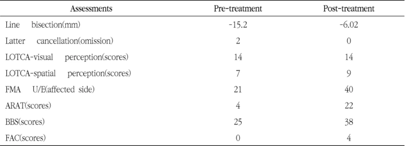
1. Visuospatial impairment
Line bisection scores increased from -15.2(pre-treat- ment) to -6.02(post-treatment). The number of omis-
sions on latter cancellation was from 2(pre-treatment) to 0(post-treatment). LOTCA- spatial perception scores in- creased from 7(pre-treatment) to 9(post-treatment).
MMSE score increased from 13(pre-treatment) to 21(post-treatment).
2. Upper motor function
The score of affected FMA U/E increased 21(pre-treat- ment) to 40(post-treatment). ARAT scores increased 4(pre-treatment) to 22(post-treatment).
3. Ambulation and balance
BBS scores increased from 25(pre-treatment) to 38(post-treatment). FAC scores increased from 0(pre-treat-
Assessments Pre-treatment Post-treatment
Line bisection(mm) -15.2 -6.02
Latter cancellation(omission) 2 0
LOTCA-visual perception(scores) 14 14
LOTCA-spatial perception(scores) 7 9
FMA U/E(affected side) 21 40
ARAT(scores) 4 22
BBS(scores) 25 38
FAC(scores) 0 4
FMA U/E=Fugl-Meyer Motor Assessment Upper extremity; ARAT=Action Research Arm Test; BBS=Berg Balance Scale; ;FAC=Functional Ambulation Classification
Table 1. Functional assessment scores of pre and post treatment
Pre-treatment Post-treatment
FIM total 54 70
FIM-motor 34 61
FIM-cognition 20 29
FIM, Functional Independent Measure Table 2. ADL scores of pre and post treatment
ment) to 4(post-treatment).
4. ADL function
Table 2 summarizes the ADL scores of pre and post treatment. FIM total scores improved from 54(pre-treat- ment) to 70(post-treatment). FIM-motor scores improved from 34(pre-treatment) to 61(post-treatment). FIM-cognition scores improved from 20(pre-treatment) to 29(post-treat- ment).
Ⅳ. Discussion
The present study highlighted the effect of prism for functional outcomes in patients following traumatic brain injury with visuospatial impairment
Pre-treatment, the patient’s visuospatial function on the right side, upper motor function, ambulation and balance, ADL function are restricted. Post-treatment, all assessments improved except the LOTCA-visual perception. A total score of LOTCA-visual perception was 16, and the patient was able to perform most of the task at both initial and discharge assessments.
Therefore, Prism adaptation could improve generally functional levels in TBI patient with visuospatial impairment. In the previous study, yoked prism and field-enhancing prisms could improve the TBI patient’s ability to function and attend to the affected side(Kapoor & Ciuffreda, 2002).
Especially, Our findings demonstrate greater improve- ment in motor functional abilities such as ADL function, upper motor, balance and ambulation after prism adapta- tion in patient following TBI with visuospatial impairments.
We suggest that the patient improve his physical per-
formance and activities of daily living such as grooming, toilet, transfer, walking and stair. He was inconsistent but observable asymmetries in the gaze direction toward right sides of space, and during self-cares, he spend quite longer time on the left side than the right.
However, this patient showed an improvement of self-care and mobility and refine visuospatial symptoms after prism adaptation. In previous study reported that Balance is an important factor in motor performance and is strongly related to spatial function and body awareness(Lang, Bland, Baile et al., 2013). Patterson et al. reported that spatial, balance, and body awareness factors predict mobility(Patterson et al., 2007)
In fact, the study of impact of visuospatial impair- ment on functional outcome in patients with traumatic brain injury, TBI patients with spatial neglect had poor- er motor function measured with the motor score of Functional Independence Measure(FIM) than Cognitive FIM score(McKenna, Cooke, Fleming, Jefferson, &
Ogden, 2006). The study suggests that visuospatial im- pairment could worsen motor dysfunction in TBI pa- tients(McKenna et al., 2006). Thus, visuospatial impair- ments may be abnormality in the affected side of space such as motor neglect(Heilman, 2004; Triggs, Gold, Adair, & Heilman, 1994; Chen, Ward, Khan, Lui, &
Hreha, 2015).
The mechanism of prism adaptation has been sug- gested that prism adaptation could influence perceptual processes through connections between the ventral and dorsal pathway located in the inferior parietal cor- tex(Striemer & Danckert, 2010). Other study reported that prism adaptation may primarily influence the visuo- motor circuit of dorsal pathway, mediating motor-re- lated processes(Striemer & Danckert, 2010; Fortis et al, 2011). Injured axon may be recovered by rehabilitation for axonal sprouting of surviving neurons(Wieloch &
Nikolich, 2006), and prism adaptation in our study may help to regenerative processes.
Prism adaptation from stroke study, the result sug- gests that prism adaptation improved motor-intentional Aiming deficit (Goedert et al., 2013) and motor func- tion(Mizuno, Tsuji, Takebayashi et al., 2011). The other study demonstrated greater improvement in motor func- tion(FIM motor score) after prism adaptation(Mizuno et al., 2011). By contrast, another study reported that prism adaptation did not effective spatial neglect in stroke patients(Rousseaux, Bemati, Saj, & Kozlowski, 2006). Because, positive effects of prism adaptation could be related to increases of vigilance or sustained attention(Rousseaux et al, 2006; Robertson, Tegner, Tham, Lo, & Nimmo-Smith, 1995).
Thus, effects of prism adaptation remain controversial. However, our data suggest that prism adaptation did improve functional level such as motor functions and ADL abilities in TBI patient.
Most of prism adaptation studies consider stroke patients. However, the present study has significance to confirm the effect of prism adaptation in patient follow- ing traumatic brain injury. A limitation of this study, it was likely that rehabilitation treatment or spontaneous recovery influence our results. Our findings support fur- ther larger and randomized trials that would determine the effectiveness of prism adaptation in improving the visuospatial functions, motor and functional level in TBI patients for generalizability of the results.
Reference
Adams, J. H., Doyle, D., Ford, I., Gennarelli, T. A., Graham, D.I., McLellan, D. R. (1989). Diffuse axonal injury in head injury: definition, diagnosis and grading.
Histopathology. 15, 49–59. doi:10.1111/j.1365-2559.
1989.tb03040.x
Arlinghaus, K. A., Shoaib, A. M., Price, T. R. P. (2005).
Neuropsychiatric assessment. In Silver JM, McAllister TW, Yudofsky SC. Textbook of Traumatic Brain Injury,
(pp. 59–62), American Psychiatric Association, Washington, DC
Blumbergs, P. C., Jones, N. R., North, J. B. (1989).
Diffuse axonal injury in head trauma. Journal of Neurology, Neurosurgery, and Psychiatry. 52, 838–841.
doi:10.1136/jnnp.52.7.838
Bruns, J., & Hauser, W. A. (2003). The epidemiology of traumatic brain injury: a review. Epilepsia, 44(s10), 2-10. doi:10.1046/j.1528-1157.44.s10.3.x
Chen, P., Ward, I., Khan, U., Liu, Y., & Hreha, K. (2015).
Spatial Neglect Hinders Success of Inpatient Rehabilitation in Individuals With Traumatic Brain Injury A Retrospective Study. Neurorehabilitation &
Neural Repair, 30(5). doi: 10.1177/1545968315604397 Comper, P., Bisschop, S. M., & Carnide, N., Tricco, A.
(2005). A systematic review of treatments for mild traumatic brain injury. Brain Injury, 19(11), 863–880.
doi:10.1080/02699050400025042
Dijkerman, H. C., Webeling, M., Ter Wal, J. M., Groet, E.,
& Van Zandvoort, M. J. E. (2004). A long-lasting im- provement of somatosensory function after prism adap- tation, a case study. Neuropsychologia, 42(12), 1697-1702. doi:10.1016/j.neuropsychologia.2004.04.004 Fortis, P., Chen, P., Goedert, K. M., & Barrett, A. M.
(2011). Effects of prism adaptation on motor-inten- tional spatial bias in neglect. Neuroreport, 22(14), 700.
doi:10.1097/WNR.0b013e32834a3e20
Frassinetti, F., Angeli, V., Meneghello, F., Avanzi, S., &
Làdavas, E. (2002). Long‐lasting amelioration of visuo- spatial neglect by prism adaptation. Brain, 125(3), 608-623. doi:10.1093/brain/awf056
Goedert, K. M., Chen, P., Boston, R. C., et al. (2013).
Presence of motor-intentional aiming deficit predicts functional improvement of spatial neglect with prism adaptation. Neurorehabilitation & Neural Repair, 28, 483–493.
Golisz, K. M., & Toglia, J. P. (2003). Perception and cognition. In: Crepeau EB, Cohn ES, Boyt BA, editors.
Willard & Spackman’s Occupational Therapy, (pp 395 –416), Williams & Wilkins, Philadelphia: Lippincott.
Granger, C. V., Hamilton, B. B., Linacre, J. M., Heinemann, A. W., & Wright, B. D. (1993). Performance profiles of the functional independence measure. American Journal of Physical Medicine & Rehabilitation, 72(2), 84-89.
Grieve J. (2000). Neuropsychology for occupational therapists, 2nd ed, Oxford: Blackwell Science Ltd.
Hall, R. C., & Chapman, M. J.. (2005). Definition, diag- nosis, and forensic implications of postconcussional syndrome. Psychosomatics, 46 (3), 195–202.
doi:10.1176/appi.psy.46.3.195
Heilman, K. M. (2004). Intentional neglect. Front Bioscience, 9, 694-705.
Itzkovich, M., Elazar, B., Averbuch, S., & Katz, N.
(2000). LOTCA manual, 2nd ed, Pequannock, NJ:
Maddak.
Jacquin-Courtois, S., Rode, G., Pisella, L., Boisson, D., &
Rossetti, Y. (2008). Wheel-chair driving improvement following visuo-manual prism adaptation. Cortex, 44(1), 90-96. doi:10.1016/j.cortex.2006.06.003 Kapoor, N., & Ciuffreda, K. J. (2002). Vision dis-
turbances following traumatic brain injury. Current Treatment Options in Neurology, 4(4), 271-280.
doi:10.1007/s11940-002-0027-z
Keller, I., Schindler, I., Kerkhoff, G., Von Rosen, F., &
Golz, D. (2005). Visuospatial neglect in near and far space: dissociation between line bisection and letter cancellation. Neuropsychologia, 43(5), 724-731.
doi:10.1016/j.neuropsychologia.2004.08.003
Kerkhoff, G. (1998). Rehabilitation of visuospatial cog- nition and visual exploration in neglect: a cross-over study. Restorative Neurology & Neuroscience, 12(1), 27-40.
Kushner, D. (1998). Mild traumatic brain injury: toward understanding manifestations and treatment. Archives of Internal Medicine, 158(15), 1617-1624.
doi:10.1001/archinte.158.15.1617
Marnane, M., Duggan, C. A., Sheehan, O. C., Merwick, A., Hannon, N., Curtin, D., ... & McCormack, P. M.
(2010). Stroke subtype classification to mecha- nism-specific and undetermined categories by TOAST, ASCO, and Causative Classification System Direct Comparison in the North Dublin Population Stroke Study. Stroke, 41(8), 1579-1586. doi:10.1161 /STROKEAHA.109.575373
McDonnell, M. (2008). Action research arm test.
Australian Journal of Physiotherapy, 54(3), 220.
McKenna, K., Cooke, D. M., Fleming, J., Jefferson, A., & Ogden, S. (2006). The incidence of visual perceptual impairment in patients with severe traumatic brain injury. Brain Injury,
20(5), 507-518. doi:10.1080/02699050600664368 Mizuno, K., Tsuji, T., & Takebayashi, T,. Fujiwara, T.,
Hase, K., Liu, M. (2011). Prism adaptation therapy enhances rehabilitation of stroke patients with uni- lateral spatial neglect: a randomized, controlled trial.
Neurorehabilitation & Neural Repair, 25(8), 711–720.
Morris, A. P., Kritikos, A., Berberovic, N., Pisella, L., Chambers, C. D,, Mattingley. J. B. (2004). Prism adaptation and spatial attention: a study of visual search in normals and patients with unilateral
neglect. Cortex. 40, 703–721.
doi:10.1016/S0010- 9452(08)70166-7
Lang, C. E,, Bland, M. D., & Bailey, R. R., et al. (2013).
Assessment of upper extremity impairment, function, and activity after stroke. Journal of Hand Therapy, 26, 104–114. doi:10.1016/j.jht.2012.06.005
Patterson, S. L., Forrester, L. W., & Rodgers, M., M, et al. (2007). Determinants of walking function after stroke: differences by deficit severity. Archives of Physical Medicine & Rehabilitation, 88, 115–119.
doi:10.1016/j.apmr.2006.10.025
Pisella, L., Rode, G., Farne, A., Tilikete, C., & Rossetti, Y. (2006). Prism adaptation in the rehabilitation of patients with visuo-spatial cognitive disorders.
Current Opinion in Neurology, 19(6), 534-542.
doi:10.1097/WCO.0b013e328010924b
Robertson, I. H., Tegner, R., Tham, K., Lo, A., Nimmo-Smith, I. (1995). Sustained attention training for unilateral neglect: theoretical and rehabilitation implications. Journal of Clinical Experimental
Neuropsychology, 17, 416–430.
doi:10.1080/01688639508405133
Rode, G., Rossetti, Y., & Boisson, D. (2001). Prism adaptation improves representational neglect.
Neuropsychologia, 39(11), 1250-1254.
doi:10.1016/S0028-3932(01)00064-1
Rousseaux, M., Bernati, T., Saj, A., & Kozlowski, O.
(2006). Ineffectiveness of prism adaptation on spatial neglect signs. Stroke, 37(2), 542-543. doi:10.1161/01.STR.0000198877 .09270.e8
Sanford, J., Moreland, J., Swanson, L. R., Stratford, P.
W., & Gowland, C. (1993). Reliability of the Fugl-Meyer assessment for testing motor perform- ance in patients following stroke. Physical Therapy, 73(7), 447-454. doi:10.1093/ptj/73.7.447
Serino, A., Barbiani, M., Rinaldesi, M. L., & Làdavas, E.
(2009). Effectiveness of prism adaptation in neglect rehabilitation a controlled trial study. Stroke, 40(4), 1392-1398. doi:10.1161/STROKEAHA.108.530485 Shiraishi, H., Muraki, T., Itou, A., Sampei, Y., &
Hirayama, K. (2010). Prism intervention helped sus- tainability of effects and ADL performances in chronic hemispatial neglect: a follow-up study.
Neurorehabilitation, 27(2), 165-172.
doi:10.3233/NRE-2010-0593
Striemer, C. L., & Danckert, J. A. (2010). Through a prism darkly: re-evaluating prisms and neglect.
Trends in Cognitive Sciences, 14, 308–316.
doi:10.1016/j.tics.2010.04.001
Wieloch, T., & Nikolich, K. (2006). Mechanisms of neu- ral plasticity following brain injury. Current Opinion in Neurobiology, 16(3), 258-264. doi:10.1016/j.conb.2006.0 5.011
Suter, P. S. (1995). Rehabilitation and management of visual dysfunction following traumatic brain injury.
In: Ashley MJ, Krych DK, editors, (pp 187–216) Traumatic brain injury rehabilitation. Boca Raton:
CRC Press
Stevenson, T., & Tsang, R. (1996). Berg Balance Test. Physical Therapy, 76(10), 1124-1126. doi:10.1093/ptj/76.10.1124 Ten Brink, A. F., Visser-Meily, J. M., & Nijboer, T. C.
(2015). Study protocol of ‘Prism Adaptation in Rehabilitation’: a randomized controlled trial in stroke patients with neglect. BMC neurology, 15(1), 5. doi:10.1186/s12883-015-0263-y
Toglia, J. P. (1991). Unilateral visual inattention:
Multidimensional components. Occupational Therapy Practice, 3, 18–34.
Triggs, W. J, Gold, M., Gerstle, G., Adair, J., Heilman, K.
M. (1994). Motor neglect associated with a discrete parietal lesion. Neurology. 44, 1164-1166.
Williams, G. (2011). Functional Ambulation Classification. In Encyclopedia of Clinical Neuropsychology, (pp.
1105-1106). Springer, New York.