INTRODUCTION
Frontotemporal dementia (FTD) is a syndrome of progres- sive changes in behavior and language due to loss of function of neurons in the frontal and temporal lobes. Around 15% of patients with FTD meet formal clinical criteria for motor neuron disease (MND).1 Patients with FTD with MND may present with behavioral changes and/or language function declines seen in other subtypes of FTD.1,2 In this syndrome, these changes are accompanied by deterioration of motor neu- rons that manifests as weakness and atrophy in the muscles, fine muscle twitches and cramps and difficulty making fine movements.2 The cause of FTD with MND is unknown.
Among many hypotheses, autoimmune mechanisms are con- sidered as a possible etiology of the disease.3,4 FTD with MND
in autoimmune pathology is very rare. Here we report about a patient who was diagnosed with FTD with MND and con- firmed antiphospholipid syndrome (APS).
CASE REPORT
A 71-year-old man presented with a 2-year history of right upper extremity weakness, a 1-year history of left upper ex- tremity weakness and a 1½-year history of weakness in both lower limbs. When this patient arrived at our hospital he was ambulating in a wheelchair and could stand for only a few sec- onds. He had other underlying diseases as well-epilepsy, an- gina pectoris and pulmonary thromboembolism (PTE). There was no family history of similar disease or exposure to any toxins or drugs. In the physical examination, weakness of up- per limbs with Medical Research Council (MRC) grade 4 and weakness of lower limbs with MRC grade 3–4 were observed.
Upper motor neuron signs were seen in the cervical region (Hoffman sign) and lower motor neuron signs were seen in bulbar (dysphagia, tongue atrophy and fasciculation) and
Frontotemporal Dementia with Motor Neuron Disease in a Patient with Antiphospholipid Syndrome: A Case Report
Yoon-Cheol Jeong, Jin-Seok Park, Seung-Hyun Kim, Hojin Choi
Department of Neurology, Hanyang University College of Medicine, Seoul, Korea
Background Frontotemporal dementia (FTD) with motor neuron disease (MND) is a syndrome of progressive changes in behavior, lan- guage, muscle weakness and atrophy due to loss of function of neurons in the frontal and temporal lobes and in motor neurons. Etiology and pathogenesis of FTD with MND are still uncertain.
Case Report A 71-year-old man presented with a 2-year history of progressive muscle weakness and cognitive deficits. We diagnosed this patient as FTD with MND by neurological examination, electromyography, brain imaging and neuro-psychological evaluation. We also con- firmed antiphospholipid syndrome (APS) in this patient as a way to rule out secondary causes of MND.
Conclusions This was a very rare case of FTD with MND in APS. We should focus study on the possible role of autoimmune pathogenesis in FTD with MND.
Key Words frontotemporal dementia, motor neuron disease, antiphospholipid syndrome.
Received: November 9, 2016 Revised: November 29, 2016 Accepted: November 29, 2016
Correspondence: Hojin Choi, MD, PhD, Department of Neurology, Hanyang University College of Medicine, Guri Hospital, 153 Gyeongchun-ro, Guri 11923, Korea
Tel: +82-31-560-2267, Fax: +82-31-560-2267, E-mail: [email protected]
cc This is an Open Access article distributed under the terms of the Cre- ative Commons Attribution Non-Commercial License (http://creative- commons.org/licenses/by-nc/3.0) which permits unrestricted non-com- mercial use, distribution, and reproduction in any medium, provided the ori- ginal work is properly cited.
DND
CASE REPORT
Yoon-Cheol Jeong et al.
FTD with in a Patient with APS
lumbar regions (lower extremity atrophy and fasciculation).
To confirm MND, electromyography (EMG) was performed and revealed normal neuronal conduction, but in needle EMGs there were frequent fasciculations and fibrillations in the upper and lower extremities (biceps, triceps, abductor pollicis brevis, vastus medialis, tibialis anterior muscles), the thoracic paraspi- nal muscles and bulbar muscles (Table 1). These findings were compatible with motor neuron involvement.
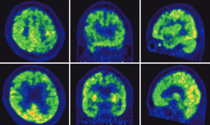
He also had memory impairment, decreased speech, inap- propriate affect, apathy and irritability. He was a middle school graduate. He scored 16 on the Korean version of Mini- Mental State Examination. On the Seoul Neuropsychological Screening Battery, his verbal and visual memory functions showed impairment and he got particularly low scores on frontal executive function. Scores on stroop test color reading, semantic word fluency, and phonemic word fluency all fell be- low normal limits, suggestive of frontotemporal dysfunction (Table 2). In brain magnetic resonance imaging, cortical atro- phy in both parietal and anterior temporal lobes was seen (Fig. 1). Fluorodeoxyglucose positron emission tomopraphy imaging demonstrated hypometabolism in the bilateral fron- to-temporo-parietal cortex (Fig. 2). He was diagnosed with FTD with MND based on the neuropsychological and elec-
trophysiological background.
To rule out secondary causes of the disease further labora- tory tests were done. Lupus anticoagulant was positive and anti-cardiolipin Ab and anti-B2 glycoprotein 1 were negative.
A follow-up lupus anticoagulant was done after three months and also showed a positive result. The patient was diagnosed with APS because he had a clinical episode of PTE and two laboratory lupus anticoagulant occasions at 12 weeks apart.
He was treated as anticoagulant because he had APS and PTE.
DISCUSSION
FTD with MND is considered important because it may help to identify the pathophysiology of FTD, a highly hetero- geneous disorder. Despite numerous pathological and genetic discoveries, much remains uncertain in FTD with MND.2 One of the most significant features of the disease is the substantial clinical heterogeneity and variability in disease prognosis.
Around 15% of patients meet the criteria for both FTD and MND, the combination associated with a worse prognosis and reduction in survival time of around 1 year.5,6
In the previous studies, an inflammatory pathogenesis to Table 1. Needle EMG findings in the patient
Muscle Side Insertional activity Positive sharp wave Fasciculation
Bulbar
Masseter Left ↑ - +
Right ↑ - -
Paraspinal
Cervical Left ↑ + -
Right ↑ + -
Thoracic Left ↑ ++ -
Right ↑ ++ -
Lumbar Left ↑ ++ -
Right ↑ ++ -
Upper extremity
Biceps brachii Left ↑ + -
Right ↑ + -
Abductor pollicis brevis Left ↑ ++ +
Right ↑ ++ +
Lower extremity
Vastus lateralis Left ↑ + -
Right ↑ + -
Tibialis anterior Left ↑ ++ +
Right ↑ ++ +
EMG: electromyography.
DND
neurodegenerative disease has long been hypothesized. Many neurodegenerative conditions are united by pathological pro- tein misfolding and aggregation accompanied by neuronal loss
and inflammatory markers around the site of pathological in- jury. Several studies of environmental risk factors in sporadic behavioral variant FTD found a significant association with head trauma and a close-to-significant association with thy- roid disease.7 Furthermore, elevations in cerebrospinal fluid cytokines, notably TNF-α, have previously been demonstrated in FTD.8 An association between MND and autoimmune dis- eases also have been suggested previously. A familial study separately using index cases of amyotrophic lateral sclerosis (ALS) and multiple sclerosis reported increased association.
Another hospital record-linkage study demonstrated an asso- ciation of Behçet disease, ulcerative colitis, and Wegener gran- ulomatosis in the offspring of patients with ALS.9 However, the basis of these observations in relation to shared pathogenesis remains unclear. Immunologic intervention for ALS, includ- ing bone marrow transplants, has so far not been effective, de- spite evidence linking inflammatory processes to pathogenesis in ALS.10
APS is a pathological status that arose from excess accumu- lation of blood clots by antiphospholipid antibodies (aPLs).11 The syndrome may occur as a primary condition or along with the autoimmune pathophysiology. The major clinical features associated with APS include recurrent thrombosis and pregnancy losses. APS can cause arterial or venous blood clots, in any organ system, or pregnancy-related complica- tions. In APS patients, the most common venous event is deep vein thrombosis of the lower extremities, and the most common arterial event is stroke. Other common findings are low platelet count, heart valve disease, and livedo reticularis.
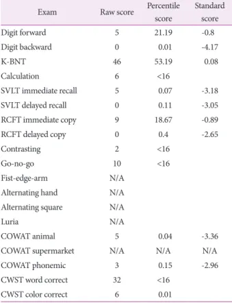
There are also associations between APS and headaches, mi- graines, and oscillopsia.12 But the presence of aPLs in the Table 2. Neuropsychological data of the patient
Exam Raw score Percentile
score
Standard score
Digit forward 5 21.19 -0.8
Digit backward 0 0.01 -4.17
K-BNT 46 53.19 0.08
Calculation 6 <16
SVLT immediate recall 5 0.07 -3.18
SVLT delayed recall 0 0.11 -3.05
RCFT immediate copy 9 18.67 -0.89
RCFT delayed copy 0 0.4 -2.65
Contrasting 2 <16
Go-no-go 10 <16
Fist-edge-arm N/A
Alternating hand N/A
Alternating square N/A
Luria N/A
COWAT animal 5 0.04 -3.36
COWAT supermarket N/A N/A N/A
COWAT phonemic 3 0.15 -2.96
CWST word correct 32 <16
CWST color correct 6 0.01
COWAT: Controlled Oral Word Association Test, CWST: Color Word Stroop Test, K-BNT: Korean version of the Boston Naming Test, RCFT:
Rey-Osterrieth Complex Figure Test, SVLT: Seoul Verbal Learning Test.
Fig. 1. Axial & sagittal T1-weighted brain MR images of the patient. Cortical atrophy was seen in both parietal and anterior temporal lobe.
Yoon-Cheol Jeong et al.
FTD with in a Patient with APS
not identify the significant inflammatory marker which sug- gested neurodegenerative pathology in serum or cerebrospinal fluid. Among several hypotheses which explain the etiology of FTD with MND, autoimmune mechanism is one. To the best of our knowledge, this is the first case of FTD with MND in APS in Korea. In this case, APS may induce its effect immuno- logically or by thrombosis of small arterioles and venules, mi- croinfarcts of neuronal cells and resultant development of clin- ical manifestations. These hypotheses lead us to elucidate the autoimmune pathogenesis and provide an explanation for FTD with MND. Further evaluation and evidence in the pathophysi- ology of the disease is needed.
Conflicts of Interest
The authors have no financial conflicts of interest.
REFERENCES
1. Rodríguez de Rivera FJ, Rambold HA, Díez-Tejedor E. Assessment of cognitive impairment in amyotrophic lateral sclerosis. J Neurol Sci 2014;337:1-2.
2. Burrell JR, Halliday GM, Kril JJ, Ittner LM, Götz J, Kiernan MC, et al. The frontotemporal dementia-motor neuron disease continuum.
Lancet 2016;388:919-931.
3. Miller ZA, Rankin KP, Graff-Radford NR, Takada LT, Sturm VE, Cleveland CM, et al. TDP-43 frontotemporal lobar degeneration and autoimmune disease. J Neurol Neurosurg Psychiatry 2013;84:956-962.
Fig. 2. Fluorodeoxyglucose positron emission tomopraphy imaging of the patient. Hypometabolism was observed in bilateral fronto-tempo- ro-parietal cortex.
blood of patients with neurodegenerative disease is very rare.
The relationship between neurodegenerative disease and APS has been proposed in several studies. Higher levels of aPLs was found in patients with dementia than in controls13 and a significant association between anticardiolipin and both vas- cular dementia and Alzheimer’s disease was noted.14 This has also been demonstrated in experimental studies. An animal study was performed with BALB/c mice using a staircase test and a T maze alternation test as cognitive assessment tools.
Mice immunized with anti-β2 glycoprotein I antibodies de- veloped a higher degree of cognitive abnormalities than those that had not been immunized.15 In another study, the impor- tance of TANK binding kinase-1 (TBK1), a multimeric kinase that modulates inflammation and autophagy, was suggested.
In human health, that had been highlighted for the first time by the recent discoveries of mutations in TBK1 that underlie ALS and FTD.16 Until now, there are several studies with evi- dence of association between MND and autoimmune disease, but further research is needed.
There are limitations to this case. First, it is still unclear whether the relationship between FTD with MND and APS was causal or simply coincidence. To clarify that unknown further search for similar cases is needed. Second, we could
DND
4. Niebroj-Dobosz I, Dziewulska D, Janik P. Auto-antibodies against pro- teins of spinal cord cells in cerebrospinal fluid of patients with amyo- trophic lateral sclerosis (ALS). Folia Neuropathol 2006;44:191-196.
5. Phukan J, Pender NP, Hardiman O. Cognitive impairment in amyo- trophic lateral sclerosis. Lancet Neurol 2007;6:994-1003.
6. Talbot K, Ansorge O. Recent advances in the genetics of amyotrophic lateral sclerosis and frontotemporal dementia: common pathways in neurodegenerative disease. Hum Mol Genet 2006;15:R182-R187.
7. Rosso SM, Landweer EJ, Houterman M, Donker Kaat L, van Duijn CM, van Swieten JC. Medical and environmental risk factors for spo- radic frontotemporal dementia: a retrospective case-control study. J Neurol Neurosurg Psychiatry 2003;74:1574-1576.
8. Sjögren M, Folkesson S, Blennow K, Tarkowski E. Increased intra- thecal inflammatory activity in frontotemporal dementia: pathophysi- ological implications. J Neurol Neurosurg Psychiatry 2004;75:1107- 1111.
9. Hemminki K, Li X, Sundquist J, Sundquist K. Familial risks for amyotrophic lateral sclerosis and autoimmune diseases. Neurogenet- ics 2009;10:111-116.
10. Philips T, Bento-Abreu A, Nonneman A, Haeck W, Staats K, Geelen
V, et al. Oligodendrocyte dysfunction in the pathogenesis of amyo- trophic lateral sclerosis. Brain 2013;136(Pt 2):471-482.
11. Atanassova PA, Dimitrov BD. Neurological and systemic disorders in antiphospholipid syndrome: novel constellations on a genetic ba- sis. Clin Neurol Neurosurg 2006;108:814.
12. Turner MR, Goldacre R, Ramagopalan S, Talbot K, Goldacre MJ.
Autoimmune disease preceding amyotrophic lateral sclerosis: an epi- demiologic study. Neurology 2013;81:1222-1225.
13. Mosek A, Yust I, Treves TA, Vardinon N, Korczyn AD, Chapman J.
Dementia and antiphospholipid antibodies. Dement Geriatr Cogn Dis- ord 2000;11:36-38.
14. Juby A, Davis P, Genge T, McElhaney J. Anticardiolipin antibodies in two elderly subpopulations. Lupus 1995;4:482-485.
15. Shrot S, Katzav A, Korczyn AD, Litvinju Y, Hershenson R, Pick CG, et al. Behavioral and cognitive deficits occur only after prolonged exposure of mice to antiphospholipid antibodies. Lupus 2002;11:736- 16. Ahmad L, Zhang SY, Casanova JL, Sancho-Shimizu V. Human TBK1: 743.
a gatekeeper of neuroinflammation. Trends Mol Med 2016;22:511-527.