Cardiovascular Disease in Women
9
0
0
전체 글
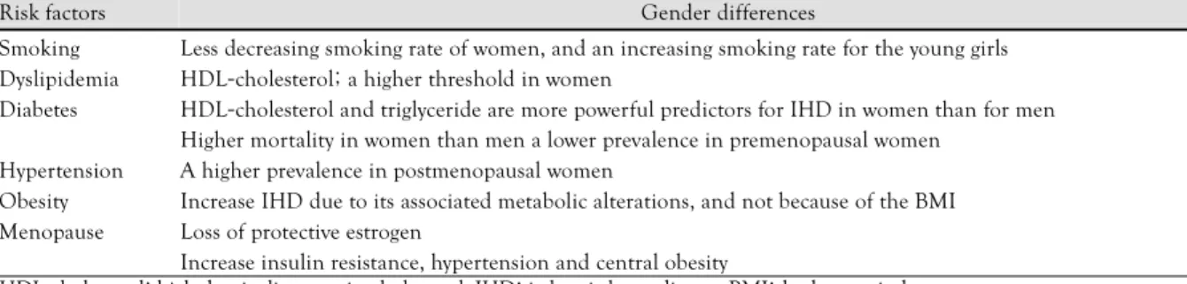
(2) 772·Korean Circulation J 2006;36:771-779 Table 1. Characteristics of the gender differences of the risk factors. Risk factors. Gender differences. Less decreasing smoking rate of women, and an increasing smoking rate for the young girls HDL-cholesterol; a higher threshold in women HDL-cholesterol and triglyceride are more powerful predictors for IHD in women than for men Higher mortality in women than men a lower prevalence in premenopausal women A higher prevalence in postmenopausal women Hypertension Increase IHD due to its associated metabolic alterations, and not because of the BMI Obesity Loss of protective estrogen Menopause Increase insulin resistance, hypertension and central obesity HDL-cholesterol: high density lipoprotein-cholesterol, IHD: ischemic heart disease, BMI: body mass index Smoking Dyslipidemia Diabetes. we wanted to recognize the current problems in applying the result of studies that have disproportionately enrolled men so as to present the issues for future studies and to improve the clinical outcomes of women who suffer with cardiovascular disease.. Gender Differences of Risk Factors for Ischemic Heart Disease Because estrogen in the premenopausal period of young women has favorable effects on the risk factors for ischemic heart disease, women are less likely to have risk factors. Yet when they occur in women, multiple risk factors tend to occur simultaneously and they act additively for ischemic heart disease, and especially in the postmenopausal period. There are some gender-related differences of the risk factors for women(Table 1).. Smoking Cigarette smoking is a major risk factor for ischemic heart disease in both sexes, and this diminishes such beneficial effects of estrogen as anti-inflammation, thrombosis inhibition and anti-oxidation of LDL-cholesterol in a dose-dependent manner.7) Recent epidemiologic reports have shown that the smoking rate has decreased dramatically in Western men and Korean men, but there has been less or no decrease in women, which affects 17% of all the deaths in U.S. women, mostly from coronary artery disease.8)9) Especially, the smoking rate of young girls has increased in the world, including Korea, which may increase the smoking-related diseases such as cancer and ischemic heart disease. For women smoking, the tendency of thrombosis is increased additively with the use of oral contraceptives and women have more problems when attempting to quit smoking such as a greater likelihood of depression and more weight gain after cessation.10) Dyslipidemia The HDL-cholesterol level is a diagnostic criterion for metabolic syndrome and a risk factor for IHD because. women’s HDL-cholesterol level is higher than men until menopause. The power of a low HDL-cholesterol level and a high triglyceride level for predicting IHD and adverse outcomes is higher for women than for men.11) Postmenopausal women who had hypertension showed the trend toward atherogenic dyslipidemia, including high triglyceride levels and low HDL-cholesterol levels in Korean women.12). Diabetes Diabetes increases the IHD by two times compared to non-diabetic women, even in the premenopausal period, and diabetic women have a worse prognosis than men due to their more frequent comorbid conditions, including hypertension, old age, poor glucose control and a higher smoking rate.13)14) Hypertension The prevalence of hypertension is highly increases after menopause due to loss of the beneficial effects of estrogen on the vascular walls.15) Gierach et al.16) said that the systolic blood pressure and pulse pressure are independent risk factors for ischemic heart disease in postmenopausal women. Obesity and metabolic syndrome The WISE study reported that being overweight increases ischemic heart disease via the associated metabolic alterations, not because of the obesity itself.17) This group of risk factors is related with insulin resistance and it is represented as metabolic syndrome, including insulin resistance(glucose intolerance), dyslipidemia (a high triglyceride level and a low HDL-cholesterol level), hypertension and abdominal obesity. It is recommended to control all the modifiable risk factors in both normal and overweight persons to prevent transition to metabolic syndrome. Estrogen and menopause The estrogen level is variable throughout a woman’s entire life, including the time of puberty, pregnancy, peripartum and menopause. The studies concerned with estrogen have reported the preventive roles of estrogen.
(3) Wook Bum Pyun, et al:Cardiovascular Disease in Women·773. for ischemic heart disease in young women. Estrogen has beneficial effects on the cardiovascular system: it reduces cellular hypertrophy, enhances vascular elasticity and has antioxidant and anti-inflammatory effects.17)18) In addition, estrogen has a role in fat distribution/deposition, insulin resistance, lipid metabolism, coagulation factors and also inflammation, as measured by hsCRP level. Women under the influence of various levels of estrogen present with different clinical features such as a lower prevalence of obstructive coronary artery disease and higher levels of endothelial dysfunction. Bailey et al. have reported that hypoestrogenemia in premenopausal women increases the risk of IHD 7.4-fold.19) Women lose the protective estrogen after menopause and they show central obesity and insulin resistance with hypertension, which increases IHD to a prevalence similar to men, at ten years after menopause.. Other novel risk factors in women Because the traditional risk factors are less effective to predict IHD, additional risk factors or markers that are more accurate have emerged.17) After Peter Libby suggested the role of inflammation in the initiation and progression of atherosclerosis, a high hsCRP level, which is a inflammatory marker, is now a novel risk factor for both sexes. Ridker et al.20) reported that a high hsCRP level was related with future cardiovascular events in their study, which was performed on a large number of women. Anemic women also have poor outcomes of myocardial infarction and heart failure. In the WISE study, women with anemia(<12 g/dl) had a higher rate of death from all causes and worse major adverse cardio-. Typical angina pectoris has three diagnostic criteria: 1) substernal chest discomfort with a characteristic quality and duration that is 2) provoked by exertion or emotional stress and 3) relieved by rest and NTG. These diagnostic criteria of typical angina, for evaluating patients with chest pain, have been mainly derived from predominantly male patient cohort studies. Although the most frequent symptom of women is chest pain, which is similar to men, more women have atypical symptoms such as fatigue, shortness of breath and sleep disturbance.21) Women have tendency to feel the symptoms during their daily activities and when under mental stress rather than when under physical stress.22)23) The first large prospective study, the Coronary Artery Surgery Study(CASS), described the differences of CAD in women; this study enrolled 24% of the women referred for coronary angiography in the 1970’s. There were some conclusions regarding the CAD gender differences as more women didn’t have obstructive CAD and the traditional risk factor assessment failed to predict CAD in women.24)25) The WISE report showed that women who have chest pain suggestive of myocardial ischemia are less likely to. 075 45. 025. 11. 16. 12. 000 Typ Ang. Atyp Ang. Age 45-55. 100. % with CAD. % with CAD. Evaluation of Women with Chest Pain. Age 35-45. 100. 050. vascular outcomes than did the non-anemic women on multivariate analysis.17) These findings may not only be relevant for understanding the mechanism of coronary artery disease (CAD) in women, but they may also have potential therapeutic implications.6). 3. 12. 075. 68. 050 21. 025 000. Typ Ang. Nonang. 84. 075 46 36 25 13. 21. 000. % with CAD. % with CAD. Nonang. Age 65-75 100. 025. Atyp Ang Chest pain type. Age 55-65. 050. 21. 17 7. Chest pain type. 100. 32. 075. 95. 60. 050. 54 36. 025. 34 17. 000 Typ Ang. Atyp Ang Chest pain type. Nonang. Typ Ang. Atyp Ang. Nonang. Chest pain type. Fig. 1. The criteria of typical angina is inadequate for women for predicting CAD.17) Typ Ang: typical angina, Atyp Ang: atypical angina, Nonang: non-angina, solid bar: WISE prevalence, open bar, predicted by Diamond probability, CAD: cardiovascular disease, WISE: women’s ischemic syndrome evaluation..
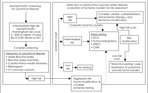
(4) 774·Korean Circulation J 2006;36:771-779. have obstructive CAD detected on thier coronary angiography than men, and this is regardless of their type of symptoms(typical chest pain and atypical chest pain, but not non-anginal chest pain)(Fig. 1).17) Women who have chest pain, but no obstructive coronary artery disease, are likely to have depression and persistent symptoms, a decreased functional capacity and even poor cardiovascular outcomes. Much money and time are expended for repeated evaluations that are simply not appropriate for women. The exercise electrocardiography test is less effective for evaluating the chest pain of women, partly because it has a high false positive rate. Women generally have a lower exercise capacity due to comorbidities and older age, and estrogen have vasodilatory and digoxin-like effects that decrease the efficacy of the test.26) In addition, women are less likely to have obstructive coronary artery disease, though they may have chest symptoms of ischemic heart disease(the incidence of insignificant CAD: 50% in women and 17% in men).27) Single-photon emission computed tomography(SPECT) is commonly used and it can detect reduction of myocardial perfusion or regional wall motion abnormalities of the left ventricle earlier than can ECG. There are some issues that should be considered when testing women; women have lower exercise capacity and Tc-99m radioisotopes are preferably used due to influence of breast tissue and obesity for improving the test specificity for women.28)29) The accuracy of this test is similar for both sexes. Because of the later changes of regional wall motion abnormality for the patient with a perfusion defect, assessment of stress-induced wall motion abnormalities has been associated with higher specificity. Yet stress echo-. cardiography required a skilled examiner and the revealed echo windows can be poor due to the large amount of breast tissue and the possible severe obesity. Because there is less likelihood of obstructive coronary artery disease in women, functional assessment with cardiovascular MR imaging has benefits in evaluating the left ventricular function and subendocardial ischemia. Panting et al.30) proved that this test was beneficial to detect subendocardial ischemia in women with chest pain, but who were without obstructive CAD. This result was repeated by WISE study, and it can explain the reason for the frequent hospitalization of women with nonobstructive coronary arteries and who have persistent and refractory chest pain.31) The men with abnormal spectroscopy and who are without CAD have outcomes similar to women with CAD, during the three years of follow up,31) and this novel test can have wide utility for evaluating women with chest pain and predicting adverse outcomes.32). Paradigms for evaluating the women with or without symptoms From a growing body of evidence, Shaw proposed new paradigms for identifying the women who are at-risk for ischemic heart disease(Fig. 2).17) Pathologic gender differences in atherosclerosis Ischemic heart disease of women is different from that of men because sex has an influence on the cardiovascular pathophysiology. There are risk factors that are confined to women, such as peripartum hypertensive disorder, diabetes, coronary or aortic root dissection, and delivering a thin baby.33) The characteristics of the risk factors of women are a higher blood pressure and LDL-. Detection of obstructive coronary artery diseaseevaluation of ischemic burden for risk assesment. Asymptomatic screening for subclinical disease. Consider cardiac catheterization, anti-ischemic therapy, and risk factor modification. High risk. Consider screening Measures of subclinical disease • Ankle-Brachial index • Brachial artery reactivity • Carotid intima-media thickness • Retinogram • CT coronary calcium High risk. Evaluate ischemic burden Symptom onset. Intermediate-high risk asymptomatic: (Framingham risk score 6-20% or higher 10-year risk of CHD death or MI)*. Intermediate risk. High risk scan. Stress testing • ECG • Echocardiography • SPECT • CMR. Risk stratification. Low risk scan Low risk. No testing. “Watchful waiting” caretreatment of symptoms and risk factor burden. Aggressive risk factor modification & consider ischemia testing. Fig. 2. Paradigm for testing the women with ischemic heart disease.17) 6% to 20% for intermediate Framingham risk scores and >20% for high Framing-. ham risk scores. CHD: coronary heart disease, CMR: cardiovascular magnetic resonance imaging, CT: computed tomography, ECG: electrocardiogram, MI: myocardial infarction, SPECT: single-photon emission computed tomography..
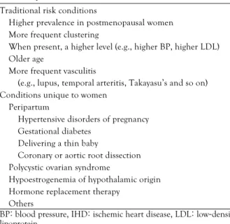
(5) Wook Bum Pyun, et al:Cardiovascular Disease in Women·775. Table 2. Specific risk factors confined to women33). Traditional risk conditions Higher prevalence in postmenopausal women More frequent clustering When present, a higher level (e.g., higher BP, higher LDL) Older age More frequent vasculitis (e.g., lupus, temporal arteritis, Takayasu’s and so on) Conditions unique to women Peripartum Hypertensive disorders of pregnancy Gestational diabetes Delivering a thin baby Coronary or aortic root dissection Polycystic ovarian syndrome Hypoestrogenemia of hypothalamic origin Hormone replacement therapy Others BP: blood pressure, IHD: ischemic heart disease, LDL: low-density lipoprotein Table 3. Anatomic and functional differences between women and. men33) Structural findings Macrovessels and microvessels Smaller size Increased stiffness (fibrosis, remodeling and so on) More diffuse disease (erosion>rupture) Microemboli, rarefaction (drop out), disarray and so on Functional findings Macro- and microvessels Endothelial dysfunction Smooth muscle dysfunction (Raynaud’s, migraine, CAS) Inflammation Plasma markers Vasculitis (Takayasu’s, rheumatoid, SLE, CNSV, giant cell and so on) CAS: coronary artery spasm, CNSV: central nervous system vasculitis, IHD: ischemic heart disease, SLE: systemic lupus erythematosus. cholesterol level, the occurrence of clustered features, older age, after menopause and a higher prevalence of vasculitis such as Takayasu’s arteritis, SLE and temporal arteritis(Table 2).33) The arteries(macrovessels and microvessels) of women are different from men; they are smaller, show increased stiffness due to fibrosis and remodeling, have more diffuse atherosclerosis with erosion but not with rupture as men show, and they display microemboli, rarefaction (drop out) and disarray. Microvascular abnormalities are evaluated by checking for retinal artery abnormalities and they can be regarded as risk factors for IHD in women, but not in men.34) The vascular functional differences between both genders are under the influence of estrogen, which affects the vascular endothelial cells and the smooth muscle. Oxidative stress impairs the endothelial function and limits myocardial perfusions. In the WISE study, more than half of the women had endothelial dysfunction,. which is an independent predictor of adverse outcomes.35) Estrogen also affects the repair mechanism of bone-marrow derived EPCs by increasing the number of circulating EPCs. The traditional risk factors, including aging, can impair the repairing capacity of EPCs. The diseases associated with altered vascular smooth muscle function(e.g. Raynaud’s phenomenon, migraine and coronary artery spasm) are more frequent in women than in men(Table 3).33). Endothelial dysfunction in women Endothelial cells have protective roles in regard to antiatherosclerosis, such as maintenance of vascular tone via multiple vasoconstrictors, inhibition of platelet aggregation and acting as a barrier against various cellular offenders. The healthy endothelium is prone to impairment by multiple risk factors, and endothelial dysfunction is a predictor of future cardiovascular disease and a good therapeutic target for maintaining the proper vascular condition. In the WISE study, 163 women who underwent coronary angiography and coronary endothelial function analysis were followed for 4 years. From that study, cardiovascular events were independently predicted by the coronary vascular endothelial function, and this was independent of the traditional risk factors and the extent of CAD.35) Restoration of abnormal endothelial function is associated with improved outcomes, and this was proven in a study of 400 hypertensive, postmenopausal women.36) As mentioned previously, the lower prevalence of obstructive coronary artery disease in women calls for alternative diagnostic approaches that can detect culprit patients, not culprit arteries.37) An optimal non-invasive risk model may include ventricular function, regional flow or perfusion, the metabolic or energy requirement according to MR spectroscopy or PET, and/or vessel wall abnormalities such as carotid intima media thickness, the findings of electron beam CT and retinograms and the markers of inflammation such as hsCRP.38) Because this abnormal vascular function is often concomitant with abnormal endothelial function, prudent treatment of these women should involve aggressive medical therapy that’s directed at improving endothelial function, their atherosclerosis and the established risk factors, and this includes administering statins, lipid-lowering drugs, angiotensin-converting enzyme inhibitors and aspirin. The future WISE and non-WISE clinical trials will be directed at testing these strategies for reducing adverse events in these women. Differences in women with obstructive coronary artery disease Older women have similar plaque pathologies like a large necrotic core surrounded by a thin fibrous cap that’s infiltrated with a large number of macrophages and in-.
(6) 776·Korean Circulation J 2006;36:771-779. flammatory cells. But in younger women, there were plaque erosions without a surrounded fibrous cap and exposure of the intima consisted of abundant smooth muscle cells and proteoglycans.39) Because of the less typical symptoms, the higher rate of non-obstructive disease and the delayed diagnosis and treatment, younger women with acute coronary syndrome display higher mortality than men. This high mortality of young women is due to not only multiple comorbid conditions, but also to the women themselves.40) Women suffering with acute coronary syndrome are likely to have non-Q myocardial infarction, which means less benefit from revascularization. In another way, women have long-term adverse outcome rates that are similar to men who undergo revascularization, despite the women’s less extensive and less severe obstructive CAD, better LV function and high rate of normal coronary arteries.41) After coronary artery bypass surgery, women have a higher rate of operative mortality due to their high rate of congestive heart failure.42) This is same situation as is noted in the percutaneous revascularization registry data. More women suffered from sustained symptoms like chest pain, evidence of ischemia and reduced functional capacity after revascularization procedure than did the men. They recovered later than the men after bypass surgery and had greater functional disability and depression.. Gender-gap in revascularization era Women with acute coronary artery disease are more likely to have small coronary artery disease, congestive heart failure and inflammation. There are some differences of biomarker between the genders; men suffering with acute coronary syndrome have higher CK-MB and troponin levels and women have higher hsCRP and BNP levels, and the latter are also risk factors for a poor outcome. There are several possible explanations for the worse outcomes of women: the higher prevalence of congestive heart failure, diastolic heart failure and vasomotor dusfunction and the lower hemoglobin levels in women than in men.37) As mentioned previously, women have higher mortality for revascularization procedures due to their older age and the gender differences of the clinical, angiographic and procedural factors.43-45) With the advances in instruments, experience and the increasing awareness of the burden of cardiovascular disease in women,46) the gap in the procedural outcomes such as mortality will decrease for various diseases regardless of the type of revascularization.47) How can we effectively assess women with IHD? The disadvantages of assessing the typical chest pain associated with obstructive coronary artery disease in men should corrected by using a gender-specific new tool for the assessment of IHD in women. Thus, a femalespecific questionnaire based on the data from the WISE. study may enhance our ability to diagnose IHD in women. To improve the diagnostic accuracy for women, scoring systems like the Duke treadmill score and exercise duration are useful for predicting future cardiac outcomes.48) The 12-item Duke Activity Status Index(DASI) questionnaire was able to stratify the risk of adverse outcomes in women and each 1-MET increase of the DASI score was independently associated with an 8%(HR: 0.92, 95% CI: 0.85 to 0.99, p=0.02) decreased risk of major adverse cardiovascular events during follow up.49) In the WISE study the DASI, which is a self questionnaire based on activities of daily living and recreational activities, can effectively estimate adverse outcomes.48) It appeared that ischemia due to vascular dysfunction plays an important role in the genesis of IHD in women. The diagnosis of coronary microvascular dysfunction or endothelial dysfunction should be considered in those women suffering with chest pain and who do not have obstructive CAD. Practitioners should no longer ignore non-obstructive coronary angiograms in women.6) 1) Consider a wide range of symptom, including atypical symptoms, functional disability and the quality-of-life 2) Add novel risk factors to the traditional factors for assessment 3) Further diagnostic tests should be done for determining the functional capacity and the presence of myocardial ischemia or vascular dysfunction. 4) Use new image techniques for detecting ischemia due to vascular dysfunction. 5) Assess of the role of sex-specific reproductive hormones.. Future research for IHD of women To improve the clinical outcomes of women with IHD, future studies should strive to better understand the clinical complexity of IHD, the appropriate diagnostic methods and the gender-specific pathophysiology.37) The vicious cycle of oxidative stress causes atherosclerosis that’s due to various risk factors, endothelial dysfunction, and vascular inflammation. Various inflammatory markers and the markers that represent the oxidative status should be evaluated as risk factors for predicting future CAD and adverse cardiovascular outcomes. The current methods for evaluating the endothelial dysfunction have remained as issues; 1) their general application to a wide range of groups, 2) the possibility of selecting targets for treatment, 3) there is a need more reproducible, noninvasive techniques like pulsatile arterial tonometry.37) The most important modifiable risk factor for preventing the CAD is obesity.50)51) The relationship of obesity and insulin resistance and such inflammatory markers as fibrinogen and CRP awaits disclosure. Finally, the current understandings about the relation-.
(7) Wook Bum Pyun, et al:Cardiovascular Disease in Women·777. ship of the injury due to endothelial function by oxidative stress and repair by peripheral bone-marrow derived EPCs should be applied to the IHD of women.. Aspirin for the primary prevention of cardiovascular events in women Aspirin is consistently effective for secondary prevention of adverse cardiovascular events for both men and women. There was a recent large randomized study and also one meta-analysis on evaluating the usefulness of aspirin as a drug for the primary prevention of cardiovascular events in women. In the study of Ridker et al,52) they enrolled 39,876 healthy women who were older than 45 years; they were given a relatively low dose of aspirin (100 mg of aspirin on every other day), and they were followed for ten years. The report showed disappointing results for preventing adverse outcomes in women, including cardiovascular deaths and myocardial infarction, with this small dose of aspirin, except for preventing ischemic stroke. The other gender-specific meta-analysis of randomized trials, which included Ridker’s study, revealed that aspirin therapy reduced the risk of composite cardiovascular events. From their analysis, among 51,342 women, aspirin therapy reduced cardiovascular events by 12% and stroke by 17% with a 24% reduction of ischemic stroke, but there was still have no beneficial effects for preventing the myocardial infarction or cardiovascular deaths.53) Gender differences in congestive heart failure(CHF) Because of the stiff increase of hypertension after menopause, and this is a bit less for women in the premenopause period, which is similar to women just after postmenopause, more women have CHF after an age > 75 years.54)55) Women are more frequently admitted for congestive heart failure symptoms and they are more likely to die with preserved LV systolic function, as compared to men, due to misunderstanding the pathophysiology of CHF in women and the inappropriate management that’s administered.1) The role of estrogen and its receptor in LV hypertrophy and congestive heart failure is postulated from the lower incidence of LV hypertrophy, the lower levels of apoptotic death signals and the protective role of exogenous estrogen for LV hypertrophy.56-58) The estrogen receptors α(ERα) and ERβ are found in human myocytes. ERα can act via the genomic or nongenomic pathway; it has protective effects against nitric oxide production and is involved in the regulation of intracellular Ca2+ accumulation.59) Nordmeyer et al.60) reported the upregulation of ERα protein and ERβ mRNA in human hypertrophied cardiomyocytes. Estrogen treatment modulates natriuretic peptide production via the mitogen-. activated protein kinase(MAPK) pathway and it increases eNOS and iNOS production via ERβ.61) Furthermore, estrogen has antiapoptotic and cardioprotective effects via the activation of the protein kinase B/Akt.62) The ERα is activated by peptide growth factors in the absence of estrogen.63) There are some difference in the clinical presentations, disease progression and long-term outcomes due to the effects of estrogen and other unknown reasons. In women and in contrast to men, hypertension and diabetes are major risk factors for congestive heart failure, and LV hypertrophy due to hypertension contributes to adverse cardiovascular outcomes more in women than in men. Women with CHF received less standard treatments such as angiotensin converting enzyme inhibitors. The effects of ACE inhibitors, beta-blockers and angiotensin receptor blockers for the treatment of CHF are similar for both sexes. Estrogen can prevent CHF by reducing the infarct size in myocardial infarction and by reducing the LV hypertrophy in hypertension, and especially during the postmenopausal period.64). Conclusions The majority of studies concerned with cardiovascular disease have disproportionately enrolled male patients because the disease is falsely regarded as a man’s disease. Recent epidemiologic data has shown the decreasing tendency of the prevalence of cardiovascular disease with exception of the women subgroups and they have displayed higher cardiovascular mortality than the men since middle 1980’s. The increasing evidence of gender differences in the clinical presentation, the pathophysiology and the cardiovascular outcomes calls for a registry study on the cardiovascular diseases in women, such as the WISE study that was sponsored by the NHLBI ten years ago. During the recent two decades, our understanding of cardiovascular diseases of women has started to improve and this has resulted some advances in the diagnostic and therapeutic strategies. The previous diagnostic tests were oriented toward detecting obstructive coronary artery disease, and they have been modified into tests for evaluating endothelial function, the extent of inflammation and the oxidative status. The traditional exercise ECG test has been improved by also employing a self questionnaire that’s based on the activities of daily living and recreational activities, by the DASI score and by functional assessment via stress echocardiography and MR spectroscopy, which have roles in predicting the culprit patients with obstructive CAD and the future adverse outcomes. We need more future research that will focus on the pathophysiology of gender differences, the significances of the new, evolving risk factors, and the determination of therapeutic targets for improving the cardiovascular outcomes in women..
(8) 778·Korean Circulation J 2006;36:771-779 REFERENCES 1) Thom T, Haase N, Rosamond W, et al. Heart disease and stroke. statistics-2006 update. Circulation 2006;113:e85-151. 2) Korea National Statistical Office statistics 2005 Update. Avail. from: http://kosis.nso.go.kr/Magazine/NEW/YD/VD0003.xls. 3) Roger VL, Jacobsen SJ, Weston SA, et al. Trends in the incidence. and survival of patients with hospitalized myocardial infarction, Olmsted County, Minnesota, 1979 to 1994. Ann Intern Med 2002; 136:341-8. 4) Chung B, Ha JW, Choi D, et al. Age-related difference in longterm prognosis of acute myocardial infarction in women. Korean Circ J 2000;30:1245-56. 5) Jha AK, Varosy PD, Kanaya AM, et al. Differences in medical care and disease outcomes among black and white women with heart disease. Circulation 2003;108:1089-94. 6) Lerman A, Sopko G. Women and cardiovascular heart disease: clinical implications from the Women’s Ischemia Syndrome Evaluation (WISE) Study: are we smarter? J Am Coll Cardiol 2006; 47:S59-62. 7) Ambrose JA, Barua RS. The pathophysiology of cigarette smoking and cardiovascular disease: an update. J Am Coll Cardiol 2004; 43:1731-7. 8) Hulley S, Grady D, Bush T, et al. Randomized trial of estrogen plud progestin for secondary prevention of coronary heart disease in postmenopausal women. JAMA 1998;280:605-13. 9) Korea National Statistical Office statistics 2003 Update. Avail from: http://kosis.nso.go.kr/Magazine/NEW/KP/KS0608.xls 10) Douglas PS. Cardiovascular health and disease in women. Avail from: http://www3.us.elsevierhealth.com/HS/promo/Douglas/home/ frame.htm. 11) Hokanson JE, Austin MA. Plasma triglyceride level is a risk factor for cardiovascular disease independent of high-density lipoprotein level: a meta-analysis of population-based prospective studies. J Cardiovasc Risk 1996;3:213-9. 12) Park JE, Park SR, Hwang CK, et al. The association of hypertension and dyslipidemia in postmenopausal women. Korean Circ J 1999;29:1195-200. 13) Barrett-Connor EL, Cohn BA, Wingard DL, Edelstein SL. Why is diabetes mellitus a stronger risk factor for fatal ischemic heart disease in women than in men? JAMA 1991;265:627-31. 14) Kanaya AM, Grady D, Barrett-Connor E. Explaining the sex difference in coronary heart disease mortality among patients with type 2 diabetes mellitus: a meta-analysis. Arch Intern Med 2002; 162:1737-45. 15) Franklin SS. Definition and epidemiology of hypertensive cardiovascular disease in women: the size of problem. J Hypertens Suppl 2002;20:S3-5. 16) Gierach GL, Johnson BD, Bairey Merz CN, et al. Hypertension, menopause, and coronary artery disease risk in the Women’s Ischemia Syndrome Evaluation (WISE) Study. J Am Coll Cardiol 2006;47:S50-8. 17) Shaw LJ, Bairey Merz CN, Pepine CJ, et al. Insights From the NHLBI-Sponsored Women’s Ischemia Syndrome Evaluation (WISE) Study: part I. gender differences in traditional and novel risk factors, symptom evaluation, and gender-optimized diagnostic strategies. J Am Coll Cardiol 2006;47:S4-20. 18) Paoletti R, Cosignani PG, Kenemans P, et al. Menopause: problems and interventions in the United States. In: Paoletti R, Cosignani PG, Kenemans P, Samsoe G, Soma M, Jackson AS, editors. Women’s Health and Menopause. Norwell, MA: Kluwer Academic Publishers; 1997. p.9-14. 19) Bairey Merz CN, Johnson BD, Sharaf BL, et al. Hypoestrogenemia of hypothalamic origin and coronary artery disease in pre-. menopausal women: a report from the NHLBI-sponsored WISE study. J Am Coll Cardiol 2003;41:413-9. 20) Ridker PM, Rifai N, Rose L, Buring JE, Cook NR. Comparison of C-reactive protein and low-density lipoprotein cholesterol levels in the prediction of first cardiovascular events. N Engl J Med 2002;347:1557-65. 21) McSweeney JC, Cody M, O’Sullivan P, Elberson K, Moser DK, Garvin BJ. Women’s early warning symptoms of acute myocardial infarction. Circulation 2003;108:2619-23. 22) Sheps DS, Kaufmann PG, Sheffield D, et al. Sex differences in chest pain in patients with documented coronary artery disease and exercise induced ischemia: results from the PIMI study. Am Heart J 2001;142:864-71. 23) Bybee KA, Prasad A, Barsness GW, et al. Clinical characteristics and thrombolysis in myocardial infarction frame counts in women with transient left ventricular apical ballooning syndrome. Am J Cardiol 2004;94:343-6. 24) Vlietstra RE, Frye RL, Kronmal RA, Sim DA, Tristani FE, Killip T 3rd. Risk factors and angiographic coronary artery disease: a report from the Coronary Artery Surgery Study (CASS). Circulation 1980;62:254-61. 25) Weiner DA, Ryan TJ, McCabe CH, et al. Exercise stress testing: correlations among history of angina, ST-segment response and prevalence of coronary-artery disease in the Coronary Artery Surgery Study (CASS). N Engl J Med 1979;301:230-5. 26) Alexander KP, Shaw LJ, Shaw LK, Delong ER, Mark DB, Peterson ED. Value of exercise treadmill testing in women. J Am Coll Cardiol 1998;32:1657-64. 27) Merz CN, Kelsey SF, Pepine CJ, et al. The Women’s Ischemia Syndrome Evaluation (WISE) study: protocol design, methodology and feasibility report. J Am Coll Cardiol 1999;33:1453-61. 28) Mieres JH, Shaw LJ, Hendel RC, et al. American Society of Nuclear Cardiology consensus statement. J Nucl Cardiol 2003;10: 95-101. 29) Taillefer R, DePuey EG, Udelson JE, Beller GA, Latour Y, Reeves F. Comparative diagnostic accuracy of Tl-201 and Tc-99m sestamibi SPECT imaging (perfusion and ECG gated SPECT) in detecting coronary artery disease in women. J Am Coll Cardiol 1997;29:69-77. 30) Panting JR, Gatehouse PD, Yang GZ, et al. Abnormal subendocardial perfusion in cardiac syndrome X detected by cardiovascular magnetic resonance imaging. N Engl J Med 2002;346:1948-53. 31) Johnson BD, Shaw LJ, Buchtal S, et al. Prognosis in women with myocardial ischemia in the absence of obstructive coronary disease: results from the NIH-NHLBI-sponsored Women’s Ischemia Syndrome Evaluation (WISE). Circulation 2004;109:2993-9. 32) Buchthal SD, den Hollander JA, Merz CN, et al. Abnormal myocardial phosphorus-31 nuclear magnetic resonance spectroscopy in women with chest pain but normal coronary angiograms. N Engl J Med 2000;342:829-35. 33) Pepine CJ, Kerensky RA, Lambert CR, et al. Some thoughts on the vasculopathy of women with ischemic heart disease. J Am Coll Cardiol 2006;47:S30-5. 34) Wong TY, Klein R, Sharrett AR, et al. Retinal arteriolar narrowing and risk of coronary heart disease in men and women. JAMA 2002;287:1153-9. 35) von Mering GO, Arant CB, Wessel TR, et al. Abnormal coronary vasomotion as a prognostic indicator of cardiovascular events in women: results from the National Heart, Lung, and Blood Institute Institute Sponsored Women’s Ischemia Syndrome Evaluation (WISE). Circulation 2004;109:722-5. 36) Modena MG, Bonetti L, Coppi F, Bursi F, Rossi R. Prognostic role of reversible endothelial dysfunction in hypertensive postmenopausal women. J Am Coll Cardiol 2002;40:505-10..
(9) Wook Bum Pyun, et al:Cardiovascular Disease in Women·779. 37) Jacobs AK. Women, ischemic heart disease, revascularization, and. the gender gap: what are we missing? J Am Coll Cardiol 2006; 47:S63-5. 38) Bairey Merz CN, Shaw LJ, Reis SE, et al. Insights from the NHLBI-Sponsored Women’s Ischemia Syndrome Evaluation (WISE) Study: part II. gender differences in presentation, diagnosis, and outcome with regard to gender-based pathophysiology of atherosclerosis and macrovascular and microvascular coronary disease. J Am Coll Cardiol 2006;47:S21-9. 39) Burke AP, Farb A, Malcom G, Virmani R. Effect of menopause on plaque morphologic characteristics in coronary atherosclerosis. Am Heart J 2001;141:S58-62. 40) Vaccarino V, Parsons L, Every NR, Barron HV, Krumholz HM. Sex-based differences in early mortality after myocardial infarction. N Engl J Med 1999;341:217-25. 41) Lagerqvist B, Safstrom K, Stahle E, Wallentin L, Swahn E. Is early invasive treatment of unstable coronary artery disease equally effective for both women and men? J Am Coll Cardiol 2001;38:41- 8. 42) Hartz RS, Rao AV, Plomondon ME, Grover FL, Shroyer AL. Effects of race, with or without gender, on operative mortality after coronary artery bypass grafting: a study using the Society of Thoracic Surgeons national database. Ann Thorac Surg 2001;71: 512-20. 43) Fox AA, Nussmeier NA. Coronary artery bypass graft surgery in women. J Cardiothorac Vasc Anesth 2004;18:344-52. 44) Jacobs AK. Coronary revascularization in women in 2003: sex revisited. Circulation 2003;107:375-7. 45) Malenka DJ, Wennberg DE, Quinton HA, et al. Gender-related changes in the practice and outcomes of percutaneous coronary interventions in northern New England 1994 to 1999. J Am Coll Cardiol 2002;40:2092-101. 46) Ferguson TB Jr, Hammill BG, Peterson ED, et al. A decade of change: risk profile and outcomes for isolated coronary artery bypass grafting procedures, 1990-1999. Society of Thoracic Surgeons. Ann Thorac Surg 2002;73:480-9. 47) Glaser R, Herrmann HC, Murphy SA, et al. Benefit of an early invasive management strategy in women with acute coronary syndromes. JAMA 2002;288:3124-9. 48) Shaw LJ, Olson MB, Kip K, et al. The value of estimated functional capacity in estimating outcome: the NHLBI-sponsored Women’s Ischemia Syndrome Evaluation (WISE) study. J Am Coll Cardiol 2006;47:S36-43. 49) Wessel TR, Arant CB, Olson MB, et al. Relationship of physical fitness vs body mass index with coronary artery disease and cardiovascular events in women. JAMA 2004;292:1179-87. 50) Ogden CL, Flegal KM, Carroll MD, Johnson CL. Prevalence and. trends in overweight among U.S. children and adolescents, 19992000. JAMA 2002;288:1728-32. 51) Sowers JR. Obesity as a cardiovascular risk factor. Am J Med 2003;115:37S-41S. 52) Ridker PM, Cook NR, Lee IM, et al. A randomizes trials of lowdose aspirin in the primary prevention of cardiovascular disease in women. N Engl J Med 2005;352:1293-304. 53) Berger JS, Roncaglioni MC, Avanzini F, Pangrazzi I, Tognoni G, Brown DL. Aspirin for the primary prevention of cardiovascular events in women and men: a sex-specific meta-analysis of ramdomized controlled trials. JAMA 2006;295:306-13. 54) Hayes SN, Taler SJ. Hypertension in women: current understanding of gender differences. Mayo Clin Proc 1998;73:157-65. 55) Gasse C, Hense HW, Stieber J, Doring A, Liese AD, Keil U. Assessing hypertension management in the community: trends of prevalence, detection, treatment, and control of hypertension in the MONICA Project, Augsburg 1984-1995. J Hum Hypertens 2001;15:27-36. 56) Modena MG, Molinari R, Muia N Jr, Castelli A, Pala F, Rossi R. Double-blind randomized placebo-controlled study of transdermal estrogen replacement therapy on hypertensive postmenopausal women. Am J Hypertens 1999;12:1000-8. 57) Carroll JD, Carroll EP, Feldman T, et al. Sex-associated differences in left ventricular function in aortic stenosis of the elderly. Circulation 1992;86:1099-107. 58) Guerra S, Leri A, Wang X, et al. Myocyte death in the failing human heart is gender dependent. Circ Res 1999;85:856-66. 59) Zhai P, Eurell TE, Cotthaus R, et al. Effect of estrogen on global myocardial ischemia-reperfusion injury in female rats. Am J Physiol Heart Circ Physiol 2000;279:H2766-75. 60) Nordmeyer J, Eder S, Mahmoodzadeh S, et al. Upregulation of myocardial estrogen receptors in human aortic stenosis. Circulation 2004;110:3270-5. 61) Nuedling S, Karas RH, Mendelsohn ME, et al. Activation of estrogen receptor beta is a prerequisite for estrogen-dependent upregulation of nitric oxide synthases in neonatal rat cardiac myocytes. FEBS Lett 2001;502:103-8. 62) Camper-Kirby D, Welch S, Walker A, et al. Myocardial Akt activation and gender: increased nuclear activity in females versus males. Circ Res 2001;88:1020-7. 63) Smith CL, Conneely OM, O’Malley BW. Modulation of the ligand-independent activation of the human estrogen receptor by hormone and antihormone. Proc Natl Acad Sci U S A 1993;90: 6120-4. 64) Regitz-Zagrosek V, Lehmkuhl E. Heart failure and its treatment in women: role of hypertension, diabetes, and estrogen. Herz 2005; 30:356-67..
(10)
수치

관련 문서
Dual antiplatelet therapy (DAPT) using aspirin and P2Y 12 receptor inhibitor has been proven to reduce recurrent ischemic events, such as stent thrombosis and myocardial infarction
CARDIOVASCULAR RISK FACTORS AND DISEASE IN WOMEN ON OVERACTIVE BLADDER AND STRESS URINARY INCONTINENCE. Hyo Ryun Lee, Soo Rim Kim, Yeo Jung Moon, Sei Kwang Kim, Sang Wook Bai
Abstract The purpose of this study is to identify physical activity levels of the basic livelihood security recipients elderly women, to investigate relation
PICASSO-COG, PreventIon of CArdiovascular events in iSchemic Stroke patients with high risk of cerebral hemOrrhage for reducing COGnitive decline; MMSE, Mini-Mental
허혈성 심혈관 질환 선별을 위한 Calcium-scoring CT의 유용성 ― The Value of Calcium-scoring CT for Ischemic Cardiovascular Disease Screening ― 가천의과학대학교
statin therapy in maintenance dialysis patients. A meta-analysis of the effects of statin treatment on cardiovascular events and all-cause mortality in diabetic dialysis
achieved with primary endothelial therapy for prevention of healthy endothelial func- tion by controlling cardiovascular risk factors and secondary endothelial therapy for to
In Asia, a recent Japanese multicenter study in 2018 reported that PA pa- tients had a higher incidence of cardiovascular and cerebrovas- cular events, including stroke
