• 검색 결과가 없습니다.

Extracorporeal Life Support After Prolonged Resuscitation for In-Hospital Cardiac Arrest due to Refractory Ventricular Fibrillation: Two Cases Resulting in a Full Recovery

N/A
N/A
Protected

Academic year: 2022

Share "Extracorporeal Life Support After Prolonged Resuscitation for In-Hospital Cardiac Arrest due to Refractory Ventricular Fibrillation: Two Cases Resulting in a Full Recovery"

Copied!
4
0
0

로드 중.... (전체 텍스트 보기)

전체 글

(1)

423 Copyright © 2012 The Korean Society of Cardiology

Korean Circulation Journal

Introduction

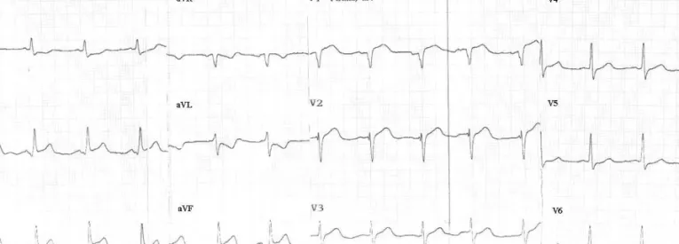
Ventricular tachycardia or fibrillation (VT/VF) in patients with acute myocardial infarction (AMI) is associated with a poor prognosis.

1)

Despite a better prognosis for early VT/VF compared to late VT/VF, early refractory VT/VF is a serious obstacle for the definitive treat- ment with primary percutaneous coronary intervention (PCI). One of the recent advances in the field of critical care is the extracorpo- real life support (ECLS) system, which is portable, readily applicable and easy to maintain. Before its advent, patients with AMI compli- cated by refractory VF/VT before commencing primary PCI would

Case Report

http://dx.doi.org/10.4070/kcj.2012.42.6.423 Print ISSN 1738-5520 • On-line ISSN 1738-5555

Extracorporeal Life Support After Prolonged Resuscitation for In-Hospital Cardiac Arrest due to Refractory Ventricular Fibrillation: Two Cases Resulting in a Full Recovery

Jin Wook Chung, MD 1 , Won Ho Chang, MD 2 , Min Su Hyon, MD 1 , and Wook Youm, MD 2

1

Departments of Internal Medicine and

2

Chest Surgery, Soonchunhyang University Hospital, Seoul, Korea

Extracorporeal life support (ECLS) has well demonstrated its efficacy in treating in-hospital cardiac arrest and is being used for broader indications. However, ECLS after prolonged cardiopulmonary resuscitation (CPR) has been traditionally contraindicated and is now chal- lenging. Here, we introduce two cases of successful ECLS after prolonged CPR, resulting in a immediate and full recovery. Both these acute ST elevation myocardial infarction patients waiting for primary percutaneous coronary intervention (PCI) suddenly collapsed due to ventricular fibrillation (VF), which was refractory to conventional treatment. After 2 hours of conventional CPR, the ECLS had been imple- mented and primary PCI could be performed. Subsequent to successful revascularization, the VF was stopped with a single electric shock.

In our second case, normal sinus rhythm was spontaneously restored after ECLS implementation, which was completed after 45 minutes of conventional resuscitation. Both patients made a full neurological recovery on the day of the event and were discharged with only mi- nor complications. (Korean Circ J 2012;42:423-426)

KEY WORDS: Cardiopulmonary resuscitation; Extracorporeal membrane oxygenation.

Received: September 1, 2011 Revision Received: October 3, 2011 Accepted: October 26, 2011

Correspondence: Jin Wook Chung, MD, Department of Internal Medicine, Soonchunhyang University College of Medicine, 59 Daesagwan-ro, Yong- san-gu, Seoul 140-743, Korea

Tel: 82-2-709-9345, Fax: 82-2-709-9083 E-mail: [email protected]

• The authors have no financial conflicts of interest.

This is an Open Access article distributed under the terms of the Creative Commons Attribution Non-Commercial License (http://creativecommons.

org/licenses/by-nc/3.0) which permits unrestricted non-commercial use, distribution, and reproduction in any medium, provided the original work is properly cited.

rarely survive or recover without ischemic brain damage. Such pa- tients are among those who can derive the most benefit from ECLS at present. In fact, ECLS has well demonstrated its efficacy in treat- ing in-hospital cardiopulmonary arrest and is being used for broa- der indications then before.

2)

The use of ECLS after prolonged car- diopulmonary resuscitation (CPR), a traditional contraindication, is particularly challenging.

Here, we present two cases of emergency ECLS after prolonged CPR for in-hospital cardiac arrest caused by refractory VF in pa- tients with AMI waiting for primary PCI.

Cases

Case 1

A 55-year old man visited the emergency room due to acute ch- est pain. His vital signs were stable but his electrocardiogram (ECG) showed ST-elevation in leads II, III and aVF as well as Q waves in leads V 1 and 2 (Fig. 1). The patient had a history of repeated AMIs, for which he had received multiple stents in two different hospitals.

Despite the higher risk of recurrent cardiovascular events, his com-

pliance seemed poor and he was still a heavy smoker. His current me-

dication included triple antiplatelet therapy, a lipid-lowering agent, a

beta-blocker, and an angiotensin-converting enzyme inhibitor. The

(2)

424 Extracorporeal Life Support After Prolonged CPR

http://dx.doi.org/10.4070/kcj.2012.42.6.423 www.e-kcj.org

pain-to-door time was about 30 minutes and the PCI team was call- ed after diagnosis of an acute inferior wall ST-elevation myocardial in- farction (STEMI).

During the stay in the emergency room, the patient suddenly col- lapsed from VF. CPR was initiated and repeated shocks at maximum energy were administered. Throughout about 30 minutes of conven- tional CPR, repeated shocks only stopped the VF for several sec- onds (Fig. 2). At this point, we decided to implement ECLS (CAPIOX EBS®, Terumo, Tokyo, Japan). The right femoral vessels were access- ed guided by anatomical landmarks and ECLS was finally imple-

mented, about 2 hours from the initiation of CPR. With full support, we achieved a flow rate of 4.8 L/min and mean arterial pressure of 70 mm Hg, but VF persisted.

Now the patient could be transferred to the catheterization room to undergo primary PCI, without the need for chest compressions for his fibrillating heart. In the catheterization room, the left femoral artery accessed also guided by anatomical landmarks and a coro- nary angiogram (CAG) was performed. The left CAG, including stent- ed segments, showed no significant stenosis. The right CAG revealed one stent in the proximal portion that was totally occluded by a

Fig. 1. Initial electrocardiogram (case 1).

Fig. 2. Ventricular fibrillation refractory to defibrillation (case 1).

Fig. 3. Right coronary angiogram before (A) and after (B) primary percutaneous coronary intervention (case 1).

A   B  

(3)

425 Jin Wook Chung, et al.

http://dx.doi.org/10.4070/kcj.2012.42.6.423 www.e-kcj.org

thrombus (Fig. 3A). Despite some difficulty in wiring, the coronary flow was successfully restored after ballooning, about three hours from the onset of chest pain. We implanted a long drug-eluting stent on the previous stent because the residual stenosis after balloon- ing was more than 80% (Fig. 3B). After the PCI, a single 360 J shock proved sufficient for successful defibrillation. We finished the pro- cedure by installing an intra-aortic balloon pulsation (IABP) cathe- ter to assist recovery.

The patient regained full consciousness without any neurologi- cal deficit later that day. The IABP and ECLS could be weaned on the third day and the patient was transferred to general ward on the fourth day. He was discharged with chest wall pain, caused by the CPR, and a large hematoma around the ECLS cannula insertion site, which had required a transfusion.

Case 2

A 43-year old man visited our emergency room with acute chest pain. Vital signs were stable but his ECG showed ST-elevation in leads V 1-4 and isolated VPC. He had hypertension, hypercholestero- lemia and acquired immunodeficiency syndrome, for which he re- ceived regular medications including anti-viral agents. The pain- to-door time was about 60 minutes and primary PCI was promptly arranged after diagnosis of an acute anterior wall STEMI.

During transfer to the catheterization room, one run of non-sus- tained VT appeared which could be suppressed with intravenous li- docane. However, during preparation for the procedure in the ca- theterization room, a sustained VT developed and turned into VF. CPR was initiated and repeated shocks at maximum energy were deliv- ered. We immediately called the ECLS team and continued CPR. At about 45 minutes of continuous CPR, normal sinus rhythm sponta- neously restored with the implementation of ECLS (CAPIOX EBS®, Terumo, Tokyo, Japan) via the right femoral vessels.

Restoration of sinus rhythm resulted in a stable patient with a flow rate of 2.5 L/min and mean arterial pressure of 80 mm Hg. Sub- sequently, the patient underwent primary PCI via the left femoral artery. His right CAG showed no significant stenosis, however, the left CAG revealed a thrombotic occlusion of proximal left anterior descending coronary artery. Coronary flow was successfully re- stored after ballooning, about two and half hours from the onset of chest pain. We implanted a drug-eluting stent in the culprit le- sion with no further complications.

He regained full consciousness the same day, in the absence of any neurological deficit. The ECLS could be weaned the second day and the patient was transferred to general ward on the third day. Fi- nally, he was discharged with no complications except for chest wall pain caused by the CPR.

Discussion

Our cases were not traditionally indicated for the application of ECLS. Prolonged CPR (>30 minutes) or unwitnessed cardiac arrest are poor prognostic markers for the implementation of ECLS.

3)

Ac- cording to Rhee et al.

4)

the time to ECLS for in-hospital arrest or shock patients was 43±23 minutes in failed weaning cases versus 25±8 minutes in successful weaning cases in Korea. With this in mind, the favorable results of our cases were beyond our expecta- tions. Although the precise impact of CPR duration on predicting survival or neurologic recovery seems to be unclear, our experienc- es suggest that well organized CPR can protect the major organs, including the brain, for up to 2 hours in witnessed in-hospital cardi- ac arrest. With advancements in ECLS systems, patients with in- hospital arrest may benefit from it, even after an extended period of CPR. More experience with the use of ECLS will help define its most appropriate time of deployment. Because primary PCI (the treatment of choice for STEMI) is readily available and highly suc- cessful in current practice, patients with refractory cardiac arrest are likely candidates for ECLS. However, conclusions from our lim- ited experiences should not be generalized to other causes of car- diac arrest.

According to recent epidemiological study, sustained VT/VF oc- curred in 5.7% of acute STEMI patients undergoing primary PCI, which mostly happened before the end of cardiac catheterization.

1)

Although the frequency of refractory cardiac arrest is not well documented, it is a potential threat in all patients with AMI. Our ex- perience of the past two years suggests refractory VF/VT accounts for about 2.5% of patients with STEMI waiting for primary PCI (un- published data).

The importance of chest compression during CPR is increasingly emphasized.

5-8)

We think the favorable results of our cases are based on the endeavored CPR, conducted with minimal interruption of chest compression. Also, the CPR in both cases was fully directed by the same experienced cardiologist. To improve the outcome of ECLS, rapid and accurate cannulation of the ECLS system is important. In one of our two cases we spent about 90 minutes on ECLS implan- tation, mostly due to difficulty in cannulation of the pulseless ves- sel without interrupting chest compression. This was further com- plicated by formation of a large hematoma at the cannulation site, requiring a transfusion.

We hope our experiences may contribute to improving the prog- nosis of patients with cardiac arrest.

References

1. Mehta RH, Starr AZ, Lopes RD, et al. Incidence of and outcomes associ-

ated with ventricular tachycardia or fibrillation in patients undergoing

(4)

426 Extracorporeal Life Support After Prolonged CPR

http://dx.doi.org/10.4070/kcj.2012.42.6.423 www.e-kcj.org

primary percutaneous coronary intervention. JAMA 2009;301:1779-89.

2. Chen YS, Lin JW, Yu HY, et al. Cardiopulmonary resuscitation with as- sisted extracorporeal life-support versus conventional cardiopulmo- nary resuscitation in adults with in-hospital cardiac arrest: an obser- vational study and propensity analysis. Lancet 2008;372:554-61.

3. Kurusz M, Zwischenberger JB. Percutaneous cardiopulmonary bypass for cardiac emergencies. Perfusion 2002;17:269-77.

4. Rhee I, Gwon HC, Choi J, et al. Percutaneous cardiopulmonary support for emergency in-hospital cardiac arrest or cardiogenic shock. Korean Circ J 2006;36:11-6.

5. Ramaraj R, Ewy GA. Rationale for continuous chest compression car-

diopulmonary resuscitation. Heart 2009;95:1978-82.

6. Ong ME, Ng FS, Anushia P, et al. Comparison of chest compression only and standard cardiopulmonary resuscitation for out-of-hospital cardi- ac arrest in Singapore. Resuscitation 2008;78:119-26.

7. SOS-KANTO study group. Cardiopulmonary resuscitation by bystanders with chest compression only (SOS-KANTO): an observational study.

Lancet 2007;369:920-6.

8. Ristagno G, Gullo A, Tang W, Weil MH. New cardiopulmonary resusci-

tation guidelines 2005: importance of uninterrupted chest compres-

sion. Crit Care Clin 2006;22:531-8.

수치

Fig. 1. Initial electrocardiogram (case 1).

참조

관련 문서

Risk factors related to fixed airway obstruction in patients with asthma after antiasthma treatment. Identification of sub- types of refractory asthma in Korean patients by

Zao Wou-Ki generated the best H1 result for the entire Asian continent in Hong Kong; 53% of Zhang Daqian’s turnover was hammered there, and lots of new

Long-term outcome after an early invasive versus selective invasive treatment strategy in patients with non.ST-elevation acute coronary syndrome and elevated

In addition, Wallace and Froum 20) reported that the bone formation rate was superior in the cases using a barrier membrane in comparison with the

After listening to the opinions of firemen in field activities, we confirmed that the firepower can be secured by reducing the loss of firepower caused

The purpose of this study was to identify the frequency and related factors of advanced airway management for patients with cardiac arrest by the

 인공신경망 기법을 활용한 노심보호계통 축방향 출력분포 합성 방법은 축방향 출력분포 합성

Here we compute the closed-form dynamic equations for the two-link planar manipulator shown in Fig. For simplicity, we assume that the mass distribution is extremely simple: