Immunoglobulin G4-Related Kidney Disease: A Comprehensive Pictorial Review of the Imaging Spectrum, Mimickers, and Clinicopathological Characteristics
12
0
0
전체 글
(2) Imaging Spectrum of IgG4-Related Kidney Disease. A. B. C. D. E Fig. 1. 72-year-old man who underwent left nephrectomy due to IgG4-KD mimicking urothelial carcinoma.. A. Contrast-enhanced CT image shows ill-defined, homogeneous, soft-tissue mass (arrow) which encases left renal pelvis causing hydronephrosis. B. He underwent left nephrectomy with diagnosis of having urothelial carcinoma. Photograph of surgical specimen demonstrates lobulated, yellowish, firm mass (arrows) around renal pelvis. C, D. Lower-power (x 100) (C) and higher-power (x 400) (D) photomicrographs of histological specimen (hematoxylin and eosin staining) show dense plasma cell infiltration (arrows) with fibrous background (Fi) and obliterative phlebitis (arrowheads). E. IgG4 immunostaining (x 100) of histologic specimen shows numerous, brown, IgG4-positive plasma cells. IgG4-KD = immunoglobulin G4-related kidney disease. kjronline.org. Korean J Radiol 16(5), Sep/Oct 2015. 1057.
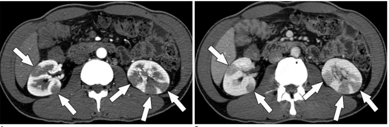
(3) Seo et al.. usually accompanied by other organ involvement in IgG4SD, typically AIP, and approximately 1/4 or 1/3 of AIP patients have IgG4-KD (11-13). Isolated cases of IgG4-KD without other organ involvement, is very rare. In our series, only 3 (6%) of 48 IgG4-KD patients had renal lesions alone. The clinical manifestations are usually mild or even asymptomatic. As the disease progresses, renal dysfunction or abnormalities are seen in imaging studies; only then it becomes clinically evident. Renal function at the time of the initial diagnosis varies from normal function to renal failure. Mild proteinuria occurs in approximately half the patients (7, 10). Hypergammaglobulinemia, either IgG4 or total IgG, is the serologic hallmark of IgG4-KD as well as IgG4-SD. Although up to 30% of patients with IgG4-SD have a normal range of serum IgG4 level, almost all IgG4-KD patients. have an elevated serum IgG4 level (7, 10). Other serologic findings include an elevated serum immunoglobulin E level, eosinophilia, and hypocomplementemia (1, 5). Immunoglobulin G4-related kidney disease manifests most commonly as tubulointerstitial nephritis, followed by glomerular disease such as membranous glomerulonephritis (1). Other less frequent conditions, including IgG4related chronic sclerosing pyelitis, IgG4-related plasmacell arteritis, and IgG4-related inflammatory pseudotumors of the ureter, have also been reported (1, 14-16). The key histologic findings of IgG4-KD are dense lymphoplasmacytic infiltration with increased IgG4-positive plasma cells, and storiform fibrosis, both of which are identical to the findings of IgG4-SD involving other organs (Fig. 1) (3, 10). In addition, the characteristic microscopic features of IgG4-. A B Fig. 2. 49-year-old man with IgG4-KD manifesting as multiple, renal parenchymal nodules.. A, B. Contrast-enhanced arterial (A) and portal (B) phase CT images show multiple, small, round or wedge-shaped, hypodense nodules (arrows) in both kidneys, predominantly in cortex. Renal nodules progressively enhance in portal phase. IgG4-KD = immunoglobulin G4-related kidney disease. A B Fig. 3. IgG4-KD manifesting as diffuse patchy infiltrative parenchymal lesions.. A. 66-year-old man. Contrast-enhanced portal phase CT image shows patchy infiltrative hypodense lesions (arrows) in both kidneys. B. 56-yearold man. T2-weighted MR image shows diffuse patchy infiltrative lesions appearing noticeably hypointense (arrows) in both kidneys. IgG4-KD = immunoglobulin G4-related kidney disease. 1058. Korean J Radiol 16(5), Sep/Oct 2015. kjronline.org.
(4) Imaging Spectrum of IgG4-Related Kidney Disease. related tubulointerstitial nephritis are nests of inflammatory cells with irregular fibers surrounding them, and immune complex deposition in the tubular basement membrane (5, 17, 18).. crucial to be aware of the imaging spectrum of IgG4-KD in order to obtain a timely and accurate diagnosis. Distribution of IgG4-KD Immunoglobulin G4-related kidney disease can be divided into three types, based on their location: renal parenchymal, renal pelvic, and a perinephric lesion. A renal parenchymal lesion is the most common type, followed by a renal pelvic and a perinephric lesion (9, 12, 19). Two types can sometimes appear together. Among the 48 IgG4-KD patients seen at our institution, 36 (75%) had only renal parenchymal lesions, 5 (10%) had only renal pelvic lesions, and 1 (2%) had only perinephric lesions. Two types of IgG4KD co-existed in 6 (13%) patients: 5 with renal parenchymal. Imaging Spectrum of IgG4-KD The diagnosis of IgG4-KD is based on a combination of imaging, serologic and histologic findings, as well as other organ involvement (5, 7). Among these findings, the imaging findings may be the most important component as they are usually the first recognized abnormal findings of IgG4-KD, which enables clinicians or radiologists to consider the occurrence of this disease. Therefore, it is. A. B. C. D. Fig. 4. MR imaging features of IgG4-KD in 64-year-old man.. A. On precontrast T1-weighted image, isointense renal lesions are not perceptible. B. T2-weighted image shows three, small, hypointense cortical nodules (arrows) in both kidneys. C, D. DWI (b value, 1000 s/mm2) (C) and ADC map (D) reveal renal nodules (arrows) which appear as having obvious hyperintensity and hypointensity, respectively, and representing diffusion restriction. ADC = apparent diffusion coefficient, DWI = diffusion-weighted MR imaging, IgG4-KD = immunoglobulin G4-related kidney disease. kjronline.org. Korean J Radiol 16(5), Sep/Oct 2015. 1059.
(5) Seo et al.. and renal pelvic lesions, and 1 with renal parenchymal and perinephric lesions. Because of the systemic nature of the disease, IgG4-KD typically shows multiplicity and bilaterality (9, 19, 20). In our series, 45 (94%) and 37 (77%) patients had multiple and bilateral lesions, respectively.. lesion mimicking a solid renal neoplasm (9, 12, 19). On contrast-enhanced CT, renal parenchymal lesions are mostly hypodense to the normal renal cortex in the early phase, i.e., the arterial or corticomedullary phase, and progressively get enhanced in the portal or delayed phase (Fig. 2) (19-21). These lesions are usually not visible on precontrast CT scans (12, 19). It can often be challenging to perceive early, subtle renal lesions depending solely on CT scans. MR imaging can improve detection of the lesions in such cases. On T2-weighted MR images, renal lesions typically appear hypointense relative to the normal renal parenchyma, and are easily detectable due to the obvious contrast between the dark lesions and the bright, normal parenchyma (Figs. 3, 4) (9, 12, 22). On the other hand,. Renal Parenchymal Lesions A renal parenchymal lesion in IgG4-KD can show three patterns: a single nodule, multiple nodules (Fig. 2), and diffuse patchy infiltrative lesions (Fig. 3). Among these, multiple nodules are the most common pattern (12, 19, 20). Renal parenchymal nodules are located predominantly in the renal cortex. They are usually small and round, or wedge-shaped. It rarely appears as a single, large, mass-like. A. B. C. D. Fig. 5. MR imaging features of IgG4-KD in 72-year-old man.. A-C. Dynamic contrast-enhanced MR images show small cortical nodule (arrows) in right kidney, and which appears hypointense in arterial phase (A) and progressively enhanced in portal (B) and delayed (C) phases, gradually becoming indistinct as phase passes. D. On DWI (b value, 1000 s/mm2), right renal lesion exhibits striking hyperintensity (arrow). DWI = diffusion-weighted MR imaging, IgG4-KD = immunoglobulin G4-related kidney disease. 1060. Korean J Radiol 16(5), Sep/Oct 2015. kjronline.org.
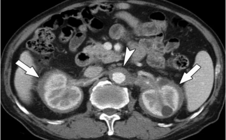
(6) Imaging Spectrum of IgG4-Related Kidney Disease. renal lesions usually appear isointense on T1-weighted images, and they are barely visible or are invisible (Fig. 4). On dynamic contrast-enhanced MR images, most renal lesions are hypointense to the normal renal cortex in the arterial phase, and progressively enhance in the portal and delayed phases, thus becoming indistinct as the phase passes; this is similar to the dynamic enhancement pattern seen on CT (Fig. 5) (9, 12, 22). Diffusion-weighted MR imaging (DWI) has recently been reported to be particularly useful for detecting IgG4-KD (9). Kim et al. (9) reported that the sensitivity of DWI (b value, 1000 s/mm2) for detecting IgG4-KD was significantly higher than that of T2-weighted images (100% vs. 77%). The renal lesions. Fig. 6. 64-year-old man with IgG4-KD manifesting as unilateral renal pelvic lesion. Contrast-enhanced arterial phase CT image shows ill-defined, mildly enhancing, soft-tissue mass (arrow) encasing right renal pelvis and without hydronephrosis. IgG4-KD = immunoglobulin G4-related kidney disease. have marked hyperintensity on DWI at a high b value and, conversely, have marked hypointensity on the apparent diffusion coefficient map (Figs. 4, 5). These DWI findings can be explained by the histopathologic background of IgG4-KD, i.e., dense lymphoplasmacytic infiltration with fibrosis. This may be supported by the hypointensity seen on T2-weighted images and the progressive enhancement pattern seen on dynamic contrast-enhanced images, which may also represent fibrosis (9, 12). DWI may also have the potential to detect IgG4-KD at an early or subclinical stage (9, 23). Ultrasonographic (US) findings of IgG4-KD are not specific and are less sensitive for lesion detection than CT or MR imaging (24). The most common US findings are illdefined, non-mass-like areas of decreased echogenicity (24). Other US findings include irregular lobular thickening of the renal parenchyma with a bulging contour and multiple, hypoechoic masses with decreased vascularity, as compared to the normal renal cortex (24). Renal Pelvic and Perinephric Lesions Renal pelvic or perinephric lesions are much rarer than renal parenchymal lesions. Renal pelvic lesions manifest as diffused wall thickening of the renal pelvis, or as a mildly enhanced, soft-tissue mass encasing the renal pelvis (12, 19). These lesions can be unilateral (Fig. 6) or bilateral (Fig. 7). Hydronephrosis seldom occurs, unless the ureter is entrapped by accompanying periaortic retroperitoneal fibrosis. In our series, only 1 of 10 patients with renal pelvic lesions had a mild, unilateral hydronephrosis without retroperitoneal fibrosis. Perinephric lesions typically. Fig. 7. 84-year-old man with IgG4-KD manifesting as bilateral renal pelvic lesions. Contrast-enhanced portal phase CT images show mildly enhanced wall thickening of both renal pelvises (arrows) and without hydronephrosis. Pancreatic head enlargement with heterogeneous attenuation (arrowheads), indicating autoimmune pancreatitis, is also seen. IgG4-KD = immunoglobulin G4-related kidney disease. kjronline.org. Korean J Radiol 16(5), Sep/Oct 2015. 1061.
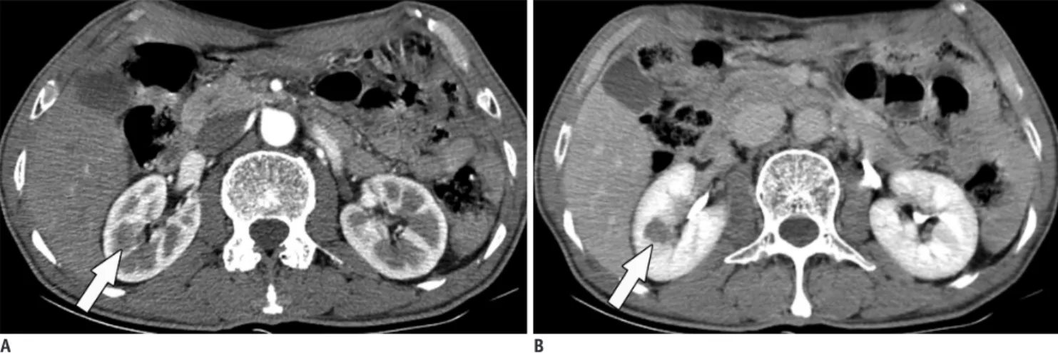
(7) Seo et al.. appear as a diffuse, rim-like, soft-tissue mass surrounding the kidneys (Fig. 8) (12). These masses usually exhibit homogeneous and mild enhancement. They can be also. unilateral or bilateral. Response of IgG4-KD to Steroid Therapy Immunoglobulin G4-related kidney disease usually shows a rapid and favorable response to steroid therapy, which similar to IgG4-SD with other organ involvement, regardless of the lesion location (i.e., renal parenchymal, renal pelvic or perinephric). According to the pathologic subtype, tubulointerstitial nephritis generally responds better than membranous glomerulonephritis (1). Approximately 90% of IgG4-KD patients show improvement of decreased renal function and abnormal imaging findings with steroid treatment (1, 10, 12, 17, 19). During the steroid treatment, the lesion size and number dramatically decreases, as seen on imaging studies (Fig. 9). After completion of the steroid therapy, varying degrees of renal atrophy can develop, especially in patients with renal parenchymal lesions, and which may be proportionate to the extent of the renal involvement before treatment (6, 10, 12). In our series, the majority of patients having renal parenchymal. Fig. 8. 74-year-old man with IgG4-KD manifesting as perinephric lesion. Contrast-enhanced arterial phase CT image shows homogeneous, rim-like, soft-tissue mass surrounding both kidneys (arrows). Peri-aortic, soft-tissue lesion, indicating retroperitoneal fibrosis, is also observed (arrowhead). IgG4-KD = immunoglobulin G4related kidney disease. A. B. C Fig. 9. Response of IgG4-KD to steroid therapy in 72-year-old man.. A. Contrast-enhanced CT image shows diffuse infiltrative lesion (arrows) in right kidney. B. Contrast-enhanced CT image obtained after completion of steroid treatment demonstrates improvement of right renal lesion. However, multifocal, tiny cortical scars remain (arrows). C. Contrast-enhanced CT image obtained two years after steroid cessation reveals nodular or infiltrative lesions in right kidney (arrows), thus indicative of disease relapse. IgG4-KD = immunoglobulin G4-related kidney disease. 1062. Korean J Radiol 16(5), Sep/Oct 2015. kjronline.org.
(8) Imaging Spectrum of IgG4-Related Kidney Disease. A. B. C. D. Fig. 10. Mimickers of IgG4-KD manifesting as multiple, renal parenchymal lesions.. A. 32-year-old woman with acute pyelonephritis. Contrast-enhanced delayed phase CT image shows multifocal, ill-defined, round and wedgeshaped, hypodense lesions (arrows) with mild swelling in left kidney. B. 53-year-old man with renal infarction. Contrast-enhanced arterial phase CT image shows large, rather well-demarcated, wedge-shaped areas of poor contrast enhancement (arrows) in both kidneys. C. 75-year-old man with renal metastases from lung cancer. Contrast-enhanced arterial phase CT image shows round, hypodense nodules (arrows) in both kidneys. D. 61-year-old man with renal lymphomas. Contrast-enhanced portal phase coronal CT image shows several, well-defined, homogeneous, round nodules/masses (arrows) in both kidneys. IgG4-KD = immunoglobulin G4-related kidney disease. lesions of a diffuse patchy infiltrative pattern, or with large, multiple nodules, experienced mild cortical scars after steroid therapy (Fig. 9). IgG4-KD as well as other organ involvement of IgG4-SD can relapse in up to 20% of patients, mainly during maintenance therapy or after cessation of the steroid therapy (Fig. 9) (6, 12). Without steroid treatment, the renal lesions can increase in size and number, or progress to diffuse cortical involvement (12).. Mimickers of IgG4-KD and the Differential Diagnosis As stated above, IgG4-KD has a broad spectrum of kjronline.org. Korean J Radiol 16(5), Sep/Oct 2015. imaging features. Therefore, various neoplastic or nonneoplastic conditions of the kidneys can mimic IgG4KD. With regard to IgG4-KD manifesting as multiple, renal parenchymal lesions, the differential diagnosis includes acute pyelonephritis, infarction, metastasis, and lymphoma (Fig. 10) (9, 12, 20). Acute pyelonephritis typically appear as unilateral or bilateral, multiple, ill-defined, and round or wedge-shaped lesions of decreased enhancement (Fig. 10A) (25, 26). Clinical findings, which include fever, pyuria or knocking tenderness in the flank area, may undoubtedly be important clues suggesting acute pyelonephritis. In addition to the aforementioned clinical findings, perilesional infiltration or fluid collection, and renal or 1063.
(9) Seo et al.. A B Fig. 11. Mimicker of IgG4-KD manifesting as single parenchymal nodule.. A, B. 78-year-old man with chromophobe-type renal cell carcinoma. Contrast-enhanced arterial (A) and delayed (B) phase CT images demonstrate single hypovascular nodule (arrows) in right kidney. IgG4-KD = immunoglobulin G4-related kidney disease. Fig. 12. Mimicker of IgG4-KD manifesting as renal pelvic lesion. 63-year-old man with transitional cell carcinoma. Contrastenhanced portal phase CT image shows diffusely enhancing wall thickening of right renal pelvis (arrow) and surrounding soft-tissue lesion (arrowhead). IgG4-KD = immunoglobulin G4-related kidney disease. perirenal abscesses seen on imaging examinations, may also help to differentiate acute pyelonephritis from IgG4KD. Well-demarcated, large, wedge-shaped areas showing poor contrast enhancement are typical of renal infarction, although they may mimic the patchy infiltrative pattern of IgG4-KD (Fig. 10B) (25, 27). A hyperenhanced cortical rim at the lesion periphery, the so-called cortical rim sign, can be seen in approximately half of the patients with renal infarction, whereas in IgG4-KD this sign has never been reported in the published medical literature, including our study (27, 28). Metastases usually appear as bilateral, multiple, and less exophytic nodules, and they are therefore capable of closely mimicking IgG4-KD (Fig. 10C) (29). The presence of primary malignancy is the most important clue 1064. to metastasis. The common sites of primary malignancy are the lung, breast, contralateral kidney, and colon (30). Renal lymphomas commonly manifest as multiple, parenchymal nodules/masses (Fig. 10D), or as a renal pelvic or perinephric mass with mild homogeneous enhancement, which can be quite similar to the IgG4-KD features (3133). Moreover, lymphomas often appear hypointense on T2-weighted MR images and have diffusion restriction (33, 34). However, renal lymphomas seem more bulky than IgG4KD and frequently accompany multiple, retroperitoneal lymphadenopathies, compared to IgG4-KD (9, 32). In rare cases, when IgG4-KD manifests as a single parenchymal nodule/mass, it can be challenging to distinguish it from renal cell carcinoma, especially when non-clear cell types, such as the papillary or chromophobe type, appear as a non-hypervascular mass (Fig. 11) (35). Diffuse wall thickening of the renal pelvis or a parapelvic mass in IgG4-KD should be differentiated from urothelial carcinoma (Fig. 12). The relatively common bilaterality and uncommon hydronephrosis in IgG4-KD may be helpful in differentiating the two diseases, and may warrant further comparative studies. In addition to the above-mentioned analyses regarding the imaging features, detection of other organ involvement of IgG4-SD, i.e., AIP (Fig. 13), sclerosing cholangitis, retroperitoneal fibrosis (Fig. 13) or sialadenitis, may be essential for differentiating IgG4-KD from the other diseases, as isolated IgG4-KD without other organ involvement is very rare, as demonstrated in our study (6%). According to recently established diagnostic criteria of IgG4-KD, the presence of other organ involvement is an important diagnostic component (5, 7). Alternatively, Korean J Radiol 16(5), Sep/Oct 2015. kjronline.org.
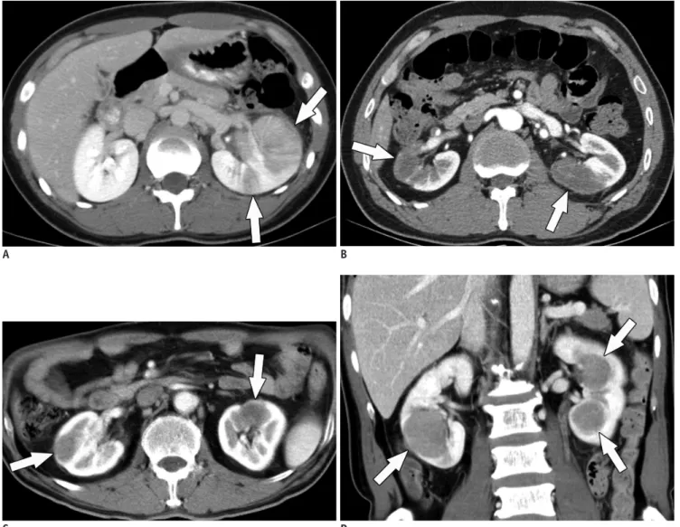
(10) Imaging Spectrum of IgG4-Related Kidney Disease. A B Fig. 13. Retroperitoneal fibrosis and autoimmune pancreatitis as clues to diagnosis of IgG4-KD in 71-year-old man.. A, B. Contrast-enhanced portal phase CT images show several, well-defined, round, hypodense nodules (arrows) in both kidneys. Differential diagnosis for these renal lesions would be lymphoma, metastasis or IgG4-KD. However, periaortic, soft-tissue lesion (arrowhead in A) suggesting retroperitoneal fibrosis and sausage-shaped pancreas swelling with peripancreatic hypodense rim (arrowheads in B) which is typical of AIP, are observed together. Accordingly, such renal lesions can confidently be diagnosed as IgG4-KD. AIP = autoimmune pancreatitis, IgG4-KD = immunoglobulin G4-related kidney disease. A. B. C. D. Fig. 14. IgG4-KD as clue for differentiating autoimmune pancreatitis from pancreatic cancer in 64-year-old man.. A, B. Precontrast T1-weighted MR image (A) shows hypointense, mass-like enlargement of pancreatic head (arrow). MR cholangiopancreatography (B) shows abrupt, severe narrowing of pancreatic and bile ducts (arrowheads) with upstream duct dilatation. These MR findings are highly suggestive of pancreatic cancer. C, D. DWIs (b value, 1000 s/mm2) show multifocal, hyperintense nodules (arrows) in right kidney. Combination of these renal lesions and pancreatic mass/enlargement strongly suggests IgG4-SD, i.e., IgG4-KD and focal type AIP. AIP = autoimmune pancreatitis, DWI = diffusion-weighted MR imaging, IgG4-KD = immunoglobulin G4-related kidney disease, IgG4-SD = IgG4-related sclerosing disease. kjronline.org. Korean J Radiol 16(5), Sep/Oct 2015. 1065.
(11) Seo et al.. identification of renal lesions can be of crucial importance for the diagnosis of IgG4-SD involving other organs, especially when imaging features of other organs are not characteristic of IgG4-SD. A typical example would be the differential diagnosis of focal-type AIP and pancreatic cancer; in this situation, the presence of renal lesions may substantially favor the former diagnosis (Fig. 14) (8). On the other hand, despite the detailed analysis using various imaging modalities, it can still be difficult to differentiate IgG4-KD from other diseases, in particular neoplasms. In such cases, CT- or US-guided biopsy of the renal lesions should be considered, in order to avoid unnecessary surgery caused by the misdiagnosis as neoplasm, in patients having IgG4-KD.. CONCLUSION Immunoglobulin G4-related kidney disease has a broad spectrum of imaging features and a variety of mimickers to be differentiated from it. Awareness of the broad imaging spectrum of IgG4-KD, differential diagnosis from its mimickers, and important clinicopathologic characteristics, will facilitate its accurate and prompt diagnosis as well as optimally timed treatment.. REFERENCES 1. Cornell LD. IgG4-related kidney disease. Semin Diagn Pathol 2012;29:245-250 2. Stone JH, Zen Y, Deshpande V. IgG4-related disease. N Engl J Med 2012;366:539-551 3. Deshpande V, Zen Y, Chan JK, Yi EE, Sato Y, Yoshino T, et al. Consensus statement on the pathology of IgG4-related disease. Mod Pathol 2012;25:1181-1192 4. Umehara H, Okazaki K, Masaki Y, Kawano M, Yamamoto M, Saeki T, et al. Comprehensive diagnostic criteria for IgG4related disease (IgG4-RD), 2011. Mod Rheumatol 2012;22:21-30 5. Raissian Y, Nasr SH, Larsen CP, Colvin RB, Smyrk TC, Takahashi N, et al. Diagnosis of IgG4-related tubulointerstitial nephritis. J Am Soc Nephrol 2011;22:1343-1352 6. Saeki T, Kawano M, Mizushima I, Yamamoto M, Wada Y, Nakashima H, et al. The clinical course of patients with IgG4related kidney disease. Kidney Int 2013;84:826-833 7. Kawano M, Saeki T, Nakashima H, Nishi S, Yamaguchi Y, Hisano S, et al. Proposal for diagnostic criteria for IgG4related kidney disease. Clin Exp Nephrol 2011;15:615-626 8. Khalili K, Doyle DJ, Chawla TP, Hanbidge AE. Renal cortical lesions in patients with autoimmune pancreatitis: a clue to differentiation from pancreatic malignancy. Eur J Radiol 2008;67:329-335. 1066. 9. Kim B, Kim JH, Byun JH, Kim HJ, Lee SS, Kim SY, et al. IgG4-related kidney disease: MRI findings with emphasis on the usefulness of diffusion-weighted imaging. Eur J Radiol 2014;83:1057-1062 10. Saeki T, Nishi S, Imai N, Ito T, Yamazaki H, Kawano M, et al. Clinicopathological characteristics of patients with IgG4related tubulointerstitial nephritis. Kidney Int 2010;78:10161023 11. Khosroshahi A, Stone JH. A clinical overview of IgG4-related systemic disease. Curr Opin Rheumatol 2011;23:57-66 12. Takahashi N, Kawashima A, Fletcher JG, Chari ST. Renal involvement in patients with autoimmune pancreatitis: CT and MR imaging findings. Radiology 2007;242:791-801 13. Kim JH, Kim MH, Byun JH, Lee SS, Lee SJ, Park SH, et al. Diagnostic strategy for differentiating autoimmune pancreatitis from pancreatic cancer: is an endoscopic retrograde pancreatography essential? Pancreas 2012;41:639-647 14. Kuroda N, Nakamura S, Miyazaki K, Inoue K, Ohara M, Mizuno K, et al. Chronic sclerosing pyelitis with an increased number of IgG4-positive plasma cells. Med Mol Morphol 2009;42:236-238 15. Kim SA, Lee SR, Huh J, Shen SS, Ro JY. IgG4-associated inflammatory pseudotumor of ureter: clinicopathologic and immunohistochemical study of 3 cases. Hum Pathol 2011;42:1178-1184 16. Sharma SG, Vlase HL, D’Agati VD. IgG4-related tubulointerstitial nephritis with plasma cell-rich renal arteritis. Am J Kidney Dis 2013;61:638-643 17. Saeki T, Kawano M. IgG4-related kidney disease. Kidney Int 2014;85:251-257 18. Yamaguchi Y, Kanetsuna Y, Honda K, Yamanaka N, Kawano M, Nagata M; Japanese study group on IgG4-related nephropathy. Characteristic tubulointerstitial nephritis in IgG4-related disease. Hum Pathol 2012;43:536-549 19. Triantopoulou C, Malachias G, Maniatis P, Anastopoulos J, Siafas I, Papailiou J. Renal lesions associated with autoimmune pancreatitis: CT findings. Acta Radiol 2010;51:702-707 20. Hedgire SS, McDermott S, Borczuk D, Elmi A, Saini S, Harisinghani MG. The spectrum of IgG4-related disease in the abdomen and pelvis. AJR Am J Roentgenol 2013;201:14-22 21. Vlachou PA, Khalili K, Jang HJ, Fischer S, Hirschfield GM, Kim TK. IgG4-related sclerosing disease: autoimmune pancreatitis and extrapancreatic manifestations. Radiographics 2011;31:1379-1402 22. Manfredi R, Frulloni L, Mantovani W, Bonatti M, Graziani R, Pozzi Mucelli R. Autoimmune pancreatitis: pancreatic and extrapancreatic MR imaging-MR cholangiopancreatography findings at diagnosis, after steroid therapy, and at recurrence. Radiology 2011;260:428-436 23. Pozdzik AA, Matos C, Rorive S, Brocheriou I, Delhaye M, Nortier JL. Diffusion-weighted magnetic resonance imaging: a non-nephrotoxic prompt assessment of kidney involvement in IgG4-related disease. Kidney Int 2014;85:981 24. Sasiwimonphan K, Gorman B, Kawashima A, Chari ST, Takahashi N. Renal involvement in patients with autoimmune Korean J Radiol 16(5), Sep/Oct 2015. kjronline.org.
(12) Imaging Spectrum of IgG4-Related Kidney Disease. pancreatitis: ultrasound findings. Eur J Radiol 2012;81:807-810 25. Saunders HS, Dyer RB, Shifrin RY, Scharling ES, Bechtold RE, Zagoria RJ. The CT nephrogram: implications for evaluation of urinary tract disease. Radiographics 1995;15:1069-1085; discussion 1086-1088 26. Kawashima A, Sandler CM, Goldman SM, Raval BK, Fishman EK. CT of renal inflammatory disease. Radiographics 1997;17:851866; discussion 867-868 27. Suzer O, Shirkhoda A, Jafri SZ, Madrazo BL, Bis KG, Mastromatteo JF. CT features of renal infarction. Eur J Radiol 2002;44:59-64 28. Wong WS, Moss AA, Federle MP, Cochran ST, London SS. Renal infarction: CT diagnosis and correlation between CT findings and etiologies. Radiology 1984;150:201-205 29. Honda H, Coffman CE, Berbaum KS, Barloon TJ, Masuda K. CT analysis of metastatic neoplasms of the kidney. Comparison with primary renal cell carcinoma. Acta Radiol 1992;33:39-44. kjronline.org. Korean J Radiol 16(5), Sep/Oct 2015. 30. Bracken RB, Chica G, Johnson DE, Luna M. Secondary renal neoplasms: an autopsy study. South Med J 1979;72:806-807 31. Ganeshan D, Iyer R, Devine C, Bhosale P, Paulson E. Imaging of primary and secondary renal lymphoma. AJR Am J Roentgenol 2013;201:W712-W719 32. Jafri SZ, Bree RL, Amendola MA, Glazer GM, Schwab RE, Francis IR, et al. CT of renal and perirenal non-Hodgkin lymphoma. AJR Am J Roentgenol 1982;138:1101-1105 33. Semelka RC, Kelekis NL, Burdeny DA, Mitchell DG, Brown JJ, Siegelman ES. Renal lymphoma: demonstration by MR imaging. AJR Am J Roentgenol 1996;166:823-827 34. Saremi F, Knoll AN, Bendavid OJ, Schultze-Haakh H, Narula N, Sarlati F. Characterization of genitourinary lesions with diffusion-weighted imaging. Radiographics 2009;29:1295-1317 35. Yamashita Y, Takahashi M, Watanabe O, Yoshimatsu S, Ueno S, Ishimaru S, et al. Small renal cell carcinoma: pathologic and radiologic correlation. Radiology 1992;184:493-498. 1067.
(13)
수치


+4

관련 문서
