45
Original Article
The Role of Focal Adhesion Kinase in the TGF-β-Induced Myofibroblast Transdifferentiation of Human Tenon’s Fibroblasts
Samin Hong, Jong Bok Lee, Yoko Iizuka, Yoo Kyung Song, Gong Je Seong, Sueng-Han Han
Institute of Vision Research, Department of Ophthalmology, Yonsei University College of Medicine, Seoul, Korea pISSN: 1011-8942 eISSN: 2092-9382
Korean J Ophthalmol 2012;26(1):45-48 http://dx.doi.org/10.3341/kjo.2012.26.1.45
Myofibroblast transdifferentiation of activated fibro- blasts is an essential step of the fibrotic process in most living tissues. Myofibroblasts have characteristics that are taken from both smooth muscle cells and fibroblasts; they express the contractile phenotype of α smooth muscle ac- tin (αSMA) and synthesize extracellular matrix proteins including collagens [1-3]. In subconjunctival fibrosis, the myofibroblast transdifferentiation of Tenon’s fibroblasts is known to be mainly caused by transforming growth factor (TGF)-β [4-6]. Many ophthalmic researchers have sought to identify an anti-fibrotic mechanism by inhibiting TGF-β signaling [7-10].
Focal adhesion kinase (FAK), a cytosolic protein ty- rosine kinase, provides scaffolding functions at sites of integrin adhesion and promotes cell migration [11,12].
The kinase modulates several basic processes in normal development and cancer metastasis. In addition, it also plays a crucial role in the transdifferentiation of fibroblasts to myofibroblasts induced by TGF-β [13-15]. However, because the precise action of the kinase is somewhat vari- able based on the tissue [14,15], the role of FAK in myofi- broblast transdifferentiation in human Tenon’s fibroblasts must be confirmed.
Using primary cultured human Tenon’s fibroblasts, the role of FAK in TGF-β-induced myofibroblast transdiffer- entiation of human Tenon’s fibroblasts was investigated in the present study.
Materials and Methods
Cell culture and exposure to transforming growth factor-β1 After the Institutional Review Board approved our pro- tocol, selected patients received comprehensive informa- tion and provided written consent for inclusion. In compli- ance with the tenets of the Declaration of Helsinki, small human Tenon’s capsule specimens were obtained during
© 2012 The Korean Ophthalmological Society
This is an Open Access article distributed under the terms of the Creative Commons Attribution Non-Commercial License (http://creativecommons.org/licenses /by-nc/3.0/) which permits unrestricted non-commercial use, distribution, and reproduction in any medium, provided the original work is properly cited.
Received: November 17, 2010 Accepted: January 1, 2011
Corresponding Author: Sueng-Han Han, MD, PhD. Department of Ophthalmology, Yonsei University College of Medicine, #211 Eonju-ro, Gangnam-gu, Seoul 135-720, Korea. Tel: 82-2-2019-3440, Fax: 82-2-3463- 1049, E-mail: [email protected]
Purpose: To investigate the role of focal adhesion kinase (FAK) in transforming growth factor (TGF)-β-induced myofibroblast transdifferentiation of human Tenon’s fibroblasts.
Methods: Primary cultured human Tenon’s fibroblasts were exposed to TGF-β1 for up to 48 hours. The mRNA levels of FAK, α smooth muscle actin (αSMA), and β-actin were determined by quantitative real time reverse transcription polymerase chain reaction. The protein levels of collagen type I, FAK, phospho-FAK, αSMA, and β-actin were determined by Western immunoblots. After the small interfering RNA targeting FAK (siRNAFAK) molecules were delivered into the cells, the expressions of αSMA proteins were determined by Western im- munoblots.
Results: In human Tenon’s fibroblasts, TGF-β1 significantly increased the mRNA and protein expressions of αSMA. However, when the action of FAK was inhibited using siRNAFAK, the TGF-β1-induced expression of αSMA was attenuated.
Conclusions: Our data suggest that FAK may be associated with the TGF-β1-induced transdifferentiation of hu- man Tenon’s fibroblasts to myofibroblasts, which is the essential step of subconjunctival fibrosis.
Key Words: Fibroblast, Focal adhesion protein-tyrosine kinases, Myofibroblast, Transforming growth factors
46
Korean J Ophthalmol Vol.26, No.1, 2012
strabismus surgery, and primary human Tenon’s fibroblasts were cultured from the explants. Cells were incubated in Dulbecco’s modified Eagle’s medium (DMEM; Life Tech- nologies, Carlsbad, CA, USA) supplemented with 10%
fetal bovine serum (Life Technologies), 100 units/mL peni- cillin, and 100 μg/mL streptomycin (Life Technologies) at 37°C in 5% CO2.
After 12 hours of incubation in serum-free media, the cells were treated with 5 ng/mL of recombinant human TGF-β1 (R&D Systems Inc., Minneapolis, MN, USA) for up to 48 hours. For the control group, the same volume of DMEM was added to the media instead of TGF-β1.
Small interfering RNAs targeting focal adhesion kinase
Small interfering RNA molecules targeting FAK mRNA (siRNAFAK) were purchased from Santa Cruz Biotechnol- ogy Inc. (Santa Cruz, CA, USA) and were delivered into cells according to the manufacturer’s instructions. Briefly, the fibroblasts were incubated in media containing 50 nM of siRNAFAK and 2 μg/mL of Lipofectamine 2000 (Life Technologies) for 16 hours at 37°C in a CO2 incubator.
Real time reverse transcription polymerase chain reaction
Total RNA was extracted using an RNeasy Mini Kit (Qiagen, Venlo, Netherlands), and cDNAs were synthe- sized using the SuperScript III First-Strand Synthesis System (Life Technologies). Real-time polymerase chain reaction (PCR) was performed with 50 ng cDNA per reac- tion using 25 μL of iQ SYBR Green Supermix (Bio-Rad Laboratories, Hercules, CA, USA) containing 500 nM of specific primers (Table 1) in the iCycler iQ Real-Time PCR Detection System (Bio-Rad Laboratories). The SYBER green data were analyzed with a relative standard curve of β-actin.
Western immunoblots
Whole cellular proteins were extracted from the cells, and equal amounts of protein (10 μg) were resolved by so- dium dodecyl sulfate polyacrylamide gel electrophoresis.
The proteins were transferred to polyvinylidene fluoride membranes and probed overnight with primary antibod- ies against human collagen type I, FAK, phospho-FAK (pFAK), αSMA, and β-actin (Santa Cruz Biotechnology).
Immunoreactive bands were detected with horseradish peroxidase-conjugated secondary antibodies (Santa Cruz Biotechnology) and visualized with an enhanced chemilu- minescent system.
Statistical analysis
Quantitative reverse transcription (RT)-PCR data is ex- pressed as mean ± SEM and compared with the Kruskal- Wallis one-way analysis of variance using the MedCalc ver. 11.4.2 (MedCalc Software bvba, Mariakerke, Bel- gium). Image analysis of the immunobands was performed using the ImageJ ver. 1.43u (National Institutes of Health, Bethesda, MD, USA). A p-value less than 0.05 was consid- Table 1. Primer sequences for reverse transcription polymerase chain reaction
Gene name Type Sequence
FAK Forward 5’-GAA GCA TTG GGT CGG GAA CTA-3’
Reverse 5’-CTC AAT GCA GTT TGG AGG TGC-3’
αSMA Forward 5’-GTG TTA TGT AGC TCT GGA CTT TGA AAA-3’
Reverse 5’-GGC AGC GGA AAC GTT CAT T-3’
β-actin Forward 5’-GCG GGA AAT CGT GCG TGA CAT T-3’
Reverse 5’-GAT GGA GTT GAA GGT AGT TTC GTG-3’
FAK = focal adhesion kinase; αSMA = α smooth muscle actin.
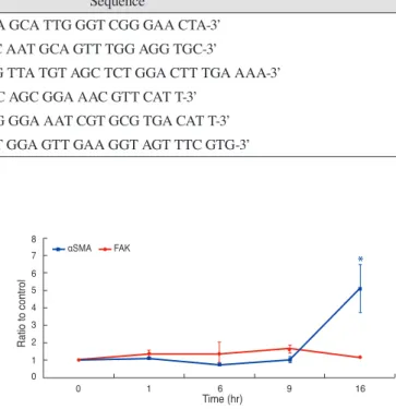
Fig. 1. Quantitative reverse transcription polymerase chain reaction for focal adhesion kinase (FAK) and α smooth muscle actin (αSMA) after exposure to 5 ng/mL of transforming growth factor-β1 for up to 16 hours. The level of target mRNA was calculated using a relative standard curve of β-actin and expressed as the mean ± SEM. *p < 0.05.
47 S Hong, et al. FAK in Myofibroblast Transdifferentiation
ered statistically significant.
Results
The quantitative data of RT-PCR for FAK and αSMA are presented in Fig. 1. In the human Tenon’s fibroblasts, the 16-hour treatment with TGF-β1 significantly increased the mRNA levels of αSMA (5.11 ± 1.37 times to control, p
= 0.023) but not of FAK (1.16 ± 0.06 times to control, p = 0.204).
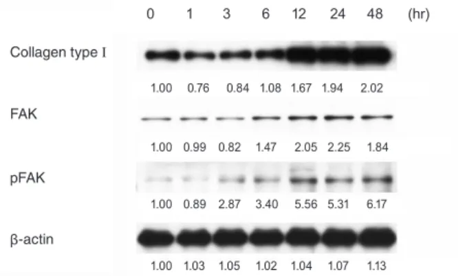
On the Western immunoblots, the TGF-β1 treatment increased the protein expression of FAK as well as that of collagen type I. Expression of pFAK, an active form of FAK, was also increased. These changes were observed after 12 hours. Representative immunobands for collagen type I, FAK, pFAK, and β-actin are shown in Fig. 2.
Regarding αSMA, though serum starvation itself slightly induced the expression of αSMA, the exposure to TGF-β1 for 48 hours stimulated αSMA expression in human Tenon’s fibroblasts. When the siRNAFAK molecules were introduced into the cells, the TGF-β1-induced αSMA expression was attenuated. Representative immunobands for αSMA in the
control group, TGF-β1 only treatment group, and siRNAFAK/ TGF-β1 treatment group are shown in Fig. 3.
Discussion
FAK, also known as protein tyrosine kinase 2, par- ticipates in the focal adhesion between the cytoskeleton and extracellular matrix and elicits intracellular signal transductions associated with cell migration and survival [11,12,16-18]. It is activated by autophosphorylation via Smad- and p38 MAPK-dependent mechanisms [19-22]
and plays a key role in cancer metastasis as well as normal development [11,12]. Even though FAK is also linked to the myofibroblast transdifferentiation of fibroblasts in re- sponse to TGF-β1 [13-15], the precise role of the kinase has not been well studied in the fibrotic process in the subcon- junctival space. Excessive subconjunctival fibrosis causes ocular morbidity in not only patients with ocularcutaneous disorders, but also patients who have undergone strabis- mus surgery and/or glaucoma-filtering surgery. A better understanding and modulation of FAK signaling in this process may result in a novel therapeutic strategy for those patients.
Using primary cultured human Tenon’s fibroblasts, we investigated the role of FAK in TGF-β-induced myofibro- blast transdifferentiation in the present study and found that silencing of FAK action using siRNAFAK duplex sig- nificantly attenuated the αSMA expression induced by TGF-β1 in human Tenon’s fibroblasts. Our data implies that FAK may participate in the myofibroblast transdifferentia- tion of those cells.
Myofibroblasts contain characteristics found in smooth muscle cells and fibroblasts in that they possess a con- tractile phenotype of αSMA and synthesize extracellular matrix proteins [1-3]. Since they play a crucial role in most fibrotic responses, myofibroblast transdifferentiation from activated fibroblasts is an essential step in the fibrotic pro- cess. In the subconjunctival space, TGF-β typically initi- ates this myofibroblast transdifferentiation of fibroblasts [4-6].
On quantitative RT-PCR, the mRNA expression of αSMA was dramatically increased by TGF-β1, but FAK was not significantly altered. However, on Western im- munoblots, the protein levels of both FAK and pFAK were minimally stimulated by TGF-β1 treatment. The increase in FAK seems to result from an increase in translation rather than transcription.
Currently, many researchers have tried to develop anti- TGF-β neutralizing antibodies to modulate postoperative scarring in patients undergoing ocular surface surgery [23- 25]. FAK, as a participant in TGF-β-associated intracel- lular signaling of myofibroblast transdifferentiation from fibroblasts, might also be valuable as a novel therapeutic Fig. 2. Representative Western immunoblots and densitomet-
ric data for collagen type I (190 kDa), focal adhesion kinase (FAK, 125 kDa), phospho-FAK (pFAK, 125 kDa), and β-actin (43 kDa) in human Tenon’s fibroblasts stimulated by 5 ng/mL of transforming growth factor-β1 for up to 48 hours.
Collagen type Ⅰ
FAK
pFAK
β-actin
0 1 3 6 12 24 48 (hr)
1.00 0.76 0.84 1.08 1.67 1.94 2.02
1.00 0.99 0.82 1.47 2.05 2.25 1.84
1.00 0.89 2.87 3.40 5.56 5.31 6.17
1.00 1.03 1.05 1.02 1.04 1.07 1.13
Fig. 3. Representative Western immunoblots and densitometric data for α smooth muscle actin (43 kDa) in human Tenon’s fi- broblasts stimulated by 5 ng/mL of transforming growth factor (TGF)-β1 for up to 48 hours with or without the treatment of siRNA targeting FAK (siRNAFAK) molecules.
No treatment
siRNAFAK + TGF-β1 (5 ng/mL) TGF-β1 (5 ng/mL)
0 1 3 6 12 24 48 (hr) 1.00 0.83 0.84 1.29 0.87 3.61 7.11 1.00 0.95 1.19 1.18 3.30 6.45 78.27 1.00 0.91 1.02 1.41 1.44 3.04 37.63
48
Korean J Ophthalmol Vol.26, No.1, 2012
strategy for anti-fibrosis.
Conflict of Interest
No potential conflict of interest relevant to this article was reported.
Acknowledgements
This work was supported by a Faculty Research Grant from the Yonsei University College of Medicine, Seoul, Republic of Korea (6-2008-0274).
References
1. Eyden B. The myofibroblast: an assessment of controversial issues and a definition useful in diagnosis and research. Ul- trastruct Pathol 2001;25:39-50.
2. Eyden B. Electron microscopy in the study of myofibro- blastic lesions. Semin Diagn Pathol 2003;20:13-24.
3. Desmouliere A, Guyot C, Gabbiani G. The stroma reaction myofibroblast: a key player in the control of tumor cell be- havior. Int J Dev Biol 2004;48:509-17.
4. Meyer-Ter-Vehn T, Grehn F, Schlunck G. Localization of TGF-beta type II receptor and ED-A fibronectin in normal conjunctiva and failed filtering blebs. Mol Vis 2008;14:136- 5. Saika S, Yamanaka O, Okada Y, et al. TGF beta in fibropro-41.
liferative diseases in the eye. Front Biosci (Schol Ed) 2009;
1:376-90.
6. Seong GJ, Hong S, Jung SA, et al. TGF-beta-induced in- terleukin-6 participates in transdifferentiation of human Tenon’s fibroblasts to myofibroblasts. Mol Vis 2009;15:2123- 7. Nakamura H, Siddiqui SS, Shen X, et al. RNA interference 8.
targeting transforming growth factor-beta type II recep- tor suppresses ocular inflammation and fibrosis. Mol Vis 2004;10:703-11.
8. Meyer-Ter-Vehn T, Gebhardt S, Sebald W, et al. p38 inhibi- tors prevent TGF-beta-induced myofibroblast transdiffer- entiation in human tenon fibroblasts. Invest Ophthalmol Vis Sci 2006;47:1500-9.
9. Yamanaka O, Saika S, Ikeda K, et al. Interleukin-7 modu- lates extracellular matrix production and TGF-beta signal- ing in cultured human subconjunctival fibroblasts. Curr Eye Res 2006;31:491-9.
10. Meyer-Ter-Vehn T, Katzenberger B, Han H, et al. Lovas- tatin inhibits TGF-beta-induced myofibroblast transdiffer- entiation in human tenon fibroblasts. Invest Ophthalmol Vis Sci 2008;49:3955-60.
11. Chatzizacharias NA, Kouraklis GP, Theocharis SE. The role of focal adhesion kinase in early development. Histol Histopathol 2010;25:1039-55.
12. Schwock J, Dhani N, Hedley DW. Targeting focal adhesion kinase signaling in tumor growth and metastasis. Expert Opin Ther Targets 2010;14:77-94.
13. Thannickal VJ, Lee DY, White ES, et al. Myofibroblast differentiation by transforming growth factor-beta1 is de- pendent on cell adhesion and integrin signaling via focal adhesion kinase. J Biol Chem 2003;278:12384-9.
14. Mimura Y, Ihn H, Jinnin M, et al. Constitutive phosphory- lation of focal adhesion kinase is involved in the myofi- broblast differentiation of scleroderma fibroblasts. J Invest Dermatol 2005;124:886-92.
15. Brenmoehl J, Miller SN, Hofmann C, et al. Transforming growth factor-beta 1 induces intestinal myofibroblast dif- ferentiation and modulates their migration. World J Gas- troenterol 2009;15:1431-42.
16. McLean GW, Carragher NO, Avizienyte E, et al. The role of focal-adhesion kinase in cancer: a new therapeutic op- portunity. Nat Rev Cancer 2005;5:505-15.
17. Cox BD, Natarajan M, Stettner MR, Gladson CL. New concepts regarding focal adhesion kinase promotion of cell migration and proliferation. J Cell Biochem 2006;99:35-52.
18. Liu S, Xu SW, Kennedy L, et al. FAK is required for TGF- beta-induced JNK phosphorylation in fibroblasts: implica- tions for acquisition of a matrix-remodeling phenotype.
Mol Biol Cell 2007;18:2169-78.
19. Horowitz JC, Rogers DS, Sharma V, et al. Combinato- rial activation of FAK and AKT by transforming growth factor-beta1 confers an anoikis-resistant phenotype to myo- fibroblasts. Cell Signal 2007;19:761-71.
20. Parsons CJ, Takashima M, Rippe RA. Molecular mecha- nisms of hepatic fibrogenesis. J Gastroenterol Hepatol 2007;22 Suppl 1:S79-84.
21. Walsh MF, Ampasala DR, Hatfield J, et al. Transforming growth factor-beta stimulates intestinal epithelial focal adhesion kinase synthesis via Smad- and p38-dependent mechanisms. Am J Pathol 2008;173:385-99.
22. Walsh MF, Ampasala DR, Rishi AK, Basson MD. TGF- beta1 modulates focal adhesion kinase expression in rat intestinal epithelial IEC-6 cells via stimulatory and in- hibitory Smad binding elements. Biochim Biophys Acta 2009;1789:88-98.
23. Cordeiro MF. Technology evaluation: lerdelimumab, Cam- bridge Antibody Technology. Curr Opin Mol Ther 2003;
5:199-203.
24. Mead AL, Wong TT, Cordeiro MF, et al. Evaluation of anti- TGF-beta2 antibody as a new postoperative anti-scarring agent in glaucoma surgery. Invest Ophthalmol Vis Sci 2003;44:3394-401.
25. CAT-152 0102 Trabeculectomy Study Group, Khaw P, Grehn F, et al. A phase III study of subconjunctival human anti-transforming growth factor beta(2) monoclonal anti- body (CAT-152) to prevent scarring after first-time trabecu- lectomy. Ophthalmology 2007;114:1822-30.