SURFACE CHARACTERISTICS AND BIOLOGICAL RESPONSES OF HYDROXYAPATITE
COATING ON TITANIUM BY HYDROTHERMAL METHOD: AN IN VITRO STUDY
Dong-Seok Kim, D.D.S., M.S.D., Chang-Whe Kim, D.D.S., M.S.D., Ph.D., Kyung-Soo Jang, D.D.S., M.S.D., Ph.D., Young-Jun Lim, D.D.S., M.S.D., Ph.D.
Department of Prothodontics, Graduate School, Seoul National University
Statement of problem.Hydroxyapatite(HA) coated titanium surfaces have not yet showed the reliable osseointegration in various conditions.
Purpose.This study was aimed to investigate microstructures, chemical composition, and surface roughness of the surface coated by the hydrothermal method and to evaluate the effect of hydrothermal coating on the cell attachment, as well as cell proliferation.
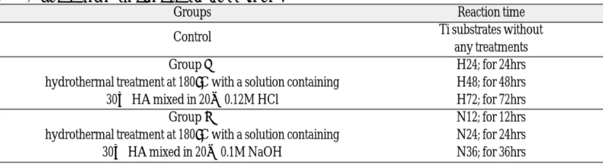
Material and Methods.Commercially pure(c.p.) titanium discs were used as substrates. The HA coating on c.p. titanium discs by hydrothermal method was performed in 0.12M HCl solu- tion mixed with HA(group I) and 0.1M NaOH solution mixed with HA(group II). GroupⅠ was heated at 180℃ for 24, 48, and 72 hours. GroupⅡ was heated at 180℃ for 12, 24, and 36 hours.
And the treated surfaces were evaluated by Scanning electron microscopy(SEM), Energy dis- persive X-ray spectroscopy(EDS), X-ray photoelectron spectroscopy(XPS), X-ray diffraction method(XRD), Confocal laser scanning microscopy(CLSM). And SEM of fibroblast and 3-(4,5- dimethylthiazol-2-yl)-2,5-diphenyl tetrazolium bromide(MTT) assay were used for cellular respons- es of the treated surfaces.
Results. The color of surface changed in both groups after the hydrothermal process. SEM images showed that coating pattern was homogeneous in group II, while inhomogeneous in group I. H72 had rosette-like precipitates. The crystalline structure grew gradually in group II, according to extending treatment period. The long needle-like crystals were prominent in N36. Calcium(Ca) and phosphorus(P) were not detected in H24 and H48 in EDS. In all spec- imens of group II and H72, Ca was found. Ca and P were identified in all treated groups through the analysis of XPS, but they were amorphous. Surface roughness did not increase in both groups after hydrothermal treatment. The values of surface roughness were not significantly differ- ent between groups I and II. According to the SEM images of fibroblasts, cell attachments were oriented and spread well in both treated groups, while they were not in the control group.
However, no substantial amount of difference was found between groups I and II.
Conclusions. In this study during the hydrothermal process procedure, coating character- istics, including the HA precipitates, crystal growth, and crystalline phases, were more satisfactory in NaOH treated group than in HCl treated group. Still, the biological responses of the modified surface by this method were not fully understood for the two tested groups did not differ significantly. Therefore, more continuous research on the relationship between the sur- face features and cellular responses seems to be in need.
Key Words
Hydrothermal method, Hydroxyapatite, Surface characteristics, Fibroblast, Cellular response J Korean Acad Prosthodont : Volume 43, Number 3, 2005
T
itanium and its alloys have been successfully used as dental implants because of their good biocom- patibility, corrosion resistance to body fluids, high strength-to-weight ratio, and low elastic modulus.1 In addition to good properties as biomaterials, reports of direct bonding to bone had been introduced in many studies. But titanium and its alloys are generally considered to be bio-inert and not likely to form direct chemical bonding to bone. Under poor bone condition and in the sites where rapid fix- ation and healing were needed, this characteris- tics were often important factor for a successful osseointegration.Therefore, to obtain faster and firmer bonding to bone tissue, much attention has been concentrated on improving the osseointegrative potentials of ti- tanium and its alloys.1It is generally accepted that it is the outermost surface of an implant that is im- portant for the biological response expressed by the recipient tissue.
According to this concept, surface modification have been suggested as one important factor for estab- lishing clinically reliable bone attachments. And several methods have been suggested to modify the surface structure of titanium implants, which all may lead to altered chemical and mechanical properties of the titanium metal surface.
Among the means of preparing bioactive titanium surfaces, surface coating methods have been wide- ly studied and tried. Surfaces can be modified by coat- ings. Results from in vitro studies suggest a positive correlation between surface modification and cellular reponses. Changes in surface chemistry and mi- crostructure correlate with implant-bone interaction.
Among the various attempts, which have been made to change titanium surface, the well-known process is calcium phosphate (e.g., hydroxyap- atite(HA)) coating on titanium.2-3
Calcium phosphate, specifically HA(Ca10
(PO4)6(OH)2), is biocompatible and bioactive in the
human body.4Phase-pure and pore-free HA has high strength and good fatigue resistance. It also displays an osseoconductivity: HA bond to and integrate with living bone spontaneously by forming a bio- logically active bone-like apatite layer on their sur- faces in the body.5Therefore, this is especially use- ful for the fabrication of dental implants where rapid healing is required.1
Despite its excellent properties as a biomaterial, the inherent mechanical properties of HA-specifically, brittleness, poor tensile strength, and poor impact resistance-restricted its application in many load-bear- ing situations.1In order to overcome the weak points of HA, the concept of applying HA onto metallic implants has been developed.6One way to take advantage of the excellent biocompatibility and bone bonding properties of HA is to coat it on- to pure titanium substrate to make a composite material with good impact resistance. Such designs are intended to take advantage of the bioactivity of HA in combination with the strength and biocom- patibility of the titanium substrate. HA coatings on titanium become of great interest because the re- sultant product was a composite mineral having good mechanical properties supplied by the metal to- gether with the biocompatibility given by the calcium phosphates.7-9 One important concern is the re- sorption and degradability of HA coatings in a bi- ological environment, which could lead to delam- ination of the coating, resulting in the loss of os- seointegration with the formation of particulate debris, including both the coating-substrate bond strength and the implant fixation.1
The most important concern is the properties of the HA coating, which has been known to influence on the major factors for both implant initial fixation and its long-term stability, such as the coating resorption, bone ingrowth, and mechanical fixation. The factors that affect the performance of the HA coating include its compositional, physical, and mechanical issues.1 To improve adhesive strength between titanium
and HA, techniques are desired to produce HA coatings with few cracks while maintaining char- acteristics like high crystallinity to provide better cel- lular responses.
Currently and commercially, plasma-spraying is the most frequently used method for HA coating on the implant materials, for its advantage with relatively tight bond to metal.2-3 However, the high temperatures and rapid cooling associated with the process yields a variety of chemical phas- es and lower HA crystallinity. HA dissolution rate increases with decreasing crystallinity, as other calcium phosphorous compounds, such as β-tri- calcium phosphate and tetracalcium phosphate, dissolve faster than HA. Also, plasma-spraying has disadvantages in terms of the difficult control of the composition and structure of the HA formed on the metal due to the high processing temperature and to the low adherence of the coating layer to the metallic substrate because of its physical nature.12
In this study, we attempted to develop a hy- drothermal process for forming more effective and reliable HA coating surface layer on commercially pure(c.p.) titanium. This study aimed to determine the surface characteristics of titanium coated by the hydrothermal coating methods and to examine the effect of the coating surfaces on the cellular re- sponses, including the in vitro attachment of fi- broblast cells on treated surfaces.
MATERIAL AND METHODS The preparation of titanium substrate
Titanium substrates was Ti-grade 4(Carpent Co., Washington, Virginia, USA). C.p. titanium discs of 2㎜ thickness and 10㎜ diameter were used as sub- strates without polishing procedure. These titani- um substrates were ultrasonically rinsed in acetone for 20 minutes in 70% ethanol solution for 20 minutes and then immerged in distilled water for 15 minutes.
HA coating by hydrothermal treatment
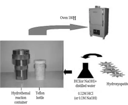
In this study, 0.12M HCl and 0.1M NaOH solution were prepared by mixing with distilled water. Into 20㎖ of this HCl and NaOH solution, 30㎎ of HA was stirred until it completely mixed. Then, two Teflon bottles were prepared by placing four titanium substrates in each container, making sure that they did not overlap. In each bottle, the prepared solutions were filled.
Each Teflon bottle was placed in the hydrothermal containers, and then these hydrothermal containers were placed in oven and heated up to 180�C. The groups of HCl mixed with HA (group Ⅰ) were heated at 180�C for 24h, 48h, and 72h. The groups of NaOH mixed with HA (group Ⅱ) were heated at 180�C for 12h, 24h, and 36h. After reaction was completed, the hydrothermal container was taken out to be cooled slowly at room temperature. After the container completely cooled down, the Teflon bot- tle that was inside hydrothermal containers were tak- en out. From this bottle, the titanium substrates were drawn and ultrasonic rinsing was performed 3 times in distilled water for 5 minutes and dried in incubator for 5 hours, until this substrates were dried completely. In final step, the prepared titanium substrates were sterilized with ethylene oxide(EO) gas. These specimens treated with different hy- drothermal conditions were summarized in table I.
The Analytical methods
Scanning electron microscopy(SEM)/ Energy dis- persive X-ray spectroscopy(EDS)
SEM(S4700, Hitachi, Japan) was used to observe the topography of the coated surface after hy- drothermal processing. The specimens were ob- served at a magnification of ×3,000 and ×50,000 us- ing 15㎸-voltage. EDS was used to identify the el- ements, such as inorganics including titanium, cal- cium(Ca), and phosphorus(P), within the precipitates
of coated surfaces. Elemental scans using EDS(IN- KA, Oxford, U.K) was performed at ×50,000 using 12㎸-voltage.
Electron Spectroscope for Chemical Analysis(ESCA) To document surface changes occurring after hydrothermal treatment, ESCA was performed us- ing SIGMA PROBE(ThermoVG, U.K) with mono- chromatic x-ray source. X-ray source was generat- ed by the Al-Kαradiation;10㎸, with the power of 50W. The diameter of the analyzed spot was 500㎛.
The analyser was set to pass energy Ep= 50eV with the step size of 1.0eV in wide scan and Ep= 20eV with the step size of 0.1eV in narrow scan. Survey X-ray photoelectron spectroscopy(XPS) spectra from 0eV to 1,000eV were acquired after hydrothermal treat- ment. The binding energy(BE) scale was calibrated by measuring the reference peak of C 1s(BE=
284.5eV) from the surface contamination.23The rel- ative atomic concentrations were determined on C 1s, O 1s, Ti 2s, Ti 2p, Ti 3s, Ti 3p, Ca 2p, P 2s, P 2p using the software supplied by the manufacturer.
X-ray diffraction method(XRD)
To obtain the crystalline phases of the coating sur- faces, XRD(High Resolution X-ray Diffractomer;
Bruker D8 DISCOVER, Germany) with Cu-Kα(λ
=1.5418A°) radiation at 40㎸and 30㎃, was used.
The scanning was set at a scan speed of 0.005�/s, a step size of 0.02, over a 2θrange of 20-80�.
Confocal laser scanning microscopy (CLSM) The samples were examined with CLSM(PASCAL LSM 5; ZEISS, Germany), equipped with an ion argon laser source and two photomultiplier tubes for examining the surface roughness(Ra).
Depth projection micrographs were visualized by 20 up to 40 horizontal image sections throughout the samples(serial optical section set), using a z-step of 256 ㎛ and 900㎛ ×900㎛ objective.
Cell culture
NIH 3T3 fibroblasts were used for evaluating bone responses to modified Ti surfaces. This cells were cultured in DMEM(Dulbecco’s modified Eagle’s medium) supplemented with 10% FBS(fetal bovine serum) and 1% AA(antibidtic-antimycotic). For routine culture, the cells were maintained at 37�C in a 5% CO2atmosphere with 10㎖ fresh medium in 100
㎜×20㎜ dish added every 2 days.
Cultures were subdivided by trypsinisation using Trypsin-EDTA solution(0.05% trypsin:0.1% ED- TA). The modified titanium discs were placed in stan- dard 32-well tissue culture plates, and then cells were seeded. One milliliter of cell suspension containing 1×10⁵ cells/㎖ added to each well. As a control substrate for cell attachment and growth, fibroblasts were plated directly onto tissue culture of poly- styrene plastic. The culture medium was changed every 3 days and cells were incubated for 4 days at 37�C in humidified air and 5% CO2.
Table I. Summary of control and treated groups
Groups Reaction time
Control Ti substrates without
any treatments
Group Ⅰ H24; for 24hrs
hydrothermal treatment at 180�C with a solution containing H48; for 48hrs
30㎎ HA mixed in 20㎖ 0.12M HCl H72; for 72hrs
Group Ⅱ N12; for 12hrs
hydrothermal treatment at 180�C with a solution containing N24; for 24hrs
30㎎ HA mixed in 20㎖ 0.1M NaOH N36; for 36hrs
Spreading and morphology of fibroblast cell at- tachment
Cellular behavior of the fibroblasts(spreading and morphology) was examined using SEM at the magnification of ×200 and ×1,000.
Growth and proliferation assay of fibroblasts (MTTT assay)
3-(4,5-dimethylthiazol-2-yl)-2,5-diphenyl tetra- zolium bromide(MTT) assay was used to evaluate cell proliferation amount and cellular responses.
A quantitative colorimetric MTT test was per- formed after 4 days of culture to characterize cellular metabolism(vitality) and, by implication, prolifer- ation. One-hundred microliters of MTT solution(M 2128, Sigma) was added to each well, and the cells were incubated at 37�C for 4 hours. The medium was then removed, and 300㎕ of dimethylsulfoxide was added to each well, followed by a 10 minutes in- cubation at room temperature on a shaker. And then 200㎕ of melted medium was place in new 96
wells. The optical density(O.D.) was measured at 540
㎚ with a micro reader(enzyme-linked im- munosorbent assay; ELISA). The samples for SEM were fixed with 1㎖ of 4% paraformaldehyde, and then it was maintained at 4�C.
RESULTS
The color change of coated surface after hy- drothermal process
After hydrothermal treatment, color change of the titanium substrates was observed. The alteration of the color was more apparent in the acidic treat- ment groups- the H24 showing a gold color, the H48 a violet, and H72 a bluish color. Although the N12 and N24 of the alkali-treated groups showed a bronze-like color, and N36 a bluish-green color, the change was not as distinct as in the acidic treat- ment groups.
Fig. 1.The experimental apparatus for the hydrothermal method.
Oven 180℃
Hydroxyapatite HCl(or NaOH)+
distilled water Hydrothemal
reaction container
Teflon bottle
0.12M HCl (or 0.1M NaOH)
SEM/EDS
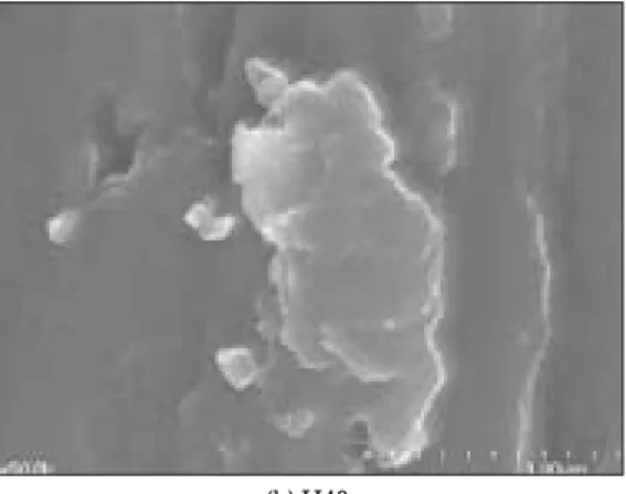
Figure 2 showed a significant variation of the texture of titanium surface after hydrothermal treat- ment, under six different conditions.
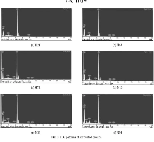
Under the magnification of ×3,000, the images of H24 did not present a coated appearance, but high- er magnification of ×50,000 revealed some cluster- like precipitates. During the EDS evaluation of this precipitates at ×50,000, Ca and P, which could be thought of as HA, were not detected.
In the ×3,000 magnification, more surface pre- cipitates were attached to the H48 than to the H24, but overall it was rather faint. When magnified to × 50,000, a cluster-like crystals which seemed to be a little more grown were observed, but as it was with the H24, Ca and P were not detected in EDS evaluation. As the last group to be carried out in the acidic condition, H72 showed a distinct, thoroughly spread appearance under both ×3,000 and ×50,000 magnifications, and under ×50,000 image, the clus- ter-like precipitates of the previous groups showed a more mature, rosette-like appearance. Also, through the EDS evaluation of this particles, Ca and P, which were not identified in the two previ- ous groups, were detected although in small quan- tity. Therefore, after 72 hours of heating time it finally showed a certain degree of coated appearance.
However, it was not coated at all, or very faintly coat- ed in general. The coated appearance observed in N12 showed a somewhat homogeneous pattern under × 3,000 magnification, and under ×50,000 magnifi- cation, coating with a definite pattern could be ob- served. This became more distinct going from N24 to N36. Also, in the EDS evaluation on N12 and N24, small amounts of Ca and P were detected in both groups. Especially in the N36, long needle-like crystals were observed, and in the EDS evaluation of these crystals, Ca and P were also detected which allowed to think of this particles as calcium phosphate.
Therefore, in the NaOH-treatment groups, Ca was
detected in all of N12, N24, and N36, and it could be thought that HA was coated on the surface.
It was also observed that an increase in the duration of hydrothermal treatments from 12 to 36 hours resulted in an increase in the formation of needle-like crystals, that is, growth of HA crystals. EDS spectrum were illustrated in figure 3.
Crystalline Phase of HA coating particles
In XRD patterns, rutile and anatase of TiO2and ti- tanium oxide form like TiO and Ti2O3were detect- ed in groups Ⅰ and Ⅱ, but crystalline structures of Ca and P were not identified(Figure 4). Therefore on the coating surface, the crystalline phase of Ca and P seemed to be amorphous.
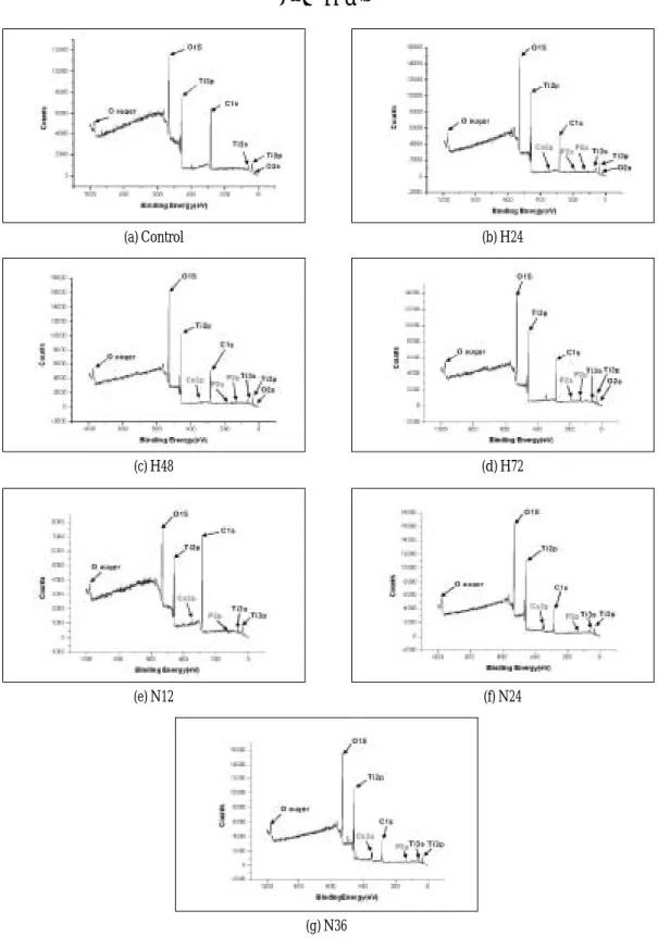
Chemical composition of coatings
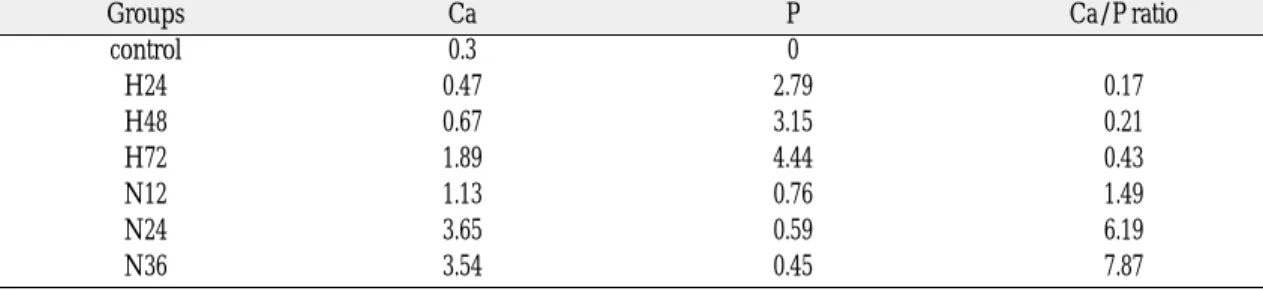
The surface composition of the control and six treat- ed groups as determined by XPS analysis was giv- en in table II. In the XPS evaluation of H24 and H48, Ca and P, which were not identified in the EDS analysis, were detected. The coating surfaces of H72 also contained the two elements. In the case of group Ⅰ, the atomic weight of Ca and P increased according to elevated heating time.
In group Ⅱ, N24 and N36 contained more amount of Ca than N12, while N12 had the highest value of P among the three treated groups. Contrary to group Ⅰ, Ca was more prominent than P in all spec- imens of group Ⅱ. XPS spectra of groups Ⅰ and Ⅱ were shown in figure 5.
Surface roughness
Confocal laser scanning microscopy results demon- strated the difference of surface roughness. In gen- eral, Ra value exhibited a lower value in both groups Ⅰ and Ⅱ than in the control group. In case of group Ⅰ, the highest value was shown in H48 af-
ter which it rather decreased in H72. In group Ⅱ, as the heating time increased, Ra value contrarily de- creased.
However in this study, we did not find significant change of surface roughness after hydrothermal treatment in both groups, as summarized in table III.
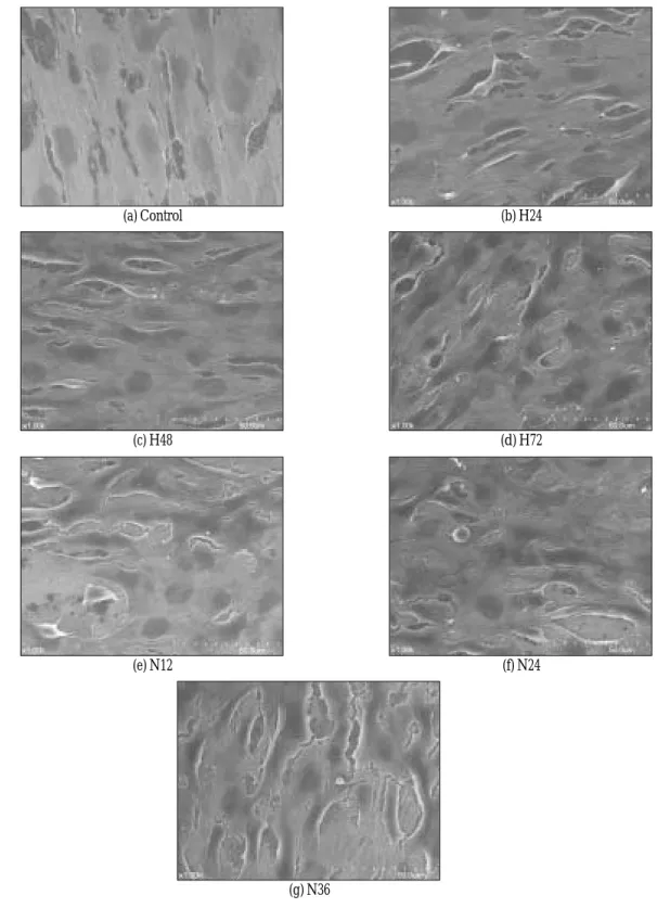
SEM of fibroblast
SEM images of fibroblast attachment showed that the cells grown on all modified surfaces displayed good adhesion and were spread well. Cellular growth also seemed to be denser on hydrother- mally treated titanium than that on the control group.
The spreading patterns of cells in groups Ⅰ and
Ⅱ showed no significant difference. But in the case of group Ⅰ, the spreading of cells on each subdivided groups were similar, while the spreading of cells on
N12 and N36 of group Ⅱ were similar and N24 group was significantly small.
Cellular morphology were varied. Fibroblasts cultured on pure titanium discs showed a wide variety of shapes. Spindle shaped, elongated cells as well as round fibroblasts were observed. In SEM images of fibroblast attachment in groups Ⅰ and Ⅱ, many cells had spindle-like and spherical shape with fine, long filopodia, and appeared intimately adherent to the surface.
Although no clear orientation of cells could be no- ticed on the surfaces of control group, the treated groups had the orientation of cells to some degree.
It was shown in figure 6.
Proliferation of fibroblast
MTT assay showed that cell proliferation in groups Ⅰ and Ⅱ was more prominent than that in
Table III. Surface roughness by Confocal laser scanning microscopy (㎛)
surface Control H24 H48 H72 N12 N24 N36
roughness
Ra* 3.769 2.802 3.417 3.150 3.777 3.231 3.051
Sa+ 2.699 2.212 3.507 2.683 3.119 2.349 2.346
*Ra: mean height deviation from peak to valley
+Sa: average height deviation value
Table II. Atomic percentage of the elements detected on the analyzed coating surfaces
Groups Ca P Ca/P ratio
control 0.3 0
H24 0.47 2.79 0.17
H48 0.67 3.15 0.21
H72 1.89 4.44 0.43
N12 1.13 0.76 1.49
N24 3.65 0.59 6.19
N36 3.54 0.45 7.87
surface concentration(atomic %)
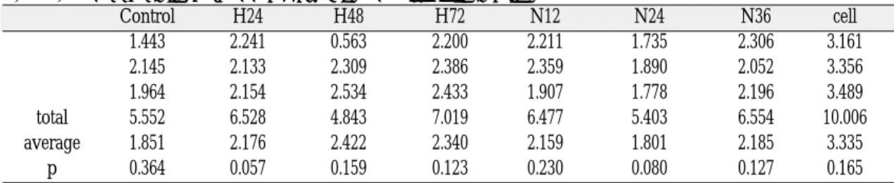
the control. The H48 group and N24 group showed significance in the result, and greatest amount of cell proliferation was observed in H48 of group Ⅰ while in H72, it seemed rather decreased. However, it was difficult to assume that the difference deserved much attention. Contrary to the prediction that there would be an increase in the cell proliferation of group Ⅱ following an increase in the heating time, rapidly decreasing rate of cell proliferation was observed in N24, and in N36 an increase again was noticed. It is thought that the errors throughout the experimental procedures should be given some consideration. The results are shown in table IV.
DISCUSSION
In order to examine the surface condition of the discs of control group, the surface features of pure titanium samples were examined by SEM at 15㎸ ac- celeration voltage. In SEM of pure titanium substrates, the microstructure of titanium substrates had not sig- nificant difference, compared with the surface of ma- chined titanium surface. The Ra of the sample sur- faces were evaluated by means of surface roughness measuring device(SV-3000, Mitutotyo, Japan). The measurements were carried out on upper and low- er surface regions of the titanium discs. The Ra of the titanium substrate surface was 0.209, which is sim- ilar to that of commercially machined titanium im- plant, which was 0.221. Titanium substrates using in the present study were regarded as machined ti-
tanium implant. Therefore, in this study, any other pre-treatments, including polishing process, were not performed.
HA coatings were first introduced in the mid-1980s for improved fixation between bone and the implant.
Various methods have been introduced to deposit HA coatings, such as dip coating-sintering, im- mersion coating, electrophoretic deposition, hot isostatic pressing(HIP), solution deposition, ion- beam sputter coating and dynamic mixing, thermal spraying techniques such as plasma spraying, flame spraying, and high-velocity oxy-fuel (HVOF) com- bustion spraying.8But, previously mentioned meth- ods have not yielded a satisfactory HA coating on titanium. Coatings can contain cracks, pores, second phases, and residual stresses that reduce their dura- bility and lead to partial or complete disintegration of the coating in body fluids.
In this study, hydrothermal modification method of HA coating was applied. As seen in the results, there existed a difference between the surface changes of HCl solution and NaOH solution.
In the study of Kuroda et al, the pH of the solution apparently influenced the kind of precipitate. In the pH 4 solution, only dicalcium phosphate an- hydrous(DCPA, CaHPO4) was formed on the sub- strate, with an appearance like piled blocks. In contrast, in the solution with pH>6, HA, which in- cluded a small amount of CO32-, was formed pre- dominantly, and the crystals were needle-like and/or thin plate-like in shape.1
Table IV. The results of MTT assay(NIH 3T3 fibroblast cell)
Control H24 H48 H72 N12 N24 N36 cell
1.443 2.241 0.563 2.200 2.211 1.735 2.306 3.161
2.145 2.133 2.309 2.386 2.359 1.890 2.052 3.356
1.964 2.154 2.534 2.433 1.907 1.778 2.196 3.489
total 5.552 6.528 4.843 7.019 6.477 5.403 6.554 10.006
average 1.851 2.176 2.422 2.340 2.159 1.801 2.185 3.335
p 0.364 0.057 0.159 0.123 0.230 0.080 0.127 0.165
Hydrothermal treatment in alkaline solution (NaOH) with HA resulted in the formation of long needle-like crystals, while cluster-like precipitates were formed in acidic groups. It was speculated from SEM and EDS that these long needle-like crystals con- tain apatite-like structures. It has also been sug- gested that apatite, once nucleated, will grow spon- taneously in the body environment.11In the present study, the coating in the NaOH solution was more homogeneous than in the HCl solution, and as a nee- dle-like crystallization was observed in the NaOH treated groups, hydrothermal treatment under alkali- treated condition was superior to one under acidic- treated condition.
There is a difference between the mechanisms of hydrothermal modification in acidic and basic so- lutions, and this can be thought of as the reason the two solutions possess varying surface properties.
When the anticipated mechanism is pondered up- on, they differ in the fact that sodium titanate gel is formed on the titanium surface in the mechanism of in vitro apatite deposition treating titanium with NaOH aqueous solution, and titania gel is formed on the surface in the mechanism of in vitro apatite de- position treating titanium with HCl aqueous solu- tion.13
Kim et al indicated that an NaOH treatment of pure titanium forms a sodium titanate hydrogel surface layer with a smooth graded interface structure to the Ti metal substrate.19
In the study of Kim et al, the sodium titanate transforms into a hydrated titania via Na+ion release to induce a bone-like apatite formation on the alloy substrate in body environment. In this process, the graded surface structure develops into one where the apatite on the top surface gradually changes into al- loy substrate through hydrated titania and titanium oxide.5Wang et al, suggested that the bioactivity of titania gels depends on the gel’s structure. includ- ing crystallographic and porous structure as well as a negative surface charge density.13In the study
of Wei et al, as far as titania gel takes an amor- phous structure, the incorporation of the Ca2+ ions in- to the titanium oxide phase does not increase the ap- atite-forming ability of the titanium oxide phase.
Important factor for the apatite-forming ability of ti- tanium oxide phase is the structure of the Ti-OH groups rather than degree of the ionic activity product of the apatite of the surrounding fluid.
The specific structure effective for the apatite nu- cleation seems to be the anatase.14
Similar mechanism can be anticipated in the hy- drothermal modification method used in this study, and for these reasons, better apatite-forming abili- ty can expected to be obtained in the basic solutions than in acidic ones. The rutile and anatase crys- talline phase of TiO2and titanium oxide of Ti2O3were detected as the surface crystalline phase of this study. However, Ca and P did not form crystalline structure and were observed in an amorphous stage. This seems to be related to the concentration of aqueous solution matter used in this study.
Another important factor in hydrothermal treat- ment is heat. In the study of Nishiguchi et al, the al- kali-treated titanium without heat treatment had no bone-bonding ability due to the unstable reactive sur- face layer of alkali-treated titanium. Both alkali and heat treatment are essential for preparing bioac- tive titanium and this bioactive titanium is thought to be useful for orthopedic implants with cementless fixation.10
Heating period and temperature are also anoth- er matter to take into consideration. Wang et al had studied the effects of various heating time and tem- perature in hydrothermal method. It was found that commercially pure titanium was treated with a H2O2/TaCl53mM solution at 80�C for various periods and a titania gel layer was formed on the surface. This gel remained amorphous when heating for 1 h below 200�C and trans- formed to anatase after heating between 300 and 600�C. The anatase titania gel layers were found
to be bioactive as to deposit carbonate ion-incor- porated apatite within 1 day of immersion in the Kokubo solution, whereas the amorphous layers did not deposit apatite within 7 days. The apatite particles were found to nucleate prefer- entially inside the cracks prevailing in the thick- er gel layers of 1-h chemically treated specimens.
After immersing for 2 days, the titanium specimens were almost completely covered by apatite.20As observed from the scanning electron microscopy, an increase in the duration of hydrothermal treatments resulted in an increase in apatite-like crystals. It has also been reported that differ- ences in crystal size have been associated with vary- ing degree of dissolution rates, with smaller, more imperfect crystals being subject to greater dissolution.
Normally HA is known to dissolve well into HCl, and not in NaOH, so the HA group dissolved completely in acid was expected to show a better coat- ed appearance. However, the result presented the NaOH groups as having a better coated appearance.
Contrary to our expectation, hydrothermal treatment in basic solutions is thought to be more advantageous to acidic solutions in acquiring a more suitable coating surface, contemplating on these various situations.
The successful incorporation of dental implants strongly depends on a firm longstanding adhesion of the tissues surrounding the implant. In the study of Groessner-Schreiber et al, deeper periodontal structures need to be protected from bacterial invasion and subsequent infection. The cellular reaction is in- fluenced by the properties of the bulk material as well as the properties of the surface, that is, the chemical composition and the topography.15In the study of Rodriguez et al, highly crystalline surfaces have been reported to promote cell growth, and the presence of calcium ions have been considered to be advantageous for cell growth.21An HA coating of higher crystallinity is more desirable in providing
durability and maintaining osseoconductive prop- erties.22
As it is mentioned above, various articles should show different cellular responses according to sur- face characteristics, but in this study, SEM and MTT assay revealed that fibroblasts on all modified surfaces tested adhered and spread well but did not show variation in cellular morphology. The ma- jority of the fibroblasts cultured on NaOH-treated titanium surfaces had a flat morphology and appeared intimately adherent to the surface, indicating good attachment, but this was not significant. Cellular growth seemed to be denser on NaOH treated groups than on HCl treated groups, but this was al- so not evident.
Lauer G et al addressed that cell morphology, ori- entation, proliferation and adhesion of human gin- gival epithelial cells in primary or secondary culture are dependent on the texture of the titanium surface whereas no such differences were observed for maxillary osteoblast-like cells. In conclusion, the soft tissue integration and response is more influenced by the surface texture than the process of osseoin- tegration.16In the study of Rosa AL et al, c.p. titanium would optimize osteoblastic differentiation by rat bone marrow cells, including reduced cell proliferation, and increased ALP activity and bone-like nodule for- mation, while surface roughness, within the Ra parameters used, would not affect significantly the rat bone marrow cell response.17
Although in this study the group treated with ba- sic solution(NaOH) was thought to be more ad- vantageous in biological response in the reasons that it showed a more distinct crystallization and more homogeneous coating pattern and more favorable Ca/P ratio, the NaOH group and HCl group did not show notable differences as it was observed in the results of fibroblast cell attachment and MTT assay.
This reveals that even though the groups Ⅰ and
Ⅱ showed different coating aspects, there was no sub- stantial difference in their cellular activity related to
the fact that the HA crystallinity was amorphous in both groups. But, more investigations seemed to be needed for the effect of surface characteristics of coat- ed titanium on cellular responses.
CONCLUSIONS
Through the study, the investigation of the mi- crostructure, chemical composition, and surface roughness of the surface coated by the hydrothermal method was carried out, and the evaluation of the effect this coated surface had on the cell attach- ment, as well as on cell proliferation was per- formed.
Several analytical methods were performed to identify the changed surfaces, and the following con- clusions could be made.
1. The color of surface changed in both groups af- ter the hydrothermal process.
2. SEM images showed that coating pattern was ho- mogeneous in group Ⅱ, while inhomogeneous in group Ⅰ. H72 had rosette-like precipitates. The crystalline structure grew gradually in group
Ⅱ, according to extending treatment period.
The long needle-like crystals were prominent in N36.
3. Ca and P were not detected in H24 and H48 in EDS. In all specimens of group Ⅱ and H72, Ca was found. Ca and P were identified in all treat- ed groups through the analysis of XPS, but they were amorphous.
4. Surface roughness did not increase in both groups after hydrothermal treatment. The values of surface roughness were not significantly dif- ferent between groups Ⅰ and Ⅱ.
5. According to the SEM images of fibroblasts, cell attachments were oriented and spread well in both treated groups, while they were not in the control group. However, no substantial amount of dif- ference was found between groups Ⅰ and Ⅱ.
As the results of MTT Assay revealed, cell pro-
liferation was more prominent in the two treated groups than in the control group. However, the difference between HCl and NaOH group was not significant. Therefore, more investigations on the effect of surface characteristics on cellular activity seemed to be needed.
In this study during the hydrothermal process pro- cedure, coating characteristics, including the HA precipitates, crystal growth, and crystalline phas- es, were more satisfactory in NaOH treated group than in HCl treated group. Still, the biological re- sponses of the modified surface by this method were not fully understood for the two tested groups did not differ significantly. Therefore, more con- tinuous research on the relationship between the sur- face features and cellular responses seems to be in need.
※I acknowledge Seoul national university's financial support for the new faculty.
REFERENCES
1. Kuroda K, Ichino R, Okido M, Takai O. Effects of ion concentration and pH on HA deposition from aqueous solution onto titanium by the thermal substrate method. J Biomed Mater Res 2002;61:354- 9.
2. Suh JY, Jang BC, Zhu X, Ong JL, and Kim K.
Effect of hydrothermally treated anodic oxide films on osteoblast attachment and proliferation.
Biomaterials 2003;24:347-55.
3. Hamada K, Kon M, Hanawa T, Yokoyama K, Miyamoto Y, and Kenzo A. Hydrothermal modi- fication of titanium surface in calcium solutions.
Biomaterials 2002;23:2265-72.
4. Kuroda K, Ichino R, Okido M, Takai O. HA coat- ing on titanium by thermal substrate method in aqueous solution. J Biomed Mater Res 2002;59:390- 7.
5. Kim HM, Takamada H, KoKubo T, Nishiguchi S, and Nakamura T. Formation of a bioactive grad- ed surface structure on Ti-15Mo-5Zr-3Al alloy by chemical treatment. Biomaterials 2000;21:353-8.
6. De Andrade MC, Sader MS, Filgueiras MRT, Ogasawara T, Microstructure of ceramic coating on titanium surface as a result of hydrothermal treat- ment. J Mater Sci 2000;11:751-5.
7. Nishiguchi S, Fujibayashi S, Kim HM, KoKubo T,
Nakamura T. Biology of alkali- and heat-treated ti- tanium implants. J Biomed Mater Res 2003;67:26.
8. Sun L, Berndt CC, Gross KA, Kucuk A. Material fun- damentals and clinical performance of plas- masprayed HA coatings: A review. J Biomed Mater Res 2001; 58:570-92.
9. Jenasoba L, Muller FA, Helebrant A, Strnad J, and Greil P. Biomimetic apatite formation on chemically treated titanium. Biomaterials 2004;25:1187-94.
10. Nishiguchi S, Nakamura T, Kobayashi M, Kim HM, Miyaji F, Kokubo T. The effect of heat treat- ment on bone-bonding abillity of alkali-treated titanium. Biomaterials 1999;20:491-500.
11. Neuman W, Neuman M. The chemical dynamics of bone mineral. Chicago: University of Chicago Press 1958;34.
12. Gross KS, Berndt CC. Thermal processing hy- droxyapatite for coating production. J Biomed Mater Res 1998;39;580-7.
13. Wang XX, Hayakawa S, Tsuru K, Osaka A. A comparision study in vitro apatite deposition on heat-, H2O2-, and NaOH-treated titanium sur- faces. J Biomed Mater Res 2001;172-8.
14. Wei M, Uchida M, Kim HM, Kokubo T, and Nakamura T. Apatite forming ability of CaO-con- taining titanium. Biomaterials 2002;23:167-72.
15. Groessner-Schreiber B, Neubert A, Muller W D, Hopp M, Griepentrog M, and Lange KW. Fibroblast growth on surface-modified dental implants: An in vitro study. J Biomed Mater Res 2003;64:591-9.
16. Lauer G, Wiedmann-Al-Ahmad, Otten JE, Hubner U, Schmelzeisen R, and Schilli W. The titanium sur- face texture effects adherence and growth of human gingival keratinocytes and human maxillar os- teoblast-like cells in vitro. Biomaterials 2001;22:2799- 809.
17. Rosa AL, Beloti MM. Rat bone marrow cell response to titanium and titanium alloy with different sur- face roughness. Clin Oral Implants Res 2003;14:43.
18. Massaro C, Baker MA, Cosentino F, Ramires PA, Klose S, Milella E. Surface and biological evalua- tion of HA-based coatings on titanium deposited by different techniques. J Biomed Mater Res 2001;58:651-7.
19. Kim HM, Miyaji F, Kokubo T, Nishiguchi S, and Nakamura T. Graded surface structure of bioactive titanium prepared by chemical treatment. J Biomed Mater Res 1999;45:100-7.
20. Wang XX, Hayakawa S, Tsuru K, Osaka A.
Improvement of bioactivity of H2O2/ TaCl5- treated titanium after subsequent heat treatments.
J Biomed Mater Res 2000;171-6.
21. Rodriguez R, Kim K, Ong JL. In vitro osteoblast re- sponse to anodized titanium and anodized titanium followed by hydrothermal treatment. J Biomed Mater Res 2003;65:352-8.
22. Chang YL, Lew D, Park JB, Keller JC. Biomechanical and morphometric analysis of hydroxyapatite- coated implants with varying crystallinity. J Oral Maxillofac Surg 1999;57:1096-108.
23. Leitao E, Barbosa MA, De GROOT K. XPS char- acterization of surface films formed on surface-mod- ified implant materials after cell culture. J Mater Sci Mater Med 1997;8:423-6.
Reprint request to:
YOUNG-JUNLIM D.D.S., M.S.D., Ph.D.
DEPARTMENT OFPROSTHODONTICS, GRADUATE SCHOOL,
SEOUL NATIONALUNIVERSITY,
28-1, YEONGUN-DONG,CHONGNO-GU,SEOUL,110-749, KOREA [email protected]
(a) H24 (b) H48
(c) H72 (d) N12
(e) N24 (f) N36
Fig. 2.SEM images of six treated groups (magnification of ×50,000).
FIGURES ①
Full Scale 5793 cts Cursor 0.000 keV keV Full Scale 5278 cts Cursor 0.000 keV keV
(b) H48 FIGURES ②
Full Scale 4300 cts Cursor 0.000 keV keV
Full Scale 4837 cts Cursor 0.000 keV keV
(c) H72 (a) H24
(d) N12
(a) control and group Ⅰ (b) control and group Ⅱ
Fig. 4.XRD patterns of control and group Ⅰ and Ⅱ.
H72 H48 H24 Cp-Ti
N36 N24 N12 Cp-Ti
Full Scale 6047 cts Cursor 0.000 keV keV Full Scale 4782 cts Cursor 0.000 keV keV
(e) N24 (f) N36
Fig. 3. EDS patterns of six treated groups.
FIGURES ③
(a) Control (b) H24
(c) H48 (d) H72
(e) N12 (f) N24
(g) N36
Fig. 5. XPS pattern of six treated surfaces control.
FIGURES ④
(a) Control (b) H24
(c) H48 (d) H72
(e) N12 (f) N24
(g) N36
Fig. 8. SEM of fibroblasts on six treated surfaces (magnification of ×1,000).