Letter to the Editor
Vol. 26 No. 5, 2014 651
Received August 7, 2013, Revised September 17, 2013, Accepted for publication October 8, 2013
Corresponding author: Sung Eun Chang, Department of Dermatology, Asan Medical Center, University of Ulsan College of Medicine, 88 Olympic-ro 43-gil, Songpa-gu, Seoul 138-736, Korea. Tel: 82-2-3010- 3467, Fax: 82-2-486-7831, E-mail: [email protected]
This is an Open Access article distributed under the terms of the Creative Commons Attribution Non-Commercial License (http://
creativecommons.org/licenses/by-nc/3.0) which permits unrestricted non-commercial use, distribution, and reproduction in any medium, provided the original work is properly cited.
http://dx.doi.org/10.5021/ad.2014.26.5.651
Effective Treatment of Congenital Melanocytic Nevus and Nevus Sebaceous Using the Pinhole Method with the Erbium-Doped Yttrium Aluminium Garnet Laser
Bo Young Chung, Seung Seog Han
1, Byung Wook Kim
2, Sung Eun Chang
2, Mi Woo Lee
2Department of Dermatology, College of Medicine, Hallym University Kangnam Sacred Heart Hospital,
1I Dermatology, 2Department of Dermatology, Asan Medical Center, University of Ulsan College of Medicine, Seoul, Korea
Dear Editor:
Congenital melanocytic nevus (CMN) is a melanocytic nevus that is either present at birth or appears during the latter stages of infancy1. Nevus sebaceous has been described as the hamartomatous locus of an embryologically defective pilosebaceous unit2. Here, we describe how we used the pinhole technique with an erbium-doped yttrium aluminium garnet (erbium : YAG) laser to treat nevi le- sions in different patients.
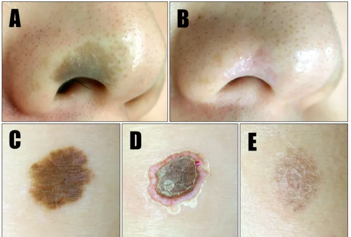
A 25-year-old male was diagnosed with CMN on his nose at birth (Fig. 1A). Emla cream (AstraZeneca AB, Sodertalje, Sweden) was applied to the treatment area prior to starting the procedure. The erbium : YAG laser (Action Er : YAG;
Lutronic Corporation, Goyang, Korea) was used in continuous- wave mode, with 1-mm spot size and output power of 0.2 mJ/cm2. Multiple small holes measuring 1 mm in diameter were made on the periphery of the CMN lesion.
A hydrocolloid dressing (DuoDERM Extra Thin CGF dressing; ConvaTec, ER Squibb & Sons, Princeton, NJ, USA) was applied to the lesions for ≥2 weeks after treatment. After performing 5 laser sessions at 4-week intervals, the cutaneous lesions resolved. There was no evidence of recurrence at the 1-year follow-up exami- nation (Fig. 1B).
A 5-month-old infant was referred to our outpatient unit
for CMN on the forearm (Fig. 1C). We performed the pinhole method of treatment using the erbium : YAG laser (Lutronic) in continuous-wave mode, with 1-mm spot size and output power of 0.2 mJ/cm2 (Fig. 1D). After receiving 6 treatment sessions at 2-month intervals, slight superficial pigmentation persisted (Fig. 1E).
A 40-year-old woman presented with nevus sebaceous lesions on the right side of her forehead (Fig. 2A). We performed the pinhole method of treatment using the erbium : YAG laser (Lutronic) in continuous-wave mode, with 1-mm spot size and output power of 0.2 mJ/cm2. After receiving 5 treatment sessions at 4-week intervals, the patient showed marked improvement with only mild hypopigmentation (Fig. 2B). No recurrence was noted at the 6-month follow-up examination (Fig. 2C).
Herein, we described how nevi lesions were effectively treated using the pinhole method. This simple method involves making multiple small holes at 2∼5-mm intervals using a CO2 laser that penetrates the epidermis and deeper dermis3. The holes produced using the pinhole method are surrounded by untreated areas that promote rapid epidermal repair, thereby reducing the downtime and likelihood of side effects4. The other advantages of performing the pinhole method with ablative lasers are that this laser is easy to use and inexpensive, as ablative lasers are usually available in most dermatology clinics.
To the best of our knowledge, this is the first report on the application of the pinhole method for the treatment of CMN and nevus sebaceous, so it is difficult to compare the advantages and disadvantages of the pinhole method with those of conventional treatment methods. Therefore, it is necessary to conduct further studies with a large sample to confirm the effectiveness and safety of the pinhole method.
In conclusion, we suggest using the pinhole method with erbium : YAG laser as an alternative therapy for treating
Letter to the Editor
652 Ann Dermatol
Fig. 1. Congenital melanocytic nevus (CMN) in a 25-year-old man. (A) Before treatment: a solitary, 1-cm brown patch is visible on the right alar area of the nose. (B) One year after the final treatment session, the skin lesions resolved with minimal scarring. (C) CMN in a 5-month-old boy. Before treatment, a solitary, 1.5∼2-cm, well-demarcated, brown patch is visible on the forearm. (D) The pinhole method was used to create 1-mm holes deep in the dermis. (E) Minimal mottled hyperpigmentation was observed 6 months after the final laser treatment.
Fig. 2. Nevus sebaceous lesions in a 40-year-old woman. (A) Before treatment, two 2.5-cm, well-demarcated, yellow, verrucous plaques were visible on the right side of the forehead. (B) Deep line- and doughnut-shaped holes were created to remove the nevus sebaceus lesions. (C) Six months after the final laser treatment, the patient showed marked improvement, with only mild hypopigmentation.
Letter to the Editor
Vol. 26 No. 5, 2014 653
Received September 5, 2013, Revised October 7, 2013, Accepted for publication October 8, 2013
Corresponding author: Sei-ichiro Motegi, Department of Dermatology, Gunma University Graduate School of Medicine, 3-39-22 Showa-Machi, Maebashi, Gunma 371-8511, Japan. Tel: 81-27-220-8284, Fax: 81-27-220-8285, E-mail: [email protected]
This is an Open Access article distributed under the terms of the Creative Commons Attribution Non-Commercial License (http://
creativecommons.org/licenses/by-nc/3.0) which permits unrestricted non-commercial use, distribution, and reproduction in any medium, provided the original work is properly cited.
nevi lesions.
ACKNOWLEDGMENT
We would like to thank Eun Jin Yeon for editing this manuscript.
REFERENCES
1. Grichnik JM, Rhodes AR, Sober AJ. Benign neoplasias and hyperplasias of melanocytes. In: Fitzpatrick TB, editor. Fitz- patrick's dermatology in general medicine. 7th ed. New
York: McGraw-Hill, 2008:1377-1410.
2. Strivastava D, Taylor RS. Appendage tumors and hamartomas of the skin. In: Fitzpatrick TB, editor. Fitzpatrick's dermatology in general medicine. 7th ed. New York: McGraw-Hill, 2008:
1337-1361.
3. Whang SW, Lee KY, Cho SB, Lee SJ, Kang JM, Kim YK, et al.
Burn scars treated by pinhole method using a carbon dioxide laser. J Dermatol 2006;33:869-872.
4. Yang JH, Han SS, Won CH, Chang SE, Lee MW, Choi JH, et al. Treatment of elastosis perforans serpiginosa with the pinhole method using a carbon dioxide laser. Dermatol Surg 2011;37:524-526.
http://dx.doi.org/10.5021/ad.2014.26.5.653
Primary Granulocytic Sarcoma of the Skin without Hematologic Disorders
Akihiko Uchiyama, Sei-ichiro Motegi, Kazuya Yamada, Hiroo Amano, Osamu Ishikawa
Department of Dermatology, Gunma University Graduate School of Medicine, Maebashi, Japan
Dear Editor:
A 78-year-old Japanese woman noticed a subcutaneous nodule on her neck. On physical examination, we found a reddish tumor, measuring 25×20 mm, on her neck and multiple small nodules on her chest and back (Fig. 1A).
There were no enlarged lymph nodes. Her white blood cell count was normal, and other routine biochemical tests also yielded normal results. The bone marrow aspiration sample showed no evidence of increased blast cell count. Computed tomographic scan of the whole body showed no lymphadenopathy. Histopathological examination of tumor in the neck showed diffuse infil- tration of histiocyte-like tumor cells with remarkable
dyskaryosis in the dermis (Fig. 1B, C). Immunohisto- chemical studies revealed that tumor cells were positive for leukocyte common antigen, CD43, CD56, CD68 (Fig.
1D), and myeloperoxidase (MPO) (Fig. 1E) and negative for keratin, terminal deoxynucleotidyl transferase, CD34, c-kit, CD3, CD4, CD5, CD7, CD8, CD20, CD21, CD138, granzyme B, S-100, and CD1a. Based on these findings, the diagnosis of primary granulocytic sarcoma (GS) without hematologic involvement was established. Patient was administered local irradiation (total, 30 Gy) and chemotherapy with etoposide. After 1 cycle of chemo- therapy, the skin tumor on her neck regressed. During 5-week follow-up period, she did not develop acute mye-