J Korean Surg Soc 2012;82:374-379 http://dx.doi.org/10.4174/jkss.2012.82.6.374
ORIGINAL ARTICLE
Journal of the Korean Surgical Society
JKSS
pISSN 2233-7903ㆍeISSN 2093-0488
Received January 9, 2012, Revised March 18, 2012, Accepted April 3, 2012 Correspondence to: Seung Huh
Division of Transplantation and Vascular Surgery, Department of Surgery, Kyungpook National University Hospital, Kyungpook National University School of Medicine, 130 Dongdeok-ro, Jung-gu, Daegu 700-721, Korea
Tel: +82-53-420-6520, Fax: +82-53-421-0510, E-mail: [email protected]
cc Journal of the Korean Surgical Society is an Open Access Journal. All articles are distributed under the terms of the Creative Commons Attribution Non-Commercial License (http://creativecommons.org/licenses/by-nc/3.0/) which permits unrestricted non-commercial use, distribution, and reproduction in any medium, provided the original work is properly cited.
Early treatment outcome of isolated calf vein thrombosis after total knee arthroplasty
Woo-Sung Yun, Kyung Keun Lee
1, Jayun Cho
1, Hyung-Kee Kim
1, Hee-Soo Kyung
2, Seung Huh
1Division of Vascular/Endovascular Surgery, Department of Surgery, Daegu Catholic University Medical Center, Catholic University of Daegu School of Medicine, Daegu, 1Division of Transplantation and Vascular Surgery, Department of Surgery, 2Department of Orthopedic Surgery, Kyungpook National University Hospital, Kyungpook National University School of Medicine, Daegu, Korea
Purpose: In contrast to proximal deep vein thrombosis (DVT), the treatment of isolated calf vein thrombosis (ICVT) remains controversial. This study aimed to investigate early treatment outcomes of ICVT after total knee arthroplasty (TKA).
Methods: Medical records of 313 patients who underwent TKA from October 2007 to December 2009 were retrospectively reviewed. A DVT-computed tomography (CT) was performed 7 days after surgery. ICVT was identified in 76 limbs of 73 patients. Of them, follow-up DVT-CT was available in 39 limbs of 37 patients. The patients with ICVTs were categorized into two groups: oral anticoagulation group (group I, 17 patients with 18 limbs) and conservative treatment group (group II, 20 patients with 21 limbs). Group I received an oral vitamin K antagonist for 3 to 6 months following low molecular weight heparin. Change of thrombus extent and development of pulmonary embolism (PE) was assessed in follow-up DVT-CT.
Results: Mean age was 68 years and 95% were female. Of 39 limbs with ICVT, 16 (41%) involved major lower leg veins (posterior tibial vein or peroneal vein), 13 (33%) involved muscular veins (soleal vein or gastrocnemius vein) and 10 (26%) in- volved both. During 1 to 6 months, follow-up DVT-CT revealed complete thrombus resolution in all limbs and there was no proximal propagation of thrombus or PE in both groups. Conclusion: There is no evidence of DVT propagation or newly de- veloped PE in the conservative treatment group. This result suggests that anticoagulation therapy for ICVT patients without PE after TKA may not be mandatory.
Key Words: Isolated calf vein thrombosis, Pulmonary embolism, Anticoagulation
INTRODUCTION
The prevalence of isolated calf vein thrombosis (ICVT) is 5 to 12% in symptomatic patients [1] and asymptomatic ICVT may occurin 15% of patients after total hip and total knee arthroplasty (TKA) [2]. However, treatment of ICVT is still controversial.
Generally, ICVT is not considered to be a source of fatal pulmonary embolism (PE) because the severity of PE de- pends on the size of the emboli or cardiopulmonary func- tion [3]. However, some authors who recommend anti- coagulation therapy are concerned that untreated ICVT may extend to proximal deep veins, which is more likely to result in PE [4].
Table 1.Risk factors for venous thromboembolism (VTE) in 37 patients with isolated calf vein thrombosis (ICVT)
Risk factor
Oral anticoagulation
(n = 17)
Conservative treatment
(n = 20)
Total (n = 37)
Prior VTE 2a) (12) 1b) (5) 3 (8) Body mass index 8 (47) 6 (30) 14 (38) > 25 kg/m2
Estrogen therapy 0 (0) 0 (0) 0 (0)
Smoking 1 (6) 1 (5) 2 (5)
Malignancy 0 (0) 1c) (5) 1 (3)
Values are presented as no. of patients (%).
a)One patient with May-Thurner syndrome previously underwent catheter-directed thrombolytic therapy with left iliac vein stenting and the other had ICVT after previous contralateral total knee arthroplasty (TKA). b)ICVT after previous contralateral TKA. c)Thy- roid cancer.
This study aimed to investigate short-term treatment outcomes (e.g., proximal thrombus propagation and in- cidence of PE) in patients with ICVT after TKA according to whether or not they were treated with anticoagulation therapies.
METHODS
Between October 2007 and December 2009, 313 TKAs were performed in Kyungpook National University Hospital. During the perioperative period, prophylactic anticoagulation was not administered and only a com- pression stocking was applied. On postoperative day 7, a deep vein thrombosis-computed tomography (DVT-CT) designed in our hospital was performed on all patients for venous thromboembolism (VTE) screening. The preva- lence of VTE was 34% (105/313). PE without DVT was 4%
(11/313) and DVT with or without PE was 30% (94/313).
DVT-CT revealed ICVT in 76 limbs of 73 patients. Of them, 39 limbs of 37 patients who underwent follow-up DVT-CT were enrolled in this study. By retrospective review of pro- spectively registered medical records, we investigated the incidence of resolution and proximal propagation of ICVT as well as the incidence of PE.
Regarding treatment, indications of 3 to 6 months oral anticoagulation therapy were as follows: 1) concomitant PE regardless of symptom, 2) concomitant proximal DVT in contralateral limb, 3) previous episode of DVT.
The patients with ICVTs were categorized into two groups according to treatment. Seventeen patients with 18 limbs (group I, oral anticoagulation group) were treated 3 to 6 months with oral anticoagulation therapy following low molecular weight heparin (LMWH) and 20 patients with 21 limbs (group II, conservative treatment group) were not. Among group II, however, 11 patients received short-term (3 to 7 days) LMWH during admission and 15 patients received antiplatelet therapy for 3 to 6 months.
Neither anticoagulation nor antiplatelet agent was ad- ministered for 4 patients.
Sixteen patients had concomitant PE and all were asymptomatic. All belonged to group I, except 1 patient who discontinued anticoagulation due to hematemesis
one day after anticoagulation therapy.
CT was performed using a Light Speed 16 CT scanner (GE Healthcare, Milwaukee, WI, USA) or Aquilion 64 CT scanner (Toshiba Medical Systems Co., Tokyo, Japan). The contrast media (Optiray 320, Tyco Healthcare, Mallin- krodt, Saint Louis, MO, USA) with a volume of 140 mL was administered through an antecubital vein using an auto- mated power injector at a rate of 3 to 4 mL/sec and was fol- lowed a 30 mL saline chaser with the same infusion rate.
Individual contrast optimization was achieved using bo- lus tracking within the main pulmonary artery. CT pulmo- nary arteriography was obtained during a shallow in- spiratory breathhold in a craniocaudal direction from the aortic arch to the heart base. At 180 seconds after injection of the contrast agent, CT venography was performed from the heart base down to the feet in the craniocaudal direction.
RESULTS
Patient mean age was 68 years (range, 48 to 86 years) and 95% were female. Two patients had bilateral ICVT and 1 patient had a contralateral proximal DVT. Sixteen patients had a concomitant PE and all were asymptomatic. The risk factors for VTE are shown in Table 1. Two patients had a previous episode of ICVT of the contralateral limb after
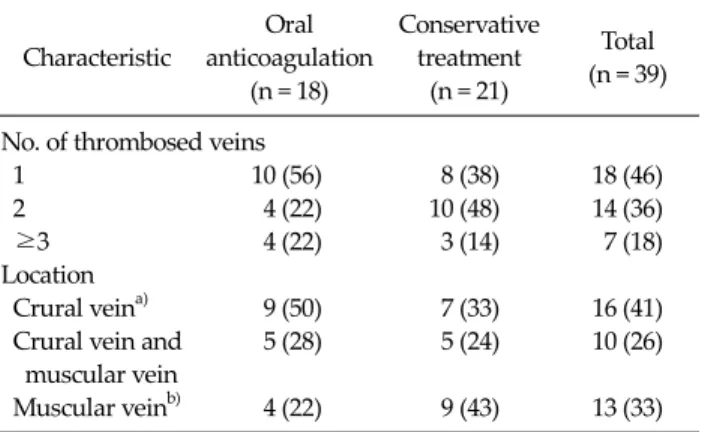
Fig. 1. Computed tomography (CT) of 66-year-old woman who underwent right total knee arthroplasty (TKA). She was treated with oral anticoagulation therapy for 3 months due to isolated calf vein thrombosis (ICVT) with pulmonary embolism (PE). (A) CT at 7 days after TKA revealed PE in right posterior basal segmental artery (arrow) and thrombosis in right muscular calf veins (arrow heads). (B) Follow-up CT after 4 months revealed complete resolution of ICVT and PE.
Table 2. Clinical characteristics of isolated calf vein thrombosis Characteristic
Oral anticoagulation
(n = 18)
Conservative treatment
(n = 21)
Total (n = 39)
No. of thrombosed veins
1 10 (56) 8 (38) 18 (46)
2 4 (22) 10 (48) 14 (36)
≥3 4 (22) 3 (14) 7 (18)
Location
Crural veina) 9 (50) 7 (33) 16 (41) Crural vein and 5 (28) 5 (24) 10 (26) muscular vein
Muscular veinb) 4 (22) 9 (43) 13 (33) Values are presented as no. of limbs (%).
a)Anterior tibial vein, posterior tibial vein and peroneal vein.
b)Gastrocnemius vein and soleal vein.
contralateral TKA and 1 patient had undergone cathe- ter-directed thrombolysis and left iliac vein stenting due to May-Thurner syndrome 5 years ago.
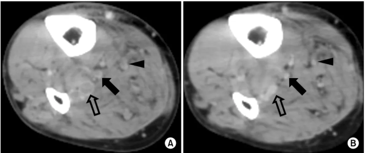
Table 2 summarizes the locations of ICVT and the num- ber of thrombosed veins. Fifty-four percent involved more than 1 vein. Among the crural veins, peroneal vein throm- bosis occurred in 16 limbs as well as the posterior tibial vein, but the anterior tibial vein was not involved in any case. Isolated muscular calf vein thrombosis (IMCVT) was seen in 33% (13/39) of patients. Follow-up DVT-CT re- vealed complete resolution of ICVT and PE in the oral anti- coagulation group (Fig. 1). In the conservative treatment group, ICVT also disappeared on follow-up DVT-CT and there was no newly developed PE (Fig. 2).
DISCUSSION
There were many reports on ICVT, but its clinical im- plication and treatment remains controversial. Tradition- ally, main concerns in DVT treatment are the risk of PE and
Fig. 2. Computed tomography (CT) of 67-year-old man who underwent right total knee arthroplasty (TKA). He was treated with low molecular weight heparin for 5 days during admission and took antiplatelet agent for 3 months. (A) CT at 7 days after TKA revealed thrombosis in right peroneal vein (open arrow), posterior tibial vein (solid arrow), and muscular vein (arrow head). (B) Previous thrombus disappeared on follow-up CT after 3 months.
post-thrombotic syndrome.
There were autopsy studies demonstrating that ICVT was a source of fatal PE [5,6]. Sevitt and Gallagher [5] per- formed autopsies in 74 fatal cases of PE and the rate of fatal PE from ICVT was 15% (11/74). In another autopsy study of 23 patients who died of PE, ICVT was detected in 3 pa- tients (13%) [6]. Although the reported incidence of PE in patients with ICVT range from 7 to 50% [3,7-9], fatal PE caused by ICVT is rarely reported in clinical studies.
However, 20 to 50% of untreated proximal DVT results in significant PE and 5% of such PE are fatal [10]. Therefore, another issue of ICVT is its propagation to the level of or above the popliteal vein.
Based on serial duplex scans of 192 patients with ICVT, Lohr et al. [11] reported that 28% had propagation of their initial thrombi. Propagation up to the proximal veins oc- curred in 21 patients (11%). They failed to identify risk fac- tors of thrombus propagation. In a randomized pro- spective studyin 51 patients with ICVT diagnosed by con- trast venography [12], 29% (8/28) of the non-warfarin group had recurrences and 18% (5/28) were recurrences with proximal extension during the first 3 months. However, there were no recurrences in the warfarin group (P < 0.01). After 1 year, there recurrence rate was 68% in the non-warfarin group, while there were no recurrences in the warfarin group, except for 1 patient who died of lung cancer after 4 months (P < 0.02). Lautz et al. [13] reported
a mean 7.5 months follow-up result of 452 limbs in 406 pa- tients with IMCVT. VTE event occurred in 18.7% of the patients. According to the treatment, the incidence of VTE events was significantly lower after therapeutic anti- coagulation (12% in the anticoagulation group vs. 27% in the prophylactic anticoagulation group vs. 30% in the no anticoagulation group, P = 0.0003).
On the contrary, there are some reports that post- operative ICVT is insignificant. Wang et al. [14] performed ascending venography after TKA and identified 51 pa- tients with ICVT. Symptomatic ICVT was treated with heparin or LMWH for 3 to 7 days and asymptomatic pa- tients received no medication. Follow-up venography was performed in 37 patients. It showed no thrombosis except in 1 patient who had residual thrombi in the muscular branches. In other study [15], a serial follow-up duplex scan was performed for 42 patients (50 limbs) with ICVT after total hip arthroplasty or TKA. Its proximal prop- agation rate was not influenced by anticoagulation (23% in treated limbs vs. 8% in untreated limbs, P = 0.43). The au- thors suggested that postoperative calf vein thrombosis need not be routinely treated and serial venous duplex scanning is useful to identify thrombotic propagation re- quiring anticoagulation.
Masuda et al. [16] reported that proximal propagation of ICVT developed in 4% of the cases within 2 weeks of di- agnosis and clinically overt PE did not occur regardless of
whether anticoagulation therapy was received or not.
Meissner et al. [17] monitored 29 limbs with ICVT with du- plex scan. Median propagation time of ICVT was 11 days.
MacDonald et al. [18] performed serial duplex scansfor 135 limbs with IMCVT that were not treated with anti- coagulation therapy. Only 3% of the patients extended their thrombi to the level of the popliteal vein and all oc- curred within 2 weeks.
Considering most thrombus propagations occur in the early period, it is reasonable that short-term anticoagula- tion may be adopted for patients with ICVT. There was a report that short-term anticoagulation in IMCVT is useful to prevent VTE complications [19], but it was not proven in a prospective randomized study [20]. The authors com- pared treatment outcomes between a short-term anti- coagulation group (treated with LMWH for 10 days) and a compression group. There was no difference in the throm- bus propagation rate (3.7% in LMWH group vs. 3.8% in compression group) and there was no clinical PE and no death.
Another issue in ICVT is post-thrombotic syndrome. In several studies, long-term symptoms and significant he- modynamic changes occur after ICVT [21-23]. Browse et al. [21] reported a 20% incidence of moderate to severe postphlebitic symptoms and signs 5 to 10 years after symptomatic ICVT. Meissner et al. [22] reported that 23%
of patients with ICVT showed persistent symptoms and the development of valvular incompetence at 1 year, al- though recanalization proceeded rapidly. In a population- based study [23], the cumulative incidence of post-throm- botic syndrome after ICVT was 10.2%, 22%, and 29.8% at 2 years, 10 years, and 20 years respectively.
We preferred 3-month oral anticoagulation therapy for patients with ICVT if a patient had a concomitant PE at ini- tial diagnosis. On follow-up DVT-CT 3 to 6 months after the first diagnosis of ICVT, thrombus propagation was not detected and all ICVTs resolved regardless of treatment.
And, there was no newly developed PE in the conservative treatment group. This result suggests that 3-month oral anticoagulation therapy for ICVT patients without PE af- ter TKA may not be necessary concerning the risk of prox- imal thrombus propagation and development of PE. It is assumed that most postoperative ICVT resolve sponta-
neously when the postoperative period passes and the pa- tients can ambulate because TKA is a transient risk factor for VTE.
The limitation of this study is that it is a retrospective design with no randomization. Further, long-term follow- up is necessary to evaluate disease recurrence and post- thrombotic syndrome. Indeed, to establish a consensus guideline for ICVT treatment, a large-scale prospective randomized study is necessary.
CONFLICTS OF INTEREST
No potential conflict of interest relevant to this article was reported.
REFERENCES
1. Gottlieb RH, Widjaja J. Clinical outcomes of untreated symptomatic patients with negative findings on sonog- raphy of the thigh for deep vein thrombosis: our experi- ence and a review of the literature. AJR Am J Roentgenol 1999;172:1601-4.
2. Oishi CS, Grady-Benson JC, Otis SM, Colwell CW Jr, Walker RH. The clinical course of distal deep venous thrombosis after total hip and total knee arthroplasty, as determined with duplex ultrasonography. J Bone Joint Surg Am 1994;76:1658-63.
3. Ohgi S, Tachibana M, Ikebuchi M, Kanaoka Y, Maeda T, Mori T. Pulmonary embolism in patients with isolated sol- eal vein thrombosis. Angiology 1998;49:759-64.
4. Lohr JM, Kerr TM, Lutter KS, Cranley RD, Spirtoff K, Cranley JJ. Lower extremity calf thrombosis: to treat or not to treat? J Vasc Surg 1991;14:618-23.
5. Sevitt S, Gallagher N. Venous thrombosis and pulmonary embolism: a clinico-pathological study in injured and burned patients. Br J Surg 1961;48:475-89.
6. Giachino A. Relationship between deep-vein thrombosis in the calf and fatal pulmonary embolism. Can J Surg 1988;31:129-30.
7. Guias B, Simoni G, Oger E, Lemire A, Leroyer C, Mottier D, et al. Calf muscle venous thrombosis and pulmonary embolism. J Mal Vasc 1999;24:132-4.
8. Hollerweger A, Macheiner P, Rettenbacher T, Gritzmann N. Sonographic diagnosis of thrombosis of the calf muscle veins and the risk of pulmonary embolism. Ultraschall Med 2000;21:66-72.
9. Philbrick JT, Becker DM. Calf deep venous thrombosis: a wolf in sheep's clothing? Arch Intern Med 1988;148:2131-8.
10. Deitcher SR, Caprini JA. Calf deep venous thrombosis
should be treated with anticoagulation. Med Clin North Am 2003;87:1157-64.
11. Lohr JM, James KV, Deshmukh RM, Hasselfeld KA, Allastair B. Karmody Award. Calf vein thrombi are not a benign finding. Am J Surg 1995;170:86-90.
12. Lagerstedt CI, Olsson CG, Fagher BO, Oqvist BW, Albrechtsson U. Need for long-term anticoagulant treat- ment in symptomatic calf-vein thrombosis. Lancet 1985;2:515-8.
13. Lautz TB, Abbas F, Walsh SJ, Chow C, Amaranto DJ, Wang E, et al. Isolated gastrocnemius and soleal vein thrombosis:
should these patients receive therapeutic anticoagulation?
Ann Surg 2010;251:735-42.
14. Wang CJ, Wang JW, Weng LH, Hsu CC, Lo CF. Outcome of calf deep-vein thrombosis after total knee arthroplasty. J Bone Joint Surg Br 2003;85:841-4.
15. Solis MM, Ranval TJ, Nix ML, Eidt JF, Nelson CL, Ferris EJ, et al. Is anticoagulation indicated for asymptomatic post- operative calf vein thrombosis? J Vasc Surg 1992;16:414-8.
16. Masuda EM, Kessler DM, Kistner RL, Eklof B, Sato DT. The natural history of calf vein thrombosis: lysis of thrombi and development of reflux. J Vasc Surg 1998;28:67-73.
17. Meissner MH, Caps MT, Bergelin RO, Manzo RA, Strandness DE Jr. Propagation, rethrombosis and new
thrombus formation after acute deep venous thrombosis. J Vasc Surg 1995;22:558-67.
18. MacDonald PS, Kahn SR, Miller N, Obrand D. Short-term natural history of isolated gastrocnemius and soleal vein thrombosis. J Vasc Surg 2003;37:523-7.
19. Schwarz T, Schmidt B, Beyer J, Schellong SM. Therapy of isolated calf muscle vein thrombosis with low-molec- ular-weight heparin. Blood Coagul Fibrinolysis 2001;12:
597-9.
20. Schwarz T, Buschmann L, Beyer J, Halbritter K, Rastan A, Schellong S. Therapy of isolated calf muscle vein thrombo- sis: a randomized, controlled study. J Vasc Surg 2010;52:
1246-50.
21. Browse NL, Clemenson G, Thomas ML. Is the post- phlebitic leg always postphlebitic? Relation between phle- bographic appearances of deep-vein thrombosis and late sequelae. Br Med J 1980;281:1167-70.
22. Meissner MH, Caps MT, Bergelin RO, Manzo RA, Strandness DE Jr. Early outcome after isolated calf vein thrombosis. J Vasc Surg 1997;26:749-56.
23. Mohr DN, Silverstein MD, Heit JA, Petterson TM, O'Fallon WM, Melton LJ. The venous stasis syndrome after deep ve- nous thrombosis or pulmonary embolism: a population- based study. Mayo Clin Proc 2000;75:1249-56.