CASE REPORT
Copyright © 2011, the Korean Surgical Society J Korean Surg Soc 2011;80:S59-62
DOI: 10.4174/jkss.2011.80.Suppl 1.S59
JKSS
Journal of the Korean Surgical Society pISSN 2233-7903ㆍeISSN 2093-0488
Received July 29, 2010, Accepted August 30, 2010 Correspondence to: Ki-Han Kim
Department of Surgery, Dong-A University College of Medicine, 1 Dongdaesin-dong 3-ga, Seo-gu, Busan 602-715, Korea Tel: +82-51-240-5146, Fax: +82-51-247-9316, E-mail: [email protected]
cc Journal of the Korean Surgical Society is an Open Access Journal. All articles are distributed under the terms of the Creative Commons Attribution Non-Commercial License (http://creativecommons.org/licenses/by-nc/3.0/) which permits unrestricted non-commercial use, distribution, and reproduction in any medium, provided the original work is properly cited.
Laparoscopic splenectomy for sclerosing angiomatoid nodular transformation of the spleen
Ki-Han Kim, Seul Lee
1, Soon Hwa Youn, Mi Ri Lee, Min Chan Kim, Seo-Hee Rha
1, Ghap Joong Jung
Departments of Surgery and 1Pathology, Dong-A University College of Medicine, Busan, Korea
Primary splenic tumors are rare and mainly found incidentally on radiologic studies. Among them, sclerosing angiomatoid nodular transformation (SANT) of the spleen is a new entity defined as a benign pathologic lesion. Most SANTs have no clin- ical symptoms and are occasionally accompanied by other splenic diseases such as malignancies. So, the exact diagnosis of the nature of the splenic tumor is mandatory for further treatment. But, preoperative diagnosis is not easy since it is difficult to obtain the tissue from the spleen for pathological study. Recently, laparoscopic splenectomy has become the more standard procedure for the spleen for diagnosis and treatment. Here, we report a rare case of SANT diagnosed following laparoscopic splenectomy.
Key Words: Laparoscopic splenectomy, Sclerosing angiomatoid nodular transformation
INTRODUCTION
Solid tumors of the spleen are relatively rare, with an in- cidence of 0.007% of all operation and autopsy specimens [1].
It is usually found incidentally on abdominal computed tomography (CT) or ultrasonography. It is difficult to rule out the malignancy by preoperative radiologic studies.
Sometimes, fine needle aspiration (FNA) biopsy is used to determine pathologic diagnosis. But this may be associated with bleeding and the risk of tumor dissemination [2].
Therefore, splenectomy is necessary for diagnosis and treatment of the splenic tumors. Recently, laparoscopic splenectomy has become the standard technique for the surgical management of hematological splenic diseases and tumors [3,4].
Sclerosing angiomatoid nodular transformation (SANT) of the spleen is a rare vascular benign lesion that is charac- terized by Martel et al. [5]. Most patients with SANT have no obvious clinical symptoms and are discovered in- cidentally through routine evaluation. Since there have been no case introduced in Korean literatures, we report a case diagnosed as SANT of the spleen after laparoscopic splenectomy.
CASE REPORT
A 23 year old female was admitted to our hospital for evaluation of splenic tumor which was incidentally diag- nosed by abdominal CT. Her previous histories and phys-
Ki-Han Kim, et al.
S60 thesurgery.or.kr
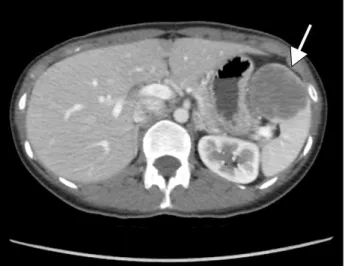
Fig. 1. Preoperative abdominal computed tomography demon- strates an expansible low density mass (white arrow).
Fig. 2. Laparoscopic view of a splenic tumor.
Fig. 3. The spleen shows a well-demarcated, unencapsulated, dark brown mass with central stellate fibrotic scar on cut section.
ical examinations were unremarkable. CT examination re- vealed about 5 × 4 cm sized solid tumor in the spleen, showing low density on pre, early phase, and progressive enhancement on delayed phase (Fig. 1). She wanted patho- logic confirmation by operation, and was undergone lapa- roscopic splenectomy.
Surgical procedure
Under general anesthesia, the patient was placed in right lateral decubitus position with the table slightly ex- tended in order to widen the operative field during splenectomy. Three trocars were used along the left sub- costal margin and one trocar for the camera port at the
umbilicus. Under the pneumoperitoneum of 12 mmHg, dissection was performed using Harmonic ACE (Ethicon Endo-Surgery, Cincinnati, OH, USA). Attending to injury of splenic tumor, we detached following areas; 1) splenic flexure, 2) anterior aspect of the hilum with short gastric vessel, 3) posterior aspect of the left gutter with separation of attachments to the kidney, and 4) some of the attach- ments to the upper pole of the spleen. After mobilization of the spleen, the vascular pedicle was divided at the level of the splenic hilum by laparoscopic stapling device (Echelon Flex, Ethicon Endo-Surgery) with a special care not to injure the pancreatic tail. Then the spleen was placed in a plastic bag, and taken out through the opening of the most lateral port with additional wound extension by about 3 cm. The normal splenic tissue was fractured using a ring clamp as much as can leave the splenic tumor intact for pathologic diagnosis (Fig. 2).
Pathologic finding
The spleen shows a well-demarcated, solitary mass, measuring 5.2 × 4.5 cm in dimension. The mass is dark brown with a central large stellate fibrotic scar (Fig. 3).
Microscopically, the spleen shows an unencapsulated but well-demarcated mass, composing multiple irregular fi- brotic nodules (Fig. 4A). The center of nodules show many small-sized vessels and extravasated red blood cells, so called angiomatoid nodule (Fig. 4B). The periphery of the nodules show abundant collagen fibers infiltrated by many plasma cells, lymphocytes and some eosinophils.
Laparoscopic splenectomy for SANT
thesurgery.or.kr S61
Fig. 4. Microscopic findings in sclerosing angiomatoid nodular transformation of spleen. (A) The tumor shows multinodular lesion surrounded by fibrosis. The nodules are irregular and composed of many small-sized vessels. The fibrotic area shows abundant collagen fibers and inflammatory cells hematoxylin-eosin (H&E, ×40). (B) The high power view of angiomatoid nodular lesion, it shows many vessels, extravasated red blood cells and inflammatory cells (H&E, ×100).
The spindle cells of the nodules show positivity for vi- mentin and α-smooth muscle actin on immunohistoche- mistry. No cells show positivity for anaplastic lymphoma kinase gene (ALK), S-100. The vascular endothelial cells show CD31 and CD34 immunoreactivity (data not shown).
Postoperative outcome
The patient started the sips of water on the post- operative 6 hours and soft diet on the following day. She was discharged on postoperative day 2 without any re- markable events.
DISCUSSION
SANT is rare and newly defined pathological disorder of the spleen with nodular vascular proliferation of the red pulp with prominent sclerosis [5]. SANT usually affects middle-aged adults and shows a female predominance.
Most of cases are asymptomatic and discovered acci- dentally, usually on imaging study. In gross examination of spleen, SANT shows a solitary, well-circumscribed nod- ule that is distinct from the surrounding splenic parenchyma. In microscopic examination, it consists of three basic cellular components (spindle cells, inflam-
matory infiltrate, and endothelial vascular proliferation).
These cellular components are mixed with each other, cre- ating the angiomatoid nodules of the SANT. These nod- ules are wrapped by collagen fibers and are intervened by inflammatory and sclerotic responses. SANT shows a dis- tinct immunophenotype. The angiomatoid nodules of the SANT are composed of several morphologically and im- munophenotypically distinct blood vessels: a cord capil- lary-like type that co-expresses CD34 and CD31 but not CD8, a sinusoid- like type that expresses CD8 and CD31 but not CD34, and small veins that express only CD31 [6].
Our pathologic results shows CD31 and CD34 immunoreactivity. Although it remains to be further stud- ied, its clinical prognosis was reported to be excellent with complete cure by splenectomy [6]. Martel et al. [5] re- ported that SANTs cases showed no evidence of re- currence, which suggests that SANT of the spleen is a com- pletely benign lesion.
Many patients presenting with solid lesions in the spleen will eventually be diagnosed with a malignant tu- mor, but it is difficult to rule out the possibility of a malig- nant neoplasm preoperatively based on conventional imaging studies. So it is mandatory to make pathologic confirmation for diagnosis and treatment of solid tumor.
Sometimes fine-needle biopsy from the spleen has been used to ascertain tissue diagnosis. However, this proce-
Ki-Han Kim, et al.
S62 thesurgery.or.kr
dure has some problems with bleeding or tumor dissem- ination [2]. Therefore, in most cases, splenectomy is rec- ommended for diagnosis and treatment. With the devel- opment of laparoscopic instruments and techniques, lapa- roscopic surgery has continued to be popularized in al- most all fields of surgery. Since the introduction of laparo- scopic splenectomy by Delaitre and Maignien [7] in 1992, now laparoscopic splenectomy has been recognized as a safe and effective treatment for hematologic disorders and other splenic diseases [3,8]. Compared with open splenec- tomy, it is a safer procedure with the advantages of less postoperative pain and complications, early recovery from the procedure, shorter hospital stay, and a much smaller wound incision [9,10]. From this point of view, lap- aroscopic splenectomy is a good procedure for the diag- nosis as well as the treatment that is safer to avoid bleeding and peritoneal dissemination of tumor cells than FNA and allows adequate histological evaluation, especially when malignancy cannot be ruled out. In conclusion, the au- thors consider that laparoscopic splenectomy can be per- formed for most splenic tumors such as SANT. SANT is a rare pathologic disease with good prognosis and accept- able for surgical diagnosis and treatment by laparoscopic splenectomy.
CONFLICTS OF INTEREST
No potential conflict of interest relevant to this article
was reported.
REFERENCES
1. Moriyama S, Inayoshi A, Kurano R. Inflammatory pseudo- tumor of the spleen: report of a case. Surg Today 2000;
30:942-6.
2. Caraway NP, Fanning CV. Use of fine-needle aspiration bi- opsy in the evaluation of splenic lesions in a cancer center.
Diagn Cytopathol 1997;16:312-6.
3. Stephens BJ, Justice JL, Sloan DA, Yoder JA. Elective lapa- roscopic splenectomy for hematologic disorders. Am Surg 1997;63:700-3.
4. Walsh RM, Brody F, Brown N. Laparoscopic splenectomy for lymphoproliferative disease. Surg Endosc 2004;18:
272-5.
5. Martel M, Cheuk W, Lombardi L, Lifschitz-Mercer B, Chan JK, Rosai J. Sclerosing angiomatoid nodular transfor- mation (SANT): report of 25 cases of a distinctive benign splenic lesion. Am J Surg Pathol 2004;28:1268-79.
6. Teng X, Yu X, Wang G, Xu L, Lai M. Sclerosing angioma- toid nodular transformation of the spleen. Anal Quant Cytol Histol 2008;30:125-32.
7. Delaitre B, Maignien B. Splenectomy by the laparoscopic approach: report of a case. Presse Med 1991;20:2263.
8. Rosen M, Brody F, Walsh RM, Tarnoff M, Malm J, Ponsky J.
Outcome of laparoscopic splenectomy based on hemato- logic indication. Surg Endosc 2002;16:272-9.
9. Lee SG, Kim MC, Kim HH, Kwon HC, Jung GJ. Treatment result of laparoscopic versus open splenectomy in benign splenic diseases. J Korean Surg Soc 2005;68:230-4.
10. Bellows CF, Sweeney JF. Laparoscopic splenectomy: pres- ent status and future perspective. Expert Rev Med Devices 2006;3:95-104.