서 론
Synucleins는 3개의 단백질, -synuclein (SNCA), -sy-
nuclein (SNCB), -synuclein (SNCG)으로 이루어진 작은 크 기의 단백질군이다.(1, 2) -synuclein 유전자는 10q23에 위치 하며 5개의 coding exon으로 이루어져 있고 1 kb의 mRNA로 전사된다. 인간의 -synuclein은 127개의 아미노산으로 이루 어져 있다.(3) Synucleins가 presynaptic terminal에서 풍부 하게 발현되고, 알츠하이머병의 amyloid plaque 형성, 파킨슨병 의 Lewy body 형성과 관련이 있지만(4) 세포내 정상 기능에 대해 Purpose: Synuclein has been identified as an important
neuroprotein for developing pathologic deposits in Alzhei- mer’s and Parkinson’s disease patients. -synuclein is also known as a breast cancer-specific gene 1 thats’s not found in normal breast tissues but it has been reported to be ove- rexpressed in breast cancer, ovarian cancer and other tu- mors. To evaluate the availability of -synuclein expression as a prognostic factor for infiltrative breast cancer, we an- alyzed its correlation with the clinical parameters and the HER-2/neu gene expression.
Methods: Two hundred fiffty samples of breast cancer ti- ssues embedded in paraffin and that were obtained from the infiltrative breast cancer patients who were operated in our institution from January 1995 to December 2000 were analyzed with employing the tissue microarray technique.
The expression of -synuclein was studied with immuno- histochemistry and with using -synulcein antibodies. One hundred thirty one cases that showed favorable staining were selected and studied retrospectively.
Results: Fiffty five% (71/131) of the patients showed -sy-
nuclein overexpression. The histopathological findings that significantly correlated with -synuclein overexpression were the number of metastatic lymph nodes (p<0.01) and the cancer stage (p<0.01). Using the same tissue mircoarray, the HER-2/neu gene expression and -synuclein expression also showed statistically significant correlation (p=0.04).
Conclusion: -synuclein overexpression showed significant correaltion with lymph node metastasis and cancer stage. It also showed significant relevance with the HER-2/neu gene expression, and that is already known to be a prognostic factor for breast cancer. Therefore -synuclein may be a useful prognostic factor for infiltrative breast cancer and fur- ther studies on the its correlation with survival, local recurren- ce, and distant metastasis should be conducted (J Breast Can- cer 2007;10:114-9)
Key Words : Invasive ductal carcinoma, -synuclein, HER-2/neu, Immunohis- tochemistry
중심단어 : 침윤성 유방 관암종, -synuclein, HER-2/neu, 면역조직화학 검사
The -Synuclein Expression in Breast Cancer and its Correlation with the Expression of the HER-2/neu Gene
Seong Rae Kim, Won Hyuk Choi, Jun Ho Park, Eun Sook Nam
1, Seong Jin Cho
1, Chan Heun Park
Departments of Surgery and 1Pathology, Hallym University College of Medicine, Seoul, Korea
Breast Cancer
O R I G N A L A R T I C L E
김성래ㆍ최원혁ㆍ박준호ㆍ남은숙1ㆍ조성진1ㆍ박찬흔 한림대학교 의과대학 외과학교실, 1병리학교실
유방암에서 -Synuclein 과발현의 예후인자로서 유용성 및 HER-2/neu 유전자 증폭과 연관성
책임저자 : 박찬흔
134-701 서울시 강동구 길1동 445, 한림대학교 강동성심병원 외과 Tel: 02-2224-2226, Fax: 02-2224-2570
E-mail : [email protected]
접수일 : 2006년 10월 26일 게재승인일 : 2007년 5월 31일
114
서는 거의 알려져 있지 않다. 유방암 특이 유전자로 밝혀진 breast cancer specific gene 1 (BCSG1)은 유방암의 cDNA library에 서 높은 빈도로 발현되고 정상 유방 조직의 cDNA library에서는 거의 발현되지 않는다.(5) BCSG1은 기존의 알려진 다른 성장 인 자나 암유전자와 상동성이 없고 신경단백인 synucleins와 염기서 열 상동성을 나타냈다(SNCA와 54%, SNCB와 56%).(6) BCSG1 동정 이후 synuclein(3)과 persyn(7)이 뇌 genomic library와 뇌 cDNA library로부터 각각 동정되었고, 세 개의 단백질은 동 일한 단백질로 밝혀졌다. 이 후 BSCG1은 -synuclein로 명명 하게 되었다. Ji 등(5)은 -synuclein이 유방암에서 비정상적으 로 발현되는 것을 최초로 발견하였다. 그들은 Northern blot과 in situ hybridization을 이용하여 악성 유방암에서 높은 빈도 로 -synuclein mRNA가 발현되고 양성 유방 종양 혹은 정상 유방 조직에서는 -synuclein mRNA가 거의 발현되지 않는 것 을 밝혀 내었다. -synuclein의 과발현이 암종의 발생 및 성장에 미치는 영향에 대해 현재까지 알려진 것은 실험적으로 세포의 운 동성과 침윤성을 증가시키고(in vitro) 생체 내에서 전이를 촉진 (in vivo)시키며 항암제 유발 세포자살에 저항성을 가지게 한다는 것이다.(8) Synucleins의 과발현은 난소암의 발생과도 관련이 있 는 것으로 알려져 있다.(9) 최근 -synuclein이 유방암과 난소암 뿐만 아니라 다른 여러 암종(간암, 식도암, 대장암, 위암, 폐암, 전 립선암, 자궁경부암)에서도 과발현되며 이들 암종의 병기가 높을 수록 그 발현빈도가 높다는 결과가 보고되었다.(10) 이전의 연구 들을 통해 유방암에서 -synuclein이 유방암의 병기에 특이적 으로 발현한다는 사실은 Western 분석(9), RT-PCR 분석(11)을 통해 확인되었다. 본 저자들은 침윤성 유방암에서 면역조직화학 염색을 이용하여 조직병리학적 소견과 -synuclein의 과발현 여부 및 환자의 임상병기와의 연관성을 조사하였다. 또한 유방암 의 예후인자인 HER-2/neu 유전자 증폭과 -synuclein의 과 발현의 상관성을 조사하여 -synuclein 과발현의 예후 인자로 서의 유용성을 평가하고자 하였다.
방 법
1. 연구대상
1995년 1월부터 2000년 12월까지 본원에서 침윤성 유방암 진 단하에 수술을 시행한 환자들의 원발성 암조직 중 파라핀 포매 조 직의 보관상태가 양호한 250예를 대상으로 조직미세배열을 시행 하였다. -synuclein의 과발현 여부는 -synuclein에 대한 항 체를 이용한 면역조직화학염색으로 측정하였으며, 동일한 조직 미세배열을 이용하여 HER-2/neu 유전자 증폭은 형광제자리 부합화(fluorescence in situ hybridization, FISH)로 확인하
였고, 에스트로겐 수용체와 프로게스테론 수용체는 면역조직화 학검사로 시행된 기존의 결과를 이용하였다. 이 중 염색상태가 양 호한 131명을 대상으로 의무 기록을 통한 후향적 연구를 시행하 였다.
2. 연구방법
1) -synuclein에대한면역조직화학 염색
면역조직화학 연구는 goat anti-human -synuclein poly- clonal antibody (clone C-20, 1:200, Santa Cruz Biote- chnology, CA, USA)을 1차 항체로 사용하여 avidin-biotin complex method로 시행하였다. 포르말린에 고정된 파라핀 포 매 조직의 조직미세배열을 4 m 두께로 절제한 면역조직화학 염 색용 슬라이드에 부착시킨 후 xylene을 이용하여 탈파라핀화하고 알코올로 수세하였다. 슬라이드를 10 mM citrate 완충액에 담근 후 항원 복구를 위해 전자렌지에서 5분간 2회 가열하였다. 내인성 과산화효소는 0.3% 과산화수소로 20분 이상 억제시킨 후 켰다. 슬 라이드는 일차 항체와 함께 phosphate buffered saline과 1%
bovine serum albumin에 4℃에서 하룻밤 동안 반응시켰다.
이차 항체로 anti-goat biotinylated antibody를 실온에서 20분간 반응시킨 후 3-3′-diaminobendizine-tetrachloride (Biogenex, San Ramon, CA, USA)를 발색제로 사용한 LSAB kit (DAKO, Carpinteria, CA, USA)를 사용하였다. 이 후 슬라 이드는 Meyer’s hematoxyline을 이용하여 대조염색을 하였다.
음성 대조군은 일차 항체 대신 식염수를 이용하여 반응시킨 것을 이용하였다. 면역염색된 슬라이드는 양성반응을 보이는 세포의 비례 점수(proportion score)와 염색 점수(intensity score)를 측정하여 검사하였다. 비례 점수는 면역반응성 세포가 관찰되지 않을 때 0, 면역반응성 세포가 5% 미만, 5-10%, 10-40%, 40- 70%, 70% 이상일 때 각각 1, 2, 3, 4, 5로 등급을 나누었다. 염색 점수는 면역반응성이 없을 때 0, 염색강도가 약, 중간, 강일 때 각 각 1, 2, 3으로 등급을 나누었다. 면역반응성의 최종 측정치는 비 례 점수와 염색 점수의 합이 4 미만일 때 음성, 4-8 사이일 때 양 성으로 평가하였다(Fig 1).
2) HER-2/neu 유전자증폭측정을위한FISH
HER-2/neu 유전자 증폭은 FISH를 이용하여 측정하였다. 녹 색의 chromosome 17 centromeric probe와 오렌지색의 HER- 2 probe (17q11.2-q12)를 이용하여 2-color FISH (Vysis inc., Downers Grove, IL, USA)를 시행하였다. 모든 사례에서 오렌 지 신호가 녹색신호에 비해 2배 이상인 경우에 증폭된 것으로 판 독하였다.
3) 통계학적 분석
SPSS 10.1을 이용하여 조직병리학적 소견과 -synuclein의 과발현 여부 및 환자의 임상병기와의 연관성을 chi-square 검정 하였으며 통계학적 유의성은p<0.05를 기준으로 하였다.
결 과
환자들의 평균연령은 50.0±11.5세였다. 조직병리학적 소견상 유방암의 병기는 I기 27예(20.6%), IIA기 58예(44.8%), IIB기 14예(10.7%), IIIA기 28예(21.4%), IIIB기 2예(1.5%), IV기는 2 예(1.5%)였다. 에스트로겐 수용체와 프로게스테론 수용체의 양성 여부는 117예에서 관찰이 가능했다. 에스트로겐 수용체는 117예 중 50예(43.1%)에서 양성이었고, 프로게스테론 수용체는 117예 중 37예(31.9%)에서 양성이었다. HER-2/neu 유전자 증폭은 131예 중 36예(27.5%)에서 관찰되었다. 대상 환자(n=131) 중 55.0
% (72/131)에서 synuclein 과발현 소견이 관찰되었다(Table 1).
조직병리학적 소견에 따른 -synuclein의 과발현의 결과는 다음과 같았다. 암종의 크기가 2 cm 미만인 경우가 35예였고 이 중 19예(54.3%)에서 -synuclein이 과발현되었으며, 암종의 크기가 2-5 cm인 경우는 85예였고 이 중 47예(55.3%)에서 - synuclein이 과발현되었다. 암종의 크기가 5 cm 이상인 경우는 11예였고 이 중 6예(54.5%)에서 -synuclein이 과발현되었다.
암종의 크기에 따른 -synuclein의 과발현 여부는 통계적으로 유의한 차이가 없었다(Table 2). 림프절 전이 상태에 따라 N0가 81예였고 이 중 35예(43.2%)에서 -synuclein이 과발현되었으
며, N1은 17예였고 이 중 11예(64.7%)에서 -synuclein이 과발 현되었다. N2가 33예였고 이 중 26예(78.8%)에서 -synuclein 이 과발현되었다. 림프절 전이의 수가 증가할수록 통계적으로 유 의하게(p=0.002) -synuclein이 과발현되었다(Table 2). 암종 의 병기에 따른 -synuclein의 과발현의 결과는 다음과 같았다.
Fig 1.Immunohistochemistry of -synuclein (A) non-expression of -synuclein (B) expression of -synuclein (brown color).
A B
LN=lymph node; ER=estrogen receptor; PR=progesterone receptor.
Pathologic tumor
T1 35 (26.7)
T2 85 (64.9)
T3 11 (8.4)
Axillary LN
N0 81 (61.9)
N1 17 (13.0)
N2 33 (25.3)
Stage
I 27 (20.6)
II 72 (55.0)
III 30 (22.9)
IV 2 (1.5)
ER status (n=116)
Negative 66 (56.9)
Positive 50 (43.1)
PR status (n=116)
Negative 79 (68.1)
Positive 37 (31.9)
HER-2/neu
Not amplified 95 (72.5)
Amplified 36 (27.5)
Table 1.Clinicopathologic findings of 131 invasive ductal car- cinomas
No. (%)
I/II기에서 99예 중 48예(48.5%)가 -synuclein의 과발현을 보 였다. III/IV기에서 32예 중 24예(78.8%)에서 -synuclein의 과 발현을 보였다. I/II기에 비해 III/IV기에서 통계적으로 유의하게 (p=0.009) -synuclein이 과발현되는 것을 확인하였다(Table 2). 에스트로겐 수용체 양성인 50예 중 28예(56.0%)에서 -sy- nuclein이 과발현되었으며 프로게스테론 수용체 양성인 37예에 서는 20예(54.1%)에서 -synuclein이 과발현되었다. 에스트로 겐 수용체와 프로게스테론 수용체가 양성인 경우와 -synuclein 의 과발현은 통계적으로 상관관계가 없었다(p>0.05) (Table 2).
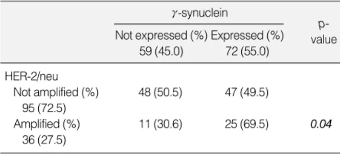
HER-2/neu 유전자가 과발현이 되지 않은 95예 중 47예(49.5
%)에서 -synuclein이 과발현되었으나, HER-2/neu 유전자가 과발현 된 36예 중에서는 25예(69.5%)에서 -synuclein이 과 발현되었다. HER-2/neu 유전자의 과발현과 -synuclein의 과발현이 통계적으로 유의한 상관관계(p=0.04)가 있음을 확인할 수 있었다(Table 3).
고 찰
유방암의 발생 및 진행은 다양한 유전적 변화에 의한 질적 그리 고 양적인 각각의 유전자 발현의 변이에 의해 이루어진다. 이러한 변이에 따른 유전자의 산물과 세포내 기능의 변화는 세포의 정상 대사를 방해하고 암 형성을 초래한다. 암종에서 과발현 혹은 미발 현되는 유전자를 동정하고 생물학적 기능을 밝히는 것은 악성 변
화의 과정을 이해하는데 중요하다. Synucleins 단백질군 중 - synuclein의 Western 분석을 통해 -synuclein이 정상 유방 조직과 I/II기 유관 상피암에서 발현되지 않고 III/IV기 유관 상피 암의 70%에서 발현된다는 연구결과가 보고된 후(9) 이러한 사실을 뒷받침하는 많은 연구들이 시행되었다. Ninkina 등(7)은 Nor- thern blotting과 Western blotting을 이용하여 유방암과 유 방암 세포주에서 -synuclein이 발현되고 정상 유방조직에서는 -synuclein이 발현되지 않는다는 것을 확인하였다. Liu 등(10) 은 79개 임상례의 RT-PCR 분석을 통해 -synuclein의 발현이 유방암의 병기에 특이적이라고 보고하였다.
본 연구는 면역조직화학염색을 통해 -synuclein의 과발현을 관찰하였다. 본 연구에서 암종의 크기와 -synuclein의 과발현 은 상관관계가 없었지만 액와 림프절 전이 상태와 -synuclein 의 과발현은 유의한 상관관계가 있었다. 또한 I/II기의 유방암보 다 III/IV기의 유방암에서 -synuclein이 과발현되는 결과를 얻 었다. 기존의 연구결과와 같이 -synuclein의 과발현이 유방암 의 병기에 특이적이라는 사실을 확인하였다.
침윤성 유방암에서 -synuclein이 높은 빈도로 발현되는 것 은 -synuclein이 유방암의 발생 및 진행에 영향을 미친다는 것 을 시사한다. 그러나 현재까지는 -synuclein이 정상 유방조직 에서 어떠한 기능을 갖고 있는지 또한 유방암에 어떻게 영향을 미 치는지는 거의 밝혀지지 않았다. Gupta 등(11)은 -synuclein 의 exon1에 위치하는 CpG island의 hypomethylation에 의해 -synuclein의 발현이 야기된다는 것을 보고하였다. Jiang 등 (12)은 -synuclein이 ER- signaling을 통하여 ligand 의존 성 세포의 성장에 영향을 미치는 것을 발견하였다. 현재까지의 연 구결과 -synuclein은 유방암의 발생과 진행에 다음과 같은 역 할을 할 것으로 생각되고 있다. 첫째, 암세포의 이동성 및 전이의 자극, 둘째, 항암제 유발 세포자살로부터 암세포의 보호이다.(13)
많은 연구를 통해 유방암에 대해 다양한 예후인자들이 제시되 었다. 이들 중 임상적으로 폭넓게 사용되는 것들은 ER, PR, Pro- liferating Cell Nuclear Antigen (PCNA), HER-2/neu 등이 다. HER-2/neu 과발현은 유방암의 악성도, 림프절 전이 등과 LN=lymph node; ER=estrogen receptor; PR=progesterone receptor.
Pathologic tumor
T1 (n=35) 16 (45.8) 19 (54.3)
T2 (n=85) 38 (44.7) 47 (55.3)
T3 (n=11) 5 (45.5) 6 (54.5) 0.9
Axillary LN
N0 (n=81) 46 (56.8) 35 (43.2)
N1 (n=17) 6 (35.3) 11 (64.7)
N2 (n=33) 7 (21.2) 26 (78.8) 0.002
Stage
I/II (n=99) 51 (51.5) 48 (48.5)
III/IV (n=32) 8 (25.0) 24 (75.0) 0.009
ER status
Negative (n=66) 27 (40.9) 39 (59.1)
Positive (n=50) 22 (44.0) 28 (56.0) 0.7 PR status
Negative (n=79) 36 (45.6) 43 (54.4)
Positive (n=37) 17 (45.9) 20 (54.1) 0.9 Table 2. -synuclein expression according to the clinicopa- thological factors
-synuclein Not expressed (%)
59 (45.0)
Expressed (%) 72 (55.0)
p- value
HER-2/neu
Not amplified (%) 48 (50.5) 47 (49.5) 95 (72.5)
Amplified (%) 11 (30.6) 25 (69.5) 0.04
36 (27.5)
Table 3.Correlation between -synuclein expression and HER- 2/neu amplification
-synuclein Not expressed (%)
59 (45.0)
Expressed (%) 72 (55.0)
p- value
관련이 있으며 불량한 예후와 강력한 상관관계가 있다.(14-16) HER-2/neu의 과발현을 측정하는 다양한 검사법이 있으며 그 방법마다 상이한 결과를 보이고 있다.(17-18) 최근에는 대부분의 연구실에서 면역조직화학염색으로 선별한 후에 FISH를 시행하 거나 단독으로 시행하고 있다.(19) HER-2/neu의 과발현은 현 재 재발성 유방암 및 전이를 동반한 유방암에서 trastuzumab (Herceptin)을 이용한 항암화학요법을 결정하는데 중요한 역할을 하고 있다.(20) Wu 등(8)은 -synuclein의 과발현이 유방암의 병기에 특이적이라는 사실을 확인한 연구에서 RT-PCR로 확인한 -synuclein의 과발현과 면역조직화학염색으로 확인한 HER- 2/neu의 과발현이 상관관계가 없다는 결과를 보고하였다.
본 연구에서는 -synuclein의 과발현과 HER-2/neu의 과발 현의 상관관계를 알아보기 위해 동일한 미세조직배열에서 -sy- nuclein에 대한 면역조직화학염색과 HER-2/neu에 대한 FISH 를 시행하였고 -synuclein의 과발현과 HER-2/neu의 과발현이 통계적으로 유의하게 상관관계가 있음을 확인하였다. -synu- clein의 과발현과 HER-2/neu의 과발현에 대한 본 연구와 Wu 등의 보고가 상이한 결과를 보였고 검사법의 차이에 의해 이러한 결과가나왔는지등에대해추가적인연구가필요할것으로보인다.
-synuclein의 과발현이 HER-2/neu의 과발현과 관련이 있 다는 본 연구의 결과는 -synuclein의 과발현이 유방암의 불량 한 예후와 연관이 있을 수 있다는 것을 시사한다. 따라서 -sy- nuclein의 과발현이 유방암에 있어서 예후 인자로서 유용성을 가질 것으로 기대된다. 현재까지 -synuclein의 과발현이 유방 암의 예후에 미치는 영향에 대한 장기간의 연구결과가 보고되지 않았으므로 향후 장기 추적 관찰을 통하여 -synuclein이 유방 암의 생존율, 국소재발 및 원격전이에 미치는 영향에 대한 연구가 더 필요할 것이다.
결 론
아직까지 국내에서 보고되지 않았던 유방암에서의 -synu- clein의 과발현과 임상병리학적 소견을 비교하여 다음과 같은 결 과를 얻을 수 있었다. -synuclein이 55%에서 과발현 되었고, 과 발현과 액와 림프절 전이 및 암종의 병기와 연관성이 있었으며 HER-2/neu 유전자 증폭과도 연관성이 있었다. -synuclein 의 과발현이 유방암의 예후에 밀접한 영향을 미칠 것으로 생각되 며 향후 장기 추적 관찰을 통한 연구가 필요할 것으로 생각된다.
참고문헌
1. Lavedan C. The synuclein family. Genome Res 1998;8:871-80.
2. George JM. The synucleins. Genome Biol 2002;3:REVIEWS3002.
3. Lavedan C, Leroy E, Dehejia A, Buchholtz S, Dutra A, Nussbaum RL, et al. Identification, localization and characterization of the hu- man -synuclein gene. Hum Genet 1998;103:106-12.
4. Takeda A, Mallory M, Sundsmo M, Honer W, Hansen L, Masliah E. Abnormal accumulation of NACP/ -synuclein in neurodegene- rative disorders. Am J Pathol 1998;152:367-72.
5. Ji H, Liu YE, Jia T, Wang M, Liu J, Joseph BK, et al. Identification of a breast cancer-specific gene, SNCG, by direct differential com- plementary DNA sequencing. Cancer Res 1997;57:759-64.
6. Clayton DF, George JM. The synucleins: a family of proteins involv- ed in synaptic function, plasticity, neurodegeneration and disease.
Trends Neurosci 1998;21:249-54.
7. Ninkina N, Alimova-Kost M, Paterson J, Delaney L, Cohen B, Imreh S, et al. Organisation, expression and polymorphism of the human persyn gene. Hum Mol Genet 1998;7:1417-24.
8. Wu K, Weng Z, Tao Q, Lin G, Wu X, Qian H, et al. Stage-specific expression of breast cancer-specific gene gamma-synuclein. Cancer Epidemiol Biomarkers Prev 2003;12:920-5.
9. Bruening W, Giasson BI, Klein-Szanto AJ, Lee VM, Trojanowski JQ, Godwin AK. Synucleins are expressed in the majority of breast and ovarian carcinomas and in preneoplastic lesions of the ovary.
Cancer 2000;88:2154-63.
10. Liu H, Liu W, Wu Y, Zhou Y, Xue R, Luo C, et al. Loss of epigene- tic control of synuclein-gamma gene as a molecular indicator of me- tastasis in a wide range of human cancers. Cancer Res 2005;65:7635- 43.
11. Gupta A, Godwin AK, Vanderveer L, Lu A, Liu J. Hypomethylation of the synuclein gamma gene CpG island promotes its aberrant exp- ression in breast carcinoma and ovarian carcinoma. Cancer Res 2003;
63:664-73.
12. Jiang Y, Liu YE, Goldberg ID, Shi YE. Gamma synuclein, a novel heat-shock protein-associated haperone, stimulates ligand-dependent estrogen receptor alpha signaling and mammary tumorigenesis. Can- cer Res 2004;64:4539-46.
13. Pan ZZ, Bruening W, Giasson BI, Lee VM, Godwin AK. -synu- clein promotes cancer cell survival and inhibits stress- and chemo- therapeutic drug-induced apoptosis by modulating MAPK athways.
J Biol Chem 2002;277:35050-60.
14. Borg A, Tandon AK, Sigurdsson H, Clark GM, Ferno M, Fuqua SA, et al. HER-2/neu amplification predicts poor survival in node-positive breast cancer. Cancer Res 1990;50:4332-7.
. .
15. Winstanley J, Cooke T, Murray GD, Platt-Higgins A, George WD, Holt S, et al. The long term prognostic significance of c-erb-2 in primary breast cancer. Br J Cancer 1991;63:447-50.
16. Paterson MC, Dietrich KD, Danyluk J, Dono K, Kanai T, Mori Tl.
Correlation between c-erb-2 amplification and risk of recurrent di- sease in node-negative breast cancer. Cancer Res 1991;51:566-7.
17. Nichols DW, Wolff DJ, Self S, Metcalf JS, Jacobs D, Kneuper-Hall R, et al. A testing lgorithm for determination of HER2 status in pa- tients with breast cancer. Ann Clin Lab Sci 2002;32:3-11.
18. Wang S, Saboorian MH, Frenkel E, Hynan L, Gokaslan ST, Ashfaq
R. Laboratory assessment of the status of Her-2/neu protein and oncogene in breast cancer specimens: comparison of immunohisto- chemistry assay with fluorescence in situ hybridisation assays. J Clin Pathol 2000;53:374-81.
19. Ross JS, Fletcher JA, Linette GP, Stec J, Clark E, Ayers M, et al.
The Her-2/neu gene and protein in breast cancer 2003: biomarker and target of therapy. Oncologist 2003;8:307-25.
20. McKeage K, Perry CM. Trastuzumab: a review of its use in the treatment of metastatic breast cancer overexpressing HER2. Drugs 2002;62:209-43.