pISSN: 0378-6471 eISSN: 2092-9374 http://dx.doi.org/10.3341/jkos.2013.54.2.357
= 증례보고 =
양측성 결막-각막 상피내 신생물의 성공적 치료 1예와 문헌고찰
최정열⋅이도형⋅김진형 인제대학교 의과대학 일산백병원 안과학교실
목적: 국내 최초로 양측성 결막‐각막 상피내 신생물(Conjunctival‐corneal Intraepithelial Neoplasia)을 진단 및 치료한 1예를 문헌고찰 과 함께 보고하고자 한다.
증례요약: 74세 남자환자가 4달전부터 시작된 양안의 이물감과 시력저하로 내원하였다. 내원시 환자의 교정시력은 우안 20/50, 좌안 20/30이었다. 세극등 검사에서 우안 7시에서 11시 방향과 좌안 3시에서 5시 방향 윤부를 포함하는 9 × 11 mm, 6 × 7.5 mm 크기의 각막‐결막 병소가 관찰되었다. 각막은 경계가 분명하고 불투명한 상피 증식이 보였고 구결막은 갈색의 색소 침착을 동반한 융기된 섬유혈관 조직이 관찰되었다. 진단 및 치료 목적의 절제생검과 결막의 절제 가장자리에 냉동치료를 시행하였다. 조직병리 소견에서 결막‐각막 상피내 신생물로 진단되었다. 술 후 교정시력은 양안 20/25로 호전되었고 8개월까지 재발 및 합병증은 관찰되지 않았다.
결론: 드물게 발생하는 양측성 결막-각막 상피내 신생물을, 최신 화학요법이 아닌 수술적 절제 및 보조적 냉동응고술의 전통적인 치료 법으로 종양의 제거 및 재발 방지에 성공한 1예를 보고하는 바이다.
<대한안과학회지 2013;54(2):357-364>
■ 접 수 일: 2012년 8월 31일 ■ 심사통과일: 2012년 10월 15일
■ 게재허가일: 2012년 12월 20일
■ 책 임 저 자: 김 진 형
경기도 고양시 일산서구 주화로 170 인제대학교 일산백병원 안과
Tel: 031-910-7240, Fax: 031-911-7241 E-mail: jhk90924@hanmail.net
* 이 논문은 인제대학교 연구기금의 보조를 받았음.
결막-각막 상피내 신생물(Conjunctival-corneal intra- epithelial neoplasia: CCIN)은 드물게 발생하나 안구 표면 의 종양 중에서는 가장 흔한 종양 중 하나이다.1,2종양세포 의 침윤 깊이에 따라 기저막을 침범하지 않은 결막-각막 상피내 신생물, 기저막을 넘어선 경우 침윤성 편평세포암 (invasive squamous cell carcinoma: SCC)이라고 하며 이 들 질환은 각각의 독립된 질환이 아니고 일련의 연속선상 에 위치한 질환으로 이들 모두를 안구표면 편평상피세포 암종(ocular surface squamous neoplasia: OSSN)이라고 한다.3 결막-각막 상피내 신생물은 서서히 진행하는 낮은 악성도를 가지는 전암성 질환으로, 원인은 불분명하며 태양 광선에 많이 노출된 60대 이상의 노년층, 사람면역결핍바이 러스(human immunodeficiency virus, HIV) 감염과 같은 면 역억제 상태, 사람유두종바이러스(human papillomavirus, HPV) 감염 등이 위험인자로 알려졌다.4,5 진단은 임상소견 을 통해 하는데 대부분 단안성으로 발생하며 양안성의 경
우는 드문 것으로 알려졌다.6주로 각막 윤부에서 호발하며 회백색의 융기물, 두꺼워진 각막 상피, 유두모양의 증식을 보이거나 색소 침착을 보인다.7진단 및 치료목적으로 병변 의 절제 및 조직병리학적 검사가 필요한데, 치료는 수술적 절제와 보조적 냉동치료를 함께 시행하는 것이 일차적 선택 법으로 알려졌으나8 최근에는 mitomycin C (MMC), 5-fluorouracil (5-FU), interferon 등을 이용한 국소적 화 학요법을 사용하는 것이 보고되고 있다.7,9,10
현재까지 국외의 보고 및 연구에서도 단안에 발생한 결 막-각막 상피내 신생물에 대한 보고는 다양하지만 양안에 발생한 경우는 드물 뿐 아니라, 그 빈도에 대한 보고도 정 확히 알려진 바가 없다. 특히 국내의 문헌보고에서 양측성 인 경우는 없었으며11-16또한 그 치료법으로 대부분의 증 례에서 수술적 절제와 보조적 화학 요법을 사용하였다. 이 에 저자들은 국내에서 처음 양측성으로 발생한 결막-각막 상피내 종양을 진단하였기에 보고하고 최신화학요법이 아 닌 수술적 절제 및 냉동치료라는 전통적인 치료법만을 사 용하여 종양의 제거와 8개월 추적 관찰 시 재발을 방지하는 데 좋은 결과를 얻었기에 국내 및 국외 문헌 고찰과 함께 이를 보고하고자 한다.
증례보고
74세 남자환자가 내원 4달전부터 시작된 양안의 이물감
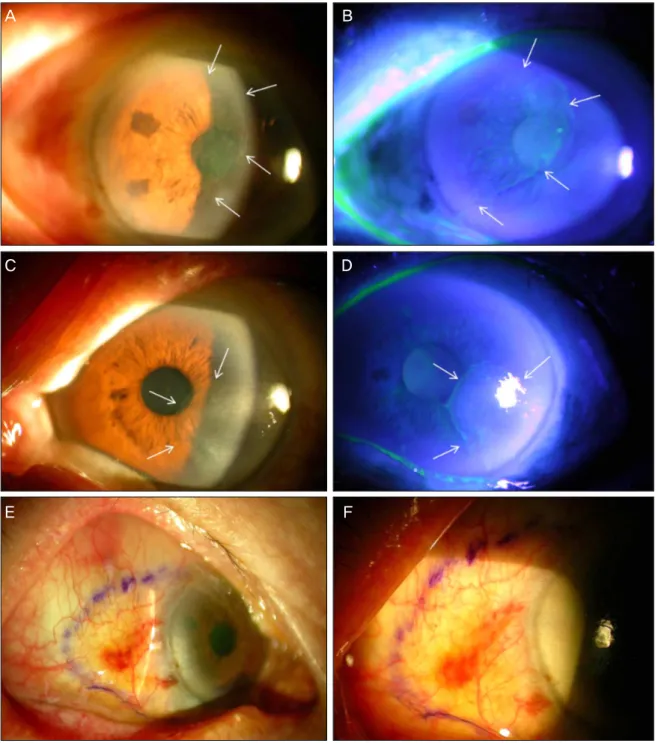
A B
C D
E F
Figure 1. (A, B) Anterior segment photograph of right eye before surgery. Well demarcated the opaque lesion covering half
of the corneal surface and extensively involving the visual axis. Note the hazy corneal component (arrows). (C, D) External photograph of the left eye showing a limbal to corneal gray and opaque lesion extending on to the visual axis from 3 to 5 O’clock position. (E, F) Relatively well demarcated yellowish elevated conjunctival lesion is seen within the violet marks.
과 시력저하를 주소로 내원하였다. 내원 당시 환자의 나안 시력은 우안 20/63, 좌안 20/30이었고 공기안압계로 측정 한 안압은 우안 13 mmHg, 좌안 12 mmHg였다. 굴절이상 은 우안 +1.50 Dsph = -1.50 Dcyl axis 90o, 좌안 +0.50 Dsph = -2.50 Dcyl axis 85o였고, 최대교정시력은 우안 20/50, 좌안 20/30이었다. 환자는 젊어서부터 오랫동안 농 부의 직업력을 갖고 있었으며 당뇨병과 고혈압을 포함한
전신적 질환은 없었으며 양안 수정체는 경도의 피질 및 핵 백내장을 보였다. 안저검사에서 특이소견은 관찰되지 않았다.
세극등 검사에서 우안의 비측 윤부를 중심으로 각막의 10시에서 7시 부위에 경계가 분명하고 불투명한 회백색의 병변이 관찰되었다. 각막의 병변은 표층에 국한되어 있었으 나 시축을 침범하고 있었고 비측 구석결막의 병변은 작은 크기의 섬유혈관조직과 함께 비교적 경계가 뚜렷하고 융기
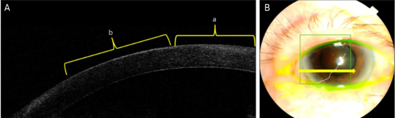
A B
Figure 2. (A) Anterior segment OCT image of the cornea between the normal cornea (a) and CIN lesion (b). Slightly thick-
ened epithelium (b) with hyperreflectivity is noticed compared with that of the normal corneal epithelium (a). (B) External photography showing corneal lesion of CCIN on the right eye. The arrow represents the direction of the OCT scan.A B
C D
Figure 3. (A) Histopathologic photograph of excised tissue from the cornea showing thickened epithelium with scattered dys-
keratotic cells and loss of polarity in the corneal epithelium (H-E stain, ×100). (B) Individual atypical cells with increased mi- totic figures, increased nucleocytoplasmic ratio within the epithelium are seen (H-E stain, ×400). (C) Immunohistochemical staining of excised corneal epithelium showing moderate dysplasia and scattered brown nuclear staining (arrows) conferred to the basal and parabasal layers (p53, ×400). (D) The excised conjunctival tissue shows loss of polarity and scattered dys- plastic cells with p53 positivity (arrows) within the epithelium (p53, ×200).되어 있었다. 좌안의 비측 각막에서 3시에서 5시 부위에 시 축의 일부를 침범한 각막 표층에 국한된 경계가 뚜렷하고 표면이 거친 병변이 관찰되었고 비측 구석 결막의 병변도 비교적 경계가 뚜렷하고 융기되어 있었다(Fig. 1). 전안부
빛간섭 단층촬영에서 양안 각막의 침범된 부위에는 정상 각막과 비교하였을 때 과반사(hyperreflective)된 상피 양 상을 보였으며 병변은 상피내에 국한되어 있었고 각막 기 질내로의 침윤은 관찰되지 않았다(Fig. 2). 수술 당일 교정
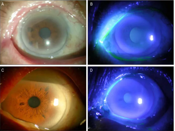
A B
C D
Figure 4. Anterior segment photograph of the right eye (A, B) and left eye (C, D) after 8 months of surgery exhibiting com-
plete remission of conjunctival and corneal intraepithelial neoplasia.시력은 우안 20/125, 좌안 20/50으로 저하되었으며 우안의 시축을 가리는 병변은 침범부위가 넓어져 있었다. 세극등 검사 시 병변의 경계를 미리 마커를 이용하여 표시한 뒤 조 직병리학적 확진 및 치료를 위해 결막과 각막의 생검 및 절 제술을 국소마취하에 시행하였다. 1:100,000 에피네프린을 혼합한 2% 리도카인으로 제거할 구결막 병변에 결막하 주 사로 국소마취한 후 Wescott scissiors를 사용하여 구결막 의 병변을 정상조직 1.5 mm를 포함하여 절제하였다. 각막 의 병변은 비버 수술칼(Beaver blade)을 사용하여 경계로 부터 정상 각막 조직 1 mm를 포함하여 단순 절제술을 시행 하였고 병변은 쉽게 각막으로부터 분리되었다. 보조적 냉동 응고술은 -85oC로 설정하여, 빠른 동결 후 냉동프로우브 (cryoprobe)에 결정이 형성되면 서서히 해동되어 얼음 결정 이 없어질 때까지 기다리는 동결-해동 주기(freeze-thaw cycle) 방식으로 하였다. 각막 윤부를 제외하고 결막 절제 경계면을 따라 각각 시행하였고, 새로운 냉동응고반을 만들 때 이전의 응고반과 교차하는 부분을 만들면서 진행시켰으 며 좌안은 5자리의 응고반, 우안은 8자리의 응고반을 각 2 회씩 시행하였다. 술 후 통증을 줄이고 결막 상피의 치유를
위해 양막 이식술을 같이 시행하였다. 양막은 시판중인 동 결보존양막(AmniSite-Cornea, Cryopreserved, Bioland, Ltd., Korea)을 사용하였으며 미리 실온에 놓아둔 양막을 좌안은 9 mm ×9 mm, 우안은 15 mm ×10 mm 크기로 잘라 구결막 노출부위에 맞추었다. 노출된 공막 위에 기질 층이 아래쪽으로 향하도록 놓은 뒤 10-0 나일론 봉합사로 단속봉합을 시행하였다.
병리조직학적 소견상 Hematoxylin & Eosin 염색에서, 각 막상피세포의 이형성과 재생성 변화(regenerative change) 가 관찰되었으며 결막의 병변은 상피세포의 이형성과 핵과 세포질간의 비율이 증가한 비전형적 세포들이 결막의 상피 층내에 국한되어 있었다. 각막및 결막 조직 모두에서 p53 면역화학염색의 양성을 보인 비전형적 세포가 상피층내에 국한되어 있는 것이 관찰되어 양안 모두 결막-각막내 신생 물로 진단되었다(Fig. 3).
술 후 2주째 각막 및 결막의 상피의 회복이 관찰되었고 술 후 1달째 굴절이상은 우안 +1.25 Dsph = -1.50 Dcyl axis 90o, 좌안 +0.50 Dsph = -2.00 Dcyl axis 90o였고, 최대교정시력은 양안 20/25로 회복되었다. 약 8개월간의
Table 1. Reports of bilateral cases of conjunctiva-corneal intraepithelial neoplasia
Authors (years) Age/Sex Number of cases Involved lesion Treatment protocol Recurr-ence Other findings Odrich et al20
(1991)
F/42 Bulbar & tarsal conjunctiva Right : Orbital exenteration Left : Surgical excision
(-) HPV* 16(+) M/58 3 cases Conjunctival-cornea Surgical excision with
cryotherapy
NA‡ HPV* 16(+)
M/57 Bulbar & tarsal conjunctiva (-) HIV†(-), HPV*
16(+) Tritten et al21
(1994)
M/46 1 case Conjunctiva & eyelid Surgical excision with cryotherapy
(-) HPV* (+)
Mahomed and Chetty25 (2002)
F/52 1 out of 41 cases NA NA‡ NA‡ HIV†(-)
Cervantes et al (2003)6
NA‡ 1 out of 287 cases Conjunctiva NA‡ NA‡ NA‡
Peksayar et al26 (2003)
NA‡ 2 out of 57 cases Conjunctiva Surgical excision with cryotherapy
NA‡ NA‡
Chen et al28 (2004)
M/73 1 case Bulbar conjunctiva Surgical excision with adjunctive perilesional interferon injection
(+) HPV* 16,18 (+)
Schechter et al7 (2008)
NA‡/72 2 out of 28 cases Conjunctiva- Cornea Topical MMC§ (-) Skin cancer (+)
NA‡/64 (-) NA‡
Gericke et al22 (2008)
NA‡/41 2 cases Conjunctiva- Cornea Surgical excision with Topical MMC§
(+) HPV* (-)
NA‡/60 Conjunctiva- Cornea Topical MMC§ (-) HIV†(-)
HPV* (+) Rundle et al19
(2010)
M/40 2 cases Conjunctiva Surgical excision with
cryotherapy and adjuvant topical MMC§
(+) HPV* : Not tested Asthma (+)
M/39 Bulbar conjunctiva Surgical excision with
cryotherapy and adjuvant topical MMC§
(+) HPV*(-)
Asthma (+)
*Human papilloma virus; †Human immunodeficiency virus; ‡Data point not available; §Mitomycin C.
경과 추적 관찰기간 동안 세극등 현미경 검사에서 재발 소 견은 관찰되지 않았다(Fig. 4).
고 찰
결막-각막 상피내 신생물은 드문 질환으로 안구 표면의 전암성 병변으로 노년에서 발생하는 질환이다. 발생 빈도는 인구 10만명당 0.03-195,17명의 발생빈도를 보인다고 알려 졌고, 대부분 편측에 발생한다.1 결막-각막 상피내 신생물 의 원인은 불분명하지만 다수의 발병요인으로 인해 발생한 다고 알려졌다. 관련된 위험 요인으로 직업과 관련된 태양 광 노출 및 자외선의 노출,18HIV 감염,4HPV 감염,3,5천식 과의 관련성,19 피부 및 내장기관의 종양과의 관련1,5 등이 발생 위험 요인으로 보고되었다. 양측성으로 발생한 결막- 각막 상피내 신생물에 대한 발생 빈도는 단안에 나타나는 것에 비해 더 드물며 이에 대한 문헌 연구는 많지 않다 (Table 1). 특히 우리나라에서는 본 증례가 양안성 결막- 각막 상피내 신생물로는 첫 문헌보고이며 지금까지 보고된 문 헌보고상의 증례는 모두 단안에 발생한 경우였다(Table 2).
따라서 양측성과 단측성의 결막-각막 상피내 신생물 발생 시 임상 특징, 위험인자, 연관 요소 등 치료에 참고할 만한 특이 사항들에 대해 제대로 알려진 바가 없어서 국내 및 국 외 문헌들을 고찰하였다. 양안성인 경우 HPV 감염과의 연 관성이 단안성에 비해 높으며,20,21젊은 환자에서 발생한 종 양의 경우 천식 및 아토피와의 관련성이 보고되었고,19 피 부신경염과 연관되어 발생한다고 알려졌다.22 Odrich et al20은 처음으로 양측성 결막 상피내 신생물의 발생에 HPV 감염과의 연관성을 보고하였고, 3명의 양측성 결막-각막 상피내 신생물의 보고에서 구결막, 각막뿐만 아니라 눈꺼풀 결막의 침범과 눈구석(fornix), 결막 조직의 섬유화 등 본 증례에 비해 광범위한 침범 소견을 보였다고 하였다. 천식 과 관련된 양측성 결막-각막 상피내 신생물 보고에서는 각 각 40세와 39세로 젊은 나이에 발생하였고,19 피부신경염 과 관련하여 발생한 양측성 결막-각막 상피내 신생물은 심 한 충혈과 검구 유착을 동반한 비전형적 형태를 보였다.22 본 증례의 경우 양측성으로 발생하였지만 천식, 아토피, 피 부신경염의 증상 및 과거력은 없었으며 HIV에 대한 면역혈 청검사에서 반응은 음성 소견을 보였다. 발생연령이 74세
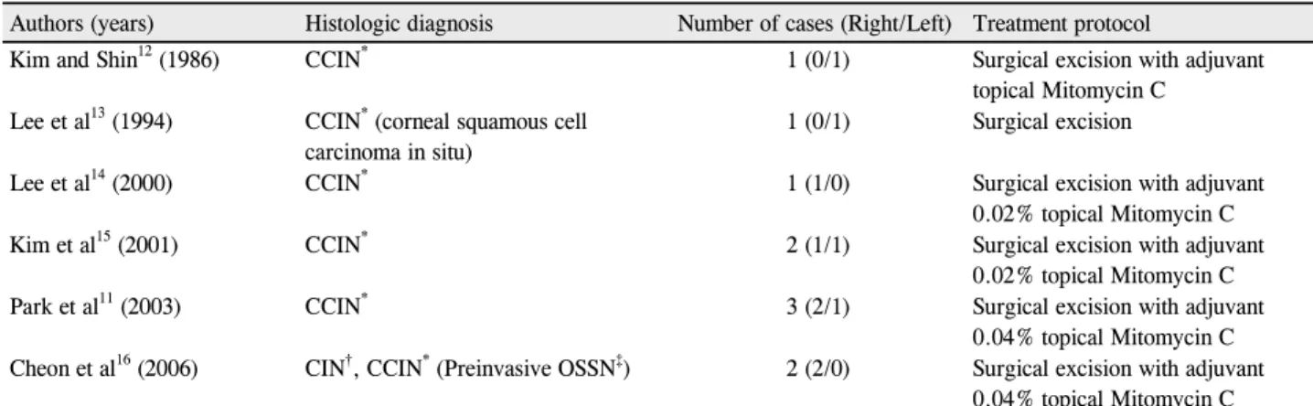
Table 2. Reported literatures of Conjunctival-Corneal Intraepithelial Neoplasia in Korea
Authors (years) Histologic diagnosis Number of cases (Right/Left) Treatment protocol
Kim and Shin12 (1986) CCIN* 1 (0/1) Surgical excision with adjuvant
topical Mitomycin C Lee et al13 (1994) CCIN* (corneal squamous cell
carcinoma in situ)
1 (0/1) Surgical excision
Lee et al14 (2000) CCIN* 1 (1/0) Surgical excision with adjuvant
0.02% topical Mitomycin C
Kim et al15 (2001) CCIN* 2 (1/1) Surgical excision with adjuvant
0.02% topical Mitomycin C
Park et al11 (2003) CCIN* 3 (2/1) Surgical excision with adjuvant
0.04% topical Mitomycin C Cheon et al16 (2006) CIN†, CCIN* (Preinvasive OSSN‡) 2 (2/0) Surgical excision with adjuvant
0.04% topical Mitomycin C
*Conjunctiva-corneal intraepithelial neoplasia; †Conjunctival intraepithelial neoplasia; ‡Ocular surface squamous neoplasia.
로 비교적 고령인 점과 결막과 각막에 걸쳐 경계가 분명하 고 혈관의 충혈이 심하지 않으며 눈꺼풀과 조직의 침범이 광범위하지 않고 국한되어 있는 점 등은 기존 단측성 결막 -각막 상피내 신생물과 별다른 임상적 차이가 없었다. 따 라서 결막-각막 상피내 신생물이 천천히 진행하고 별다른 증상이 없는 것을 감안하면, 전형적인 단안성으로 발생한 결막-각막 상피내 신생물 환자라 할 지라도 매 검사시마다 반대쪽 눈의 발병을 염두에 두고 세심한 관찰을 해야함을 알 수 있다. 다만 본 증례에서 HPV감염에 대한 검사가 진 행되지 못하여 본 증례의 양측성 발병 원인이 HPV 감염에 의한 것인 지에 대해서는 알 수는 없었다. 하지만 발생 원 인에 대한 HPV 감염과의 연관성은 일부에서는 그 연관성 을 찾을 수 없었다는 보고도 있으며23 단측성과 양측성 모 두에서 나타날 수 있다.24 환자의 직업력상 자외선과 광선 에 노출이 많은 농부인 점을 고려하면 오랜 직업력에서 오 는 자외선 노출과 외부 자극이 종양 발생원인으로 관련이 있을 것으로 생각한다. 양안성 결막-각막 상피내 신생물의 치료는 병변의 범위, 침범 깊이, 재발성 여부에 따라 결정되 었으며 단안성, 양안성에 있어 기존의 치료 방침과 차이를 보이지는 않았다.6,7,19-22,25,26
결막-각막 상피내 신생물은 actinic 형태와 미만성 (diffuse) 형태로 나눌 수 있으며 본 증례의 경우와 같은 actinic 형태는 60-70대 남성에 흔하며, 각막 윤부에 호발 하고 융기된 무경의(sessile) 병변으로 나타나는 것으로 알 려졌다.1,2미만성은 actinic 형태보다 드물지만 병변의 경계 가 분명하지 않아 절제의 범위를 정하기 어려워 완전한 수 술적 절제가 어렵다.1,2결막-각막 상피내 신생물의 치료로 서 병변의 수술적 절제만으로는 재발률이 높아 수술적 절 제 및 절제부위 변연부의 냉동치료가 전통적인 치료법으로 알려졌다.8본 증례는 actinic 형태로 좌안은 병변의 경계가 비교적 분명하였으며 윤부를 포함한 각막의 병변 크기가
작아 완전 절제와 보조적 냉동치료만으로 좋은 예후를 기 대할 수 있는 경우였다. 우안의 경우는 결막-각막 상피내 신생물의 병변이 각막의 50% 넓이에 해당하였으며 시축을 광범위하게 침범한 상태였으나 결막과 각막 병변의 경계가 명확하고 각막 윤부는 전체의 33%에 해당하여 수술 후 발 생할 수 있는 윤부 결핍, 지속적 각막 상피 결손 발생이 높 지 않았던 경우로 생각한다. 수술적 절제 후 보조요법으로 서의 냉동응고술은 종양의 절제 후 경계면에서 불완전한 제거로 인한 재발을 막는 장점이 있다. 본 증례의 경우는 냉동프로우브 끝의 직경이 2.5 mm 크기를 사용하였으므로 정상조직을 1-1.5 mm 포함하여 절제한 수술적 치료의 절 제범위를 추가적으로 1.25 mm 확장시키는 효과와 함께 국 소적으로 남아있던 종양세포들의 파괴를 유발하였을 것으 로 생각한다.
결막-각막 상피내 신생물 병변의 침범이 광범위하거나 다수의 병소를 가진 경우, 미만성 형태인 경우, 잦은 재발을 보였던 경우는 수술적 절제가 어렵거나 절제의 경계를 명 확히 알 수 없다. 종양의 재발은 변연부에서 불완전한 절제 로 인해 발생하므로 최근에는 절제 후 변연부의 상태를 고 려하지 않아도 되는(margin independent) MMC, 5-FU, interferon 등을 이용한 국소적 화학요법을 사용하여 좋은 결과를 보인 연구들이 보고되었고 실제로 임상에서 많은 경우 우선적으로 고려하는 치료법이기도 하다.7,9,10 그러나 결막-각막 신생물의 치료로서의 국소 화학요법은 종양세 포에 선택적으로 비가역적인 손상을 줄 수 있는 최소 용량 에 대해 정해진 바가 없으며, MMC의 사용에 있어서도 사 용 농도는 0.001-0.04%로 다양하게 보고되고 있다. 또한 국소 화학요법으로 사용할 점안액의 치료 중단 시점과 점 안액을 사용하는 방법에 대한 기준도 명확하지 않으며 연 구자에 따라 다르다.7,9,10,27국소 화학요법은 치료기간이 수 술적 절제 및 냉동치료에 비해 상대적으로 길고 약물사용
에 따른 안구 자극증상, 결막 충혈, 각막 미란, 유루증, 검구 유착 등의 합병증 발생에 대한 추적관찰이 자주 필요하다.
따라서 수술적 절제 및 냉동치료는 국소화학요법에 비해 치료기간이 짧고 안구 전체에 대한 부작용이 적은 장점이 있다.
국소화학요법은 과거 중심 시축을 침범하여 수술 후 중 심시력의 장애를 초래할 수 있는 경우 치료적 선택법으로 제시된 바 있다.11본 증례의 우안은 광범위한 시축을 침범 하고 있었으며 전체 각막의 약 50%에 해당하는 병변의 침 윤을 보여 국소화학요법을 통해 종양의 관해를 유도해 볼 수 있는 경우에 해당한다. 반면에 윤부에서의 병변은 범위 가 7시에서 11시 방향에 걸쳐 존재하므로 윤부 손상은 각 막침범범위에 비해 많지 않은 편에 속하므로 본 증례의 경 우처럼 수술적 절제를 시행하고 추가적 국소화학치료 혹은 냉동치료를 시도해 보는 방법 또한 가능할 것이다.3 본 증 례의 경우는 술 전 전안부 빛간섭 단층촬영을 통해 병변이 상피 내에 국한되어 있음을 알 수 있었으며 각막기질로의 침윤이 관찰되지 않았다. 따라서 전안부 빛간섭 단층촬영소 견을 참고하여 각막 상피내 병변의 제거 후 각막의 혼탁 없 이 상피의 재수복이 정상적으로 이루어 지면 시력 향상을 기대할 수 있다고 판단하였기에 화학요법대신 수술적 절제 의 방법을 사용하였고 술 후 환자의 교정시력은 20/25로 호전되었다. 병변이 중심 시축을 침범하였더라도 기질로의 침윤 없이 상피 내에 국한되며 경계가 분명하여 절제가 가 능하고, 절제 후 각막, 결막 조직의 소실이 크지 않은 경우, 수술적 절제 및 보조적 냉동치료를 여러가지 치료법 중 하 나로 고려해 볼 수 있을 것으로 생각한다. 그러나 병변이 미만성으로 완전한 절제가 불가능하거나 수술적 치료로 과 도한 각막, 결막의 상피 소실이 우려되는 경우, 여러 차례 재발한 경우 등에서는 국소적 화학 요법이 우선적으로 고 려되어야 할 것이다. 따라서 전통적 치료방법을 완전히 배 제하기보다는 최신요법과 함께 각 증례의 상태에 맞는 치 료법을 선택하는 것이 중요할 것으로 생각한다. 하지만 본 증례가 술 후 비교적 짧은 기간인 8개월 경과 관찰 결과이 고, 1예에 불과하므로 향후 재발에 대한 더 장기적인 추적 관찰과 추가적인 증례보고가 필요할 것이다.
저자들은 국내에서 처음으로 양측성으로 발생한 결막- 각막 상피내 종양을 전통적인 수술적 절제 및 냉동치료만 을 통해 종양의 제거와 술 후 시력이 호전되는 좋은 결과를 얻었기에 국내외 문헌 고찰을 통해 이를 보고하는 바이다.
참고문헌
1) Erie JC, Campbell RJ, Liesegang TJ. Conjunctival and corneal in- traepithelial and invasive neoplasia. Ophthalmology 1986;93:176-83.
2) Waring GO 3rd, Roth AM, Elkins MB. Clinical and pathologic de- scription of 17 cases of corneal intraepithelial neoplasia. AM J Ophthalmol 1984;97:547-59.
3) Basti S, Macsai MS. Ocular surface squamous neoplasia: a review.
Cornea 2003;22:687-704.
4) Waddell KM, Lewallen S, Lucas SB, et al. Carcinoma of the con- junctiva and HIV infection in Uganda and Malawi. Br J Ophthalmol 1996;80:503-8.
5) Lee Ga, Hirst LW. Retrospective study of ocular surface squamous neoplasia. Aust N Z J Ophthalmol 1997;25:269-76.
6) Cervantes G, Rodríguez AA Jr, Leal AG. Squamous cell carcinoma of the conjunctiva: clinicopathological features in 287 cases. Can J Ophthalmol 2002;37:14-9.
7) Schechter BA, Koreishi AF, Karp CL, Feuer W. Long-term fol- low-up of conjunctival and corneal intraepithelial neoplasia treated with topical interferon alfa-2b. Ophthalmology 2008;115:1291-6.
8) Krachmer, Mannis, Holland. Cornea, 2nd ed. Philadelphia: Elsevier Mosby, v. 2. 2005;1773-80.
9) Frucht-Pery J, Rozenman Y. Mitomycin C therapy for corneal in- traepithelial neoplasia. Am J Ophthalmol 1994;117:164-8.
10) Yeatts RP, Ford JG, Stanton CA, Reed JW. Topical 5-fluorouracil in treating epithelial neoplasia of the conjunctiva and cornea.
Ophthalmology 1995;102:1338-44.
11) Park HJ, Lee JE, Lee JS, Park DY. Treatment of conjunctival- cor- neal intraepithelial neoplasia with topical mitomycin C. J Korean Ophthalmol Soc 2003;44:1924-30.
12) Kim JW, Shin DE. A case of conjunctival carcinoma in situ. J Korean Ophthalmol Soc 1986;27:943-7.
13) Lee JS, On KK, Kim JD. A case of corneal squamous cell carcino- ma in situ. J Korean Ophthalmol Soc 1994;35:206-9.
14) Lee DH, Jang JH, Oh JY, Kim JS. A case of conjunctival intra- epithelial neoplasia (CIN) misdiagnosed as atypical pterygium. J Korean Ophthalmol Soc 2000;41:2750-4.
15) Kim JH, Kang HS, Kim IC. The effect of topical mitomycin C after excisional biopsy in conjunctival-corneal intraepithelial neoplasia:
two cases. J Korean Ophthalmol Soc 2001;42:1102-10.
16) Cheon HC, Lee DY, Park WC, Ahn HB. Ocular surface squamous neoplasia. J Korean Ophthalmol Soc 2006;47:1920-8.
17) Sun EC, Fears TR, Goedert JJ. Epidemiology of squamous cell conjunctival cancer. Cancer Epidemiol Biomarkers Prev 1997;6:
73-7.
18) Newton R, Ferlay J, Reeves G, et al. Effect of ambient solar ultra- violet radiation on incidence of squamous-cell carcinoma of the eye. Lancet 1996;347:1450-1.
19) Rundle P, Mudhar HS, Rennie I. Conjunctival intra-epithelial neo- plasia occurring in young patients with asthma. Eye 2010;24:1182-5.
20) Odrich MG, Jakobiec FA, Lancaster WD, et al. A spectrum of bi- lateral squamous conjunctival tumors associated with human pap- illomavirus type 16. Ophthalmology 1991;98:628-35.
21) Tritten JJ, Beati D, Sahli R, Uffer S. [Bilateral conjunctivo-palpebral tumor in an immunocompetent man cause by human papilloma vi- rus]. Klin Monbl Augenheilkd 1994;204:453-5.
22) Gericke A, Pitz S, Strempel I, Sekundo W. [Bilateral ocular surface squamous neoplasia and neurodermatitis. Two cases with different courses]. Ophthalmologe 2008;105:1142-5.
23) Guthoff R, Marx A, Stroebel P. No evidence for a pathogenic role of human papillomavirus infection in ocular surface squamous ne- oplasia in Germany. Curr Eye Res 2009;34:666-71.
=ABSTRACT=
Successful Treatment of Bilateral Conjunctival-Corneal Intraepithelial Neoplasia: Case Report and Review of the Literature
Jung Yeol Choi, MD, Doh Hyung Lee, MD, PhD, Jin Hyoung Kim, MD, PhD
Department of Ophthalmology, Inje University Ilsan Paik Hospital, Inje University College of Medicine, Goyang, Korea
Purpose: We report a case of successfully treating bilateral conjunctival-corneal intraepithelial neoplasia (CCIN) with sur- gical excision and adjunctive cryotherapy.
Case summary: A 74-year-old male visited our clinic for bilateral foreign body sensation and decreased visual acuity. His initial best corrected visual acuity was 20/50 in the right eye and 20/30 in the left eye. The right eye showed a 9 mm × 11 mm sized, gray-opaque limbal lesion from approximately the 7-o’clock position to the 11-o’clock position with spreading onto the cornea and conjunctiva. Biomicroscopy revealed a 6 mm × 7.5 mm sized minimally elevated, opaque lesion from the 3-o’clock to the 5-o’clock position extending to the central cornea in the left eye. The corneal lesion was well demar- cated, opaque, and minimally elevated with bilateral focal pigmentation. Conjunctival lesions were finely vascularized and slightly elevated with melanocytic pigmentation. An excisional biopsy was performed to confirm the diagnosis and for ther- apeutic purposes, followed by an adjunctive cryotherapy. Postoperative corrected visual acuity improved up to 20/25 bi- laterally and the patient had no recurrence 8 months after surgery.
Conclusions: Bilateral conjunctival-corneal intraepithelial neoplasia is a rare condition. We report successful treatment and control of recurrence in a patient with bilateral conjunctival-corneal intraepithelial neoplasia using conventional surgi- cal excision and adjuvant cryotherapy rather than topical chemotherapy.
J Korean Ophthalmol Soc 2013;54(2):357-364
Key Words: Bilateral, Conjunctival-corneal intraepithelial neoplasia, Cryotherapy, Surgical excision
Address reprint requests to Jin Hyoung Kim, MD, PhD
Department of Ophthalmology, Inje University Ilsan Paik Hospital
#170 Juhwa-ro, Ilsanseo-gu, Goyang 411-760, Korea
Tel: 82-31-910-7240, Fax: 82-31-911-7241, E-mail: jhk90924@hanmail.net 24) McDonnell JM, McDonnell PJ, Sun YY. Human papillomavirus
DNA in tissues and ocular surface swabs of patients with con- junctival epithelial neoplasia. Invest Ophthalmol Vis Sci 1992;
33:184-9.
25) Mahomed A, Chetty R. Human immunodeficiency virus infection, Bcl-2, p53 protein, and Ki-67 analysis in ocular surface squamous neoplasia. Arch Ophthalmol 2002;120:554-8.
26) Peksayar G, Altan-Yaycioglu R, Onal S. Excision and cryosurgery in the treatment of conjunctival malignant epithelial tumours. Eye
2003;17:228-32.
27) Rozenman Y, Frucht-Pery J. Treatment of conjunctival intra- epithelial neoplasia with topical drops of mitomycin C. Cornea 2000;19:1-6.
28) Chen HC, Chang SW, Huang SF. Adjunctive treatment with inter- ferona alpha-2b may decrease the risk of papilloma-associated conjunctival intraepithelial neoplasm recurrence. Cornea 2004;23:
726-9.